
R.M. Romeilah
Department of Biochemistry, Faculty of Agriculture, Cairo University, P.O. Box 12613, Gamma St, Giza, Egypt
Asian Journal of Biochemistry
Year: 2016 | Volume: 11 | Issue: 2 | Page No.: 104-117


ABSTRACT
Chemical compositions of hydro-distilled Myrtus communis and Origanum vulgare leaves essential oils were examined by gas chromatography-mass spectrometry (GC/MS). Myrtus communis oil was found to contain thirty-one compounds, the major components being α-Pinene (26.99%) and 1,8-Cineol (20.37%), while O. vulgare oil analysis reached fifty-five compounds, the major components being thymol (25.48%) and carvacrol (10.29%). The 2,2-diphenyl-1-picrylhydrazyl (DPPH) free radical scavenging activities of M. communis and O. vulgare essential oils at various concentrations ranging from 25-200 μg mL–1 were determined. The 50% inhibition concentration (IC50) of O. vulgare essential oil (81.99 μg mL–1) was higher than Myrtus communis essential oil (122.78 μg mL–1). Human promyelocytic leukemia cell lines (HL-60, NB4) and Ehrlish Ascites Carcinoma Cells (EACC) were incubated in vitro with different levels of two essential oils ranged from 25-200 μg mL–1 for viability test and the percentages of dead cells were determined. The 50% Lethal Concentration (LC50) values indicated that the anticancer activity of O. vulgare essential oil was higher than M. communis essential oil against HL-60 and NB4 cell lines while The LC50 values indicated that the anticancer activity of M. communis essential oil was higher than O. vulgare essential oil against EACC. In in vivo assay, pre-initiation treatments with the both essential oils were more effective than initiation and post-initiation treatments, respectively on the tumor (EACC) transplanted female mice (decrease total EACC number and increase dead cells). Biological effects of both essential oils on normal mice indicated that all the obtained values in all experimental animals were within the normal range.
PDF Abstract XML References Citation
Received: October 01, 2015;
Accepted: December 15, 2015;
Published: February 15, 2016
How to cite this article
R.M. Romeilah, 2016. Chemical Compositions, Antioxidant, Anticancer Activities and Biological Effects of Myrtus communis L. and Origanum vulgare Essential Oils. Asian Journal of Biochemistry, 11: 104-117.
DOI: 10.3923/ajb.2016.104.117
URL: https://scialert.net/abstract/?doi=ajb.2016.104.117
DOI: 10.3923/ajb.2016.104.117
URL: https://scialert.net/abstract/?doi=ajb.2016.104.117
INTRODUCTION
It is well known that Reactive Oxygen Species (ROS) have the potential to cause serious health problems, including cancer (Cozzi et al., 1997; Perera et al., 2008). Since antioxidants can pair with these ROS more quickly than body chemicals can, one way to diminish, the effect of ROS is to take antioxidants into the body. Medicinal plants are potential sources of natural compounds with biological activities and therefore attract the attention of researchers worldwide. Information on the antioxidant activity and phenolic compounds of medicinal plants associated with anticancer properties is scarce. Free radicals are active chemical species involved in biological processes whose high existence can give rise to several diseases (cancer, etc.) (Hung et al., 2006). Many people suffer from such diseases, especially from middle age onwards. Consumption of plants with high contents of antioxidant compounds such as polyphenols and flavon4oids should feature in everyone’s diet, since such species have high potential to diminish the negative effects of free radicals as a result of their electronic and molecular structures (Koldas et al., 2015).
Myrtle (Myrtus communis L., Myrtaceae) is a well-known medicinal plant that has been used worldwide in traditional medicine. Myrtaceae family includes 100 genera and 3000 species. Myrtus genus belongs to this family of evergreen shrubs or small trees, which grow up to 5-m tall spontaneously (Sumbul et al., 2011), it is widespread in Mediterranean woodlands, maquis and garrigues. Essential oil from leaves, flowers and fruits of the plant is widely used as a functional ingredient in the food, liquor and cosmetic industries. It tends to vary in composition and it is mainly used for the treatment of lung disorders. It has been found to also have antibacterial and antioxidant activities (Yadegarinia et al., 2006; Gardeli et al., 2008). The leaves of myrtle contain tannins, flavonoids such as quercetin, catechin and myricetin derivatives and volatile oils (Baytop, 1999; Romani et al., 1999). One of the main constituents of myrtle essential oil is 1,8-cineole (Bradesi et al., 1997). The essential oil obtained from the leaves by steam distillation is also important in perfumery (Baytop, 1999). The oil in leaves of M. communis growing in Turkey contains 1,8-cineole, linalool, myrtenyl acetate and myrtenol as major components (Ozek et al., 2000).
Origanum vulgare L. is a perennial herbaceous plant belonging to the family Lamiaceae whose center of differentiation is located in the Mediterranean area. The common name of this species is oregano. The scientific name of this plant is derived from two Greek terms-oros (mountain) and ganos-(joy, beauty, decoration).
Origanum vulgare is probably one of most widely used aromatic plant, whose essential oils are particularly rich in mono- and sesquiterpenes. Oregano essential oils have been shown to possess antioxidant, antibacterial, antifungal, diaphoretic, carminative, antispasmodic and analgesic activities (Sahin et al., 2004; Faleiro et al., 2005; Souza et al., 2007; Sarac et al., 2009; Da Costa et al., 2009; Tommasi et al., 2009) and among these, the antimicrobial potential is of special interest. In recent years, a large number of researches have reported the efficacy of essential oils from several Origanum species against a panel of bacterial strains (Dadalioglu and Evrendilek, 2004; Baydar et al., 2004; Vardar-Unlu et al., 2007; Bouhdid et al., 2008; Baser, 2008) identified carvacrol as the main responsible for this biological activity (Baser, 2008).
The aim of the present study was to evaluate antioxidant activity using DPPH radical scavenging assay and anticancer activity against two human promyelocytic leukemia cell lines (HL-60 and NB4) and experimental animals model cancer cells (Ehrlish ascites carcinoma cells, EACC) of Myrtus communis L. (Myrtle) leaves and Origanum vulgare (Oregano) leaves essential oils. In addition the present study aimed to investigate the cytotoxicity (if any) induced by these essential oils.
MATERIALS AND METHODS
Plant material: The dried leaves of Myrtus communis L. (Myrtle) belonging to the family Myrtaceae were collected from the garden of Agriculture Faculty, Cairo University and the dried leaves of Origanum vulgare (Oregano) belonging to the family Lamiaceae, were purchased from experimental station of medicinal plants, Faculty of Pharmacy, Cairo University, Egypt. The plant samples were kindly identified by Dr. Mohamed Osama El-Segaee, Professor of Taxonomy, Faculty of Agriculture, Cairo University.
Essential oil extraction: One hundred grams of dried leaves of M. communis L. and dried leaves of O. vulgare were hydro-distilled in a Clevenger type apparatus according to Council of Europe (1997). The essential oils were dried over anhydrous sodium sulphate, stored in a dark glass bottle and kept at 4°C until analysis. The amount of oil obtained from plant material was calculated as:
![]()
GC/MS analysis of essential oils: The essential oils were analyzed by GC-MS according to Adams (1989). The GC/MS analysis was performed on a Thermoquest-Finnigan Trace GC-MS equipped with a DB-5 (5% phenyl) methylpolysiloxane column (60 m\0.25 mm i.d., film thickness 0.25 μm). The injection temperature was 220°C and the oven temperature was raised from 40°C (3 min hold) to 250°C at a rate of 5°C/min, then held at 250°C for 2 min, transfer line temperature was 250°C. One microgram of sample was injected and helium was used as the carrier gas at a flow rate of 1.0 mL min–1. The mass spectrometer was scanned over the 40-500 m z–1 with an ionizing voltage of 70 eV and identification was based on standard mass library that National Institute of Standards and Technology (NIST Version 2.0) to detect the possibilities of essential oil components.
Antioxidant activity of essential oils using DPPH radical scavenging assay: Radical scavenging activity of plant essential oils against the stable 2,2-diphenyl-1-picrylhydrazyl (DPPH) radical was determined spectrophotometrically (Brand-Williams et al., 1995). The colorimetric changes (from deep-violet to light-yellow), when DPPH∙ is reduced, were measured at 517 nm on a UV/visible light spectrophotometer. The antioxidant activity of essential oils were measured in terms of hydrogen donating or radical scavenging ability, using the stable radical DPPH. Fifty microliters of various concentrations (25, 50, 75, 100 and 200 μg mL–1) of the essential oils in dimethyl sulphoxide (DMSO) as well as ascorbic acid (as standard antioxidant compound) were put into appropriate tubes and 5 mL of 0.004% methanolic solution of DPPH∙ was added to each tube to give final concentrations (25, 50, 75, 100, 200 μg mL–1). Absorbance measurements commenced immediately. The decrease in absorbance at 517 nm was determined after 1 h for all samples. Methanol was used to zero the spectrophotometer. Absorbance of the DPPH radical without antioxidant, i.e. the control, was measured. Special care was taken to minimize the loss of free radical activity of the DPPH radical stock solution. The DPPH radical by the samples was calculated according to the formula of Yen and Duh (1994):
![]()
where, AC(o) is the absorbance of the control at t = 0 min and As(t) is the absorbance of the antioxidant at t = 1 h.
The percentage of scavenging activity was plotted against the essential oil concentrations to obtain the inhibitory concentration (IC50), defined as the essential oil concentration necessary to cause 50% scavenging. Tests were carried out in triplicate.
Anticancer activity of essential oils
Cell growth and viability assay: Three types of cancer cell lines were used in this study, human promyelocytic leukemia cell lines (HL-60 and NB4) and experimental animals model cancer cells (Ehrlish ascites carcinoma cells, EACC).
In vitro assay for anticancer activity
Cell lines: Human promyelocytic leukemia cell lines (HL60 and NB4) obtained from American Type Culture Collection (ATCC). All of these cells were maintained in RPMI-1640 supplemented with 10% FBS, 2 mmol L–1 L-glutamine, penicillin (100 U mL–1) and streptomycin (100 mg mL–1) in a humidified atmosphere of 5% CO2 at 37°C for 24 h after that the cell counts were determined. After this period the cell viability was evaluated using trypan blue technique.
On the other hand, experimental animals model cancer cell line (EACC) was maintained in the National Cancer Institute (NCI) Cairo, Egypt in female Swiss albino mice by weekly intraperitoneal (i.p) transplantation of 2.5×10⁶ cells. Similar line was proceeded in our department for the same cells. For in vivo and in vitro assays, the cells were taken from tumor transplanted animals after 7 days of transplantation then the number of cells/mL was calculated by using appropriate microscope counting technique (≈2x10⁷ cells mL–1).
Trypan blue method: The viability percentage of cancer HL60 and NB4 cells were measured by the modified cytotoxic trypan blue-exclusion technique of Bennett et al. (1976). Before the assay, 2x10⁵ cells mL–1 were seeded on 96-well plate and after that the viability percentage of cancer cells were measured by treated with different volumes of examined essential oils to give final concentrations of 25, 50, 75, 100, 200 μg mL–1. The plate was incubated at 37°C for 24 h under 5% CO2. The final volume in each experiment was made up to 100 μL with the media containing 1% dimethyl sulphoxide (DMSO). Control cells were treated with the equivalent amount of vehicle DMSO and then an equal volume of 0.4% trypan blue were added to each experiment and left to stand for 5 min at room temperature. Ten microliter of stained cells were add in a hemocytometer slide and the number of viable (unstained) and dead (stained) cells were counted. Each experiment was carried out in triplicate. The percentage of live cells calculated according to the following equation:
![]()
On the other hand , the EACC cells were taken from tumor transplanted animals, then centrifuged at 1000 rpm for 5 min and then washed with saline. The number of cells needed to the test was prepared by suspending the cells in the appropriate volume of saline. The culture medium used was prepared using RPMI 1640 media, 10% fetal bovine serum, 2 mmol L–1 glutamine, penicillin (100 U mL–1) and streptomycin (100 μg mL–1). The viability percentage of tumor cells was measured after incubation with the essential oil as well as saline and DMSO as control. Two milliliter of medium containing EACC (2×10⁶ cells) were transferred into a set of tubes, then different volumes of examined essential oil were added into the appropriate tube as well as control to give final concentrations 25, 50, 75, 100 and 200 μg mL–1. The tubes were incubated at 37°C under 5% CO2 for 12 h, then the viability percentage of tumor cells were measured by the method of Bennett et al. (1976) as described before. Each experiment was carried out in triplicate. The percentage of dead cells of each cell line was plotted against the essential oil concentrations to obtain the "50% lethal concentration" LC50, defined as the essential oil concentration necessary to cause 50% death.
In vivo assay for anticancer activity
Animals: Forty eight healthy female Swiss albino mice weighting 20-25 g (7-8 weeks old) were used throughout this experiment. The animals were purchased from the animal house of Helwan Station for Experimental Animals, Helwan, Egypt. They were raised in the animal house of Biochemistry Department, Faculty of Agriculture, Cairo University, Giza, Egypt. The animals were housed in polyethylene cages in groups of six mice per cage in a controlled environment condition (25±2°C, 50-60% relative humidity and 12 h light-dark cycle). All animals were fed standard pellet diet and water ad libitum for two weeks (adaption period). This study was carried out in strict accordance with the recommendations in the Guide for the Care and Use of Laboratory Animals of the National Institutes of Health. The protocol was approved by the Committee on the Ethics of Animal Experiments of the University of Cairo.
Experimental design: The animals were divided into eight groups and each group contained six mice. Group I served as normal control animals and was given corn oil orally for 8 weeks. Group II animals (tumor control) were transplanted intraperitoneal cavity with EACC at 1×10⁶ cells (0.2 mL). Group III mice were treated with EACC (as in Group II) and M. communis essential oil (10 mg kg–1 b.wt., dissolved in corn oil orally). Myrtus communis treatment started after 24 h of EACC transplantation (initiation treatment) and continued daily for the end of the experiment. Animals in Group IV for the post-initiating studies were treated with EACC (as in Group II) and after one week of tumor transplantation (tumor induction period) mice were treated daily with M. communis essential oil (10 mg kg–1 b.wt., dissolved in corn oil orally) till the end of the experiment (post-initiation treatment). Animals in Group V for pre-initiating studies were treated daily with M. communis essential oil (10 mg Kg–1 b.wt., dissolved in corn oil orally) for 2 weeks followed by EACC (as in Group II). M. communis treatment continued till the end of the experiment (pre-initiation treatment). Groups VI, VII and VII were treated as in Groups III, IV and V, respectively, but with O. vulgare essential oil (20 mg kg–1 b.wt., dissolved in corn oil orally) instead of M. communis essential oil. The initiation, post-initiation and pre-initiation treatments were used to study the chemopreventive and/or chemotherapeutic efficacies of M. communis and O. vulgare essential oils in the experimental animals.
Antitumor effect of both essential oils was assessed by observing the changes with respect to EACC, tumor number (all, viable and dead cells) was counted after 12 days of EACC transplantation (Bennett et al., 1976). The Mean of Survival Time (MST) of each group consisting of 6 mice was monitored by recording the mortality daily. The MST of the treated groups were compared with that of the tumor control group to calculate the Increase in Lifespan (ILS) using the following formula according to Rajkapoor et al. (2004):
![]()
where, T is the MST of treated group and C is the MST of tumor control group.
Lactate dehydrogenase (LDH) activity was determined in the supernatant of tumor cell suspension (EACC) according to Legrand et al. (1992) and Young (2001).
Biological effects of M. communis and O. vulgare essential oils
Animals: Eighteen male Swiss albino mice weighting 20-25 g (7-8 weeks old) were used throughout this experiment. Animals were obtained and adapted as in the previous experiment. The animals were divided into three groups and each group contained six animals. Group A (normal control) was given corn oil orally. Group B (M. communis) animals were treated daily with M. communis essential oil (10 mg kg–1 b.wt., dissolved in corn oil orally) and Group C (O. vulgare) animals were treated daily with O. vulgare essential oil (20 mg kg–1 b.wt., dissolved in corn oil orally) for three months to study the cytotoxicity (if any) induced by these essential oils. At the end of the experimental period (3 months), the animals were killed by cervical decapitation. Blood was collected. Serum was separated by centrifugation at 2500 rpm at 37°C for 15 min. This study was carried out in strict accordance with the recommendations in the Guide for the Care and Use of Laboratory Animals of the National Institutes of Health. The protocol was approved by the Committee on the Ethics of Animal Experiments of the University of Cairo.
Biochemical analyses: Serum glucose was determined according to Trinder (1969), serum total soluble proteins and albumin were determined according to Hoffman (1966) and Tietz (1995), respectively, but serum globulin was calculated by the difference between total protein and albumin. Serum total cholesterol and triglycerides were determined according to Allain et al. (1974) and Fossati and Prencipe (1982), respectively.
Serum uric acid, urea and creatinine contents were determined according to Tietz (1995), Martin (2003) and Faulkner and King (1976), respectively. Aspartate aminotransferase (AST) and alanine aminotransferase (ALT) activities were measured colorimetrically in serum according to the method described by Reitman and Frankel (1957). Serum alkaline phosphatase was determined according to the method of Kind and King (1954). Lactate dehydrogenase (LDH) activity in serum was determined according to the method of Young (2001).
Statistical analysis: Statistical analyses (standard deviation "SD" and standard error "SE") was carried out according to Fisher (1970). LSD (Least significant difference) test was used to compare the significant differences between means of treatment (Waller and Duncan, 1969). The statistical package for social science (SPSS., 2014) program version was used for all analysis.
RESULTS AND DISCUSSION
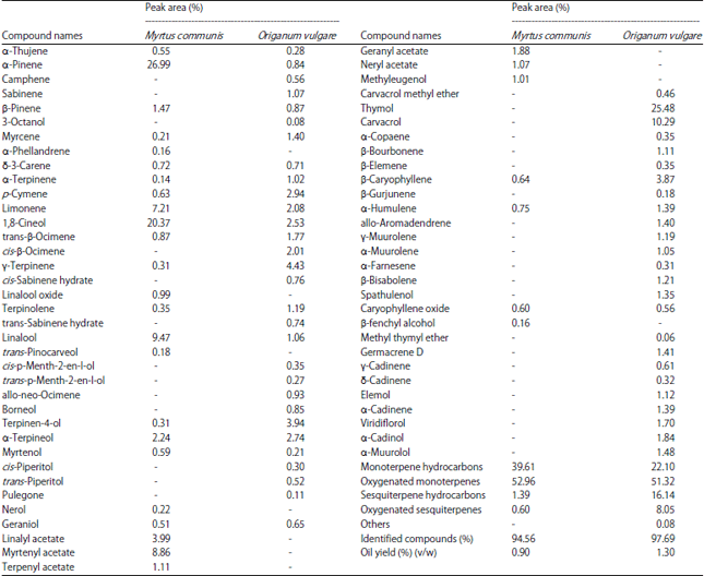
The hydro distillation of M. communis and O. vulgare dried leaves yield oil about 0.90% (v/w) and 1.30 % (v/w), respectively. The essential oils were analyzed by GC/MS for determination of their components and results are given in Table 1 as a relative peak area of each constituent.
GC/MS analysis of essential oils: More than 94% of the studied essential oils constituents were identified. It seems that there were no similarities among chemical compositions of the two essential oils. Thirty-one compounds of M. communis essential oil were identified having the total area of 94.56%, while fifty-five compounds of O. vulgare essential oil were identified having the total area of 97.69%. The major components found in M. communis essential oil were α-pinene (26.99%), 1,8-cineol (20.37%), linalool (9.47%), myrtenyl acetate (8.86%), limonene (7.21%) and linalyl acetate (3.99%), while the results show that the major components of O. vulgare oil were thymol (25.48%), carvacrol (10.29%), γ-terpinene (4.43%), terpinen-4-ol (3.94%) and β-caryophyllene (3.87%). The number of substances having quantities higher than 1% of the M. communis and O. vulgare essential oils were 5 and 24 substances, respectively. The most abundant chemical structure within components of M. communis and O. vulgare essential oils were oxygenated monoterpenes (52.96 and 51.32%, respectively), followed by monoterpene hydrocarbons (39.61 and 22.10%, respectively), sesquiterpene hydrocarbons (1.39 and 16.14%, respectively) and oxygenated sesquiterpenes (0.60 and 8.05%, respectively). The structures of the main constituents of the essential oils are reported in Fig. 1.
These results are in agreement with the percentage of α-pinene, 1,8-cineol and linalool reported by Bajalan et al. (2013), who indicated that the M. communis essential oil contain α pinene (27.87%), 1,8-cineol (20.15%) and linalool (10.26%), Also Gardeli et al. (2008) reported that the monoterpene fraction consisted mainly of oxygenated monoterpenes (70.1-73.2%), followed by monoterpene hydrocarbons (10.8-12.5%). The major compounds of essential oil extracted from Albanian leaves of myrtle (Asllani, 2000), were 1,8-cineole, α-pinene and limonene, as well as myrtenyl acetate and linalool. In the case of Turkish myrtle (Ozek et al., 2000), three major compounds were found: 1,8-cineole, linalool and myrtenyl acetate. Vazirian et al. (2015) found that the major components of O. vulgare oil were thymol (37.13%) and carvacrol (9.57%). Origanum vulgare oil was characterized by a high percentage of oxygenated monoterpenes (59.25%), followed by monoterpenes hydrocarbons (18.71%), oxygenated sesquiterpenes (12.86%) and sesquiterpene hydrocarbons (5.02%). On the other hand, results disagree with Kawase et al. (2013) and Raina and Negri (2012) who found that the carvacrol percentage in O. vulgare oil was 0.84 and 6.90%, respectively. Also, myrtenyl acetate (20.75%) and 1,8-cineol (16.55%) were found the two major compounds of M. communis oil that reported by Ben Hsouna et al. (2014).
| Table 1: | Chemical composition of dried leaves of Myrtus communis and Origanum vulgare essential oils |
 | |
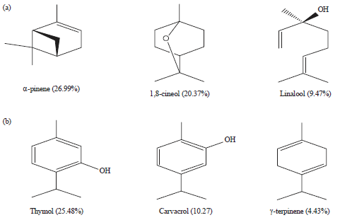 | |
| Fig. 1(a-b): | Chemical structures of the main constituents of (a) Myrtus communis and (b) Origanum vulgare essential oils |
| Table 2: | Percentage of scavenging activity of DPPH radicals induced by various concentrations of Myrtus communis, Origanum vulgare essential oils and ascorbic acid used as standard antioxidant substance |
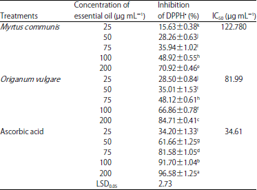 | |
The values are Mean±SE. The mean values with different small letters within a column indicate significant differences (p<0.05), DHHP: 2,2-diphenyl-1-picrylhydrazyl | |
Antioxidant activity of essential oils: Antioxidant activities of essential oils from aromatic plants are mainly attributed to the active compounds present in them. This can be due to the high percentage of main constituents, but also to the presence of other constituents in small quantities or to synergy among them. In this study, the antioxidant activities of essential oils of two plants belonging to different plant families compared with ascorbic acid as a reference anti-oxidant compound were determined by the method of DPPH∙ radical scavenging assay and the results are summarized in Table 2. It was found that the essential oils of two analyzed plants showed good antioxidant capacities compared with ascorbic acid. The results from Table 2 indicate that the radical scavenging activity (% inhibition) of the essential oil from M. communis was measured as 15.63, 28.26, 35.94, 48.92 and 70.92% with different concentrations of the essential oil 25, 50, 75, 100, 200 μg mL–1, respectively, whereas, treated with the same concentrations of O. vulgare oil reached the percentages of DPPH∙ inhibition of 28.50, 35.01, 48.12, 66.86 and 84.71%, respectively. On the other hand the radical scavenging activity of ascorbic acid was determined as 34.20, 61.66, 81.58, 91.70 and 96.58% with the same previous concentrations.
It was noticed that the scavenging activity of the essential oils were significantly increased with the increased of the essential oils concentrations. It is clear from the data that the concentration of 200 μg mL–1 of O. vulgare essential oil gave the highest percentage inhibition of DPPH∙ (84.71%) which was high significant inhibition value compared with other treatments, while the same concentration of ascorbic acid gave 96.58% inhibition. Also, radical scavenging activity of the M. communis essential oil was 70.92% at 200 μg mL–1 concentration. Both plants essential oils were able to reduce the stable, purple-colored radical DPPH into yellow-colored DPPH reaching 50% of reduction with IC50 values as follows: IC50 (M. communis) = 122.78 μg mL–1; IC50 (O. vulgare) = 81.99 μg mL–1 and IC50 (ascorbic acid) = 34.61 μg mL–1. The quantity of M. communis and O. vulgare essential oils required were about 3.55 and 2.37 fold, respectively, when compared with the standard antioxidant ascorbic acid. The antiradical scavenging activity of oils might be attributed to the replacement of hydroxyl groups in the aromatic ring systems of the phenolic compounds as a result of their hydrogen donating ability (Brand-Williams et al., 1995).
In the DPPH assay, M. communis essential oil showed high antioxidant activity according to Berka-Zougali et al. (2012). M. communis L. essential oil has a significant antioxidant effect when tested by DPPH method. The strong antioxidant activity of the studied oil can be attributed to the high content in hydrocarbon monoterpenes and oxygenated monoterpenes (Snoussi et al., 2011). Wannes et al. (2010) showed antioxidant activities of myrtle essential oil by using DPPH radical scavenging, β-carotene-linoleic acid bleaching, reducing power and metal chelating activity assays. The essential oil, Iranian O. vulgare may be categorized in "Thymol" chemotype and by its very strong antioxidant activity (Vazirian et al., 2015). Also, the antioxidant index of O. vulgare oil was better than BHA against DPPH∙ (Galego et al., 2008). DPPH radical scavenging activity of O. vulgare essential oil was very high and this was obviously related to its chemical compositions which was markedly rich in phenolic components such as thymol and carvacrol (Cosge et al., 2011).
Anticancer effect of essential oils
In vitro assay for anticancer activity: The anticancer effect of M. communis and O. vulgare essential oils were tested in vitro against two species of human promyelocytic leukemia cell lines (HL-60 and NB4) and experimental animals model cancer cells (Ehrlish ascites carcinoma cells, EACC) using viability test (trypan blue method). The viability percentages of HL-60 and NB4 cells after incubation with different concentrations of the essential oils were recorded in Table 3. Data showed that the incubation of cancer cells with different concentrations (from 25-200 μg mL–1) for 24 h of essential oils significantly reduced the viability of those cells when compared to untreated cells (control). The dead cells (HL-60 and NB4) were significantly increased with increasing essential oils concentrations. The results showed that the anticancer effects of M. communis and O. vulgare essential oils on HL-60 cells were more than the anticancer effects on NB4 cells.
| Table 3: | Effect of Myrtus communis and Origanum vulgare essential oils on cells viability of HL-60 and NB4 cells after 24 h of treatment and EACC after 12 h of treatment |
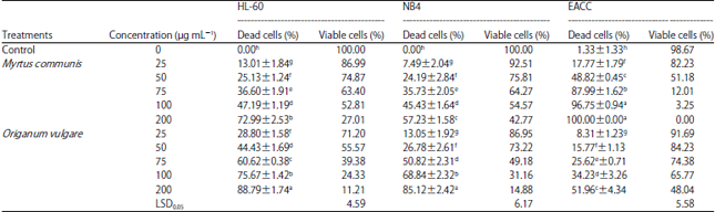 | |
| Values are Mean±SE. Mean values with different small letters within a column indicate significant differences (p<0.05), EACC: Ehrlish ascites carcinoma cells | |
On the other side the anticancer effects of O. vulgare essential oil on HL-60 and NB4 cells were more than the anticancer effects of M. communis essential oil on HL-60 and NB4 cells. The highest dead cells percentage of HL-60 was recorded by O. vulgare essential oil (88.79%) for concentration of 200 μg mL–1, while it was 72.99% for 200 μg mL–1 concentration of M. communis essential oil.
In the same trend, the highest dead cells percentage of NB4 was recorded by O. vulgare essential oil (85.12%) for concentration of 200 μg mL–1, while it was 57.23% for 200 μg mL–1 concentration of M. communis essential oil. No significant difference in the percentage of HL-60 dead cells line between M. communis at 100 μg mL–1 concentration and O. vulgare at 50 μg mL–1 concentration. In addition, no significant different in the percentage of NB4 dead cells line between M. communis at 100 μg mL–1 concentration and O. vulgare at 75 μg mL–1 concentration.
The LC50 values were determined from the graphs of the essential oils effects on HL-60 and NB4 cell lines. Myrtus communis essential oil showed potent cytotoxic effects with the LC50 values of 104.55 μg mL–1 in HL-60 cell line and 137.01 μg mL–1 in NB4 cell line, whereas O. vulgare essential oil gave the LC50 values of 60.43 μg mL–1 in HL-60 cell line and 73.65 μg mL–1 in NB4 cell line. The LC50 values indicated that the anticancer activity of O. vulgare essential oil was higher than M. communis essential oil against HL-60 and NB4 cell lines.
On the other hand, the effects of M. communis and O. vulgare essential oils on EACC viability were recorded in Table 3. The incubation of EACC with M. communis and O. vulgare essential oils at all concentrations (25-200 μg mL–1) for 12 h reduced the viability of these cells. The increase of both plant essential oils concentration increased the percentage of dead cells. The highest EACC dead cells (%) was recorded by M. communis essential oil (96.75 and 100%) for concentrations of 100 and 200 μg mL–1, respectively, while it was 51.96% for 200 μg mL–1 concentration of O. vulgare essential oil. No significant difference in dead cells (%) of EACC between concentrations 100 and 200 μg mL–1 of M. communis essential oil. In general, it was noticed that the anticancer activity of M. communis essential oil was higher than O. vulgare essential oil against EACC. The LC50 values were determined from the graphs of the essential oils on EACC. Myrtus communis and O. vulgare essential oils showed potent cytotoxic effects with the LC50 values of 58.30 and 182.52 μg mL–1, respectively in EACC. The LC50 values indicated that the anticancer activity of M. communis essential oil was higher than O. vulgare essential oil against EACC.
In vivo assay for anticancer activity: The effect of M. communis and O. vulgare essential oils initiation, post-initiation and pre-initiation treatments on survival of tumor (EACC) transplanted female mice and the number of EACC (all, dead and viable) and also LDH activity in supernatant of tumor cell suspension are shown in Table 4 and 5. The mean of survival days for the untreated tumor control group was 11.80±1.36 days, whereas different treatments showed significant increase in mean of survival days of tumor bearing mice compared with untreated tumor control. The pre-initiation treatments of M. communis and O. vulgare essential oils were increased transplanted female mice lifespan (%) more than initiation and post-initiation treatments. Pre-initiation treatment with M. communis oil gave the highest significant increase in lifespan percentage (330.44%) compared with tumor control. The second effect was by M. communis oil initiation treatment which increase lifespan by 252.73%.
| Table 4: | Effect of Myrtus communis and Origanum vulgare essential oils on survival of tumor (EACC) transplanted female mice |
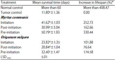 | |
| *Values are Mean±SE. Mean values with different small letters within a column indicate significant differences (p<0.05) | |
| Table 5: | Effect of Myrtus communis and Origanum vulgare essential oils on EACC number and lactate dehydrogenase activity in the tumor of transplanted female mice |
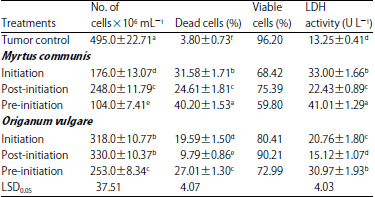 | |
| Values are Mean±SE. Mean values with different small letters within a column indicate significant differences (p<0.05) | |
Also Pre-initiation treatment with O. vulgare oil gave significant increase in lifespan percentage (174.58%) compared with tumor control followed by initiation and post-initiation treatments which were 101.88 and 76.64%, respectively.
From data recorded in Table 5 it was noticed a significant reduction in the tumor cells number by different treatments with M. communis and O. vulgare essential oils compared with tumor control. The highest significant reduction in tumor cells number was 104×106 mL–1 when pre-initiation treatment with M. communis oil followed by M. communis initiation treatment (176×106 mL–1) and M. communis post-initiation treatment (248×106 mL–1) compared with tumor control (495×106 mL–1). Also initiation, post-initiation and pre-initiation treatments of O. vulgare oil significantly reduce tumor cell numbers to 318, 330 and 253×x106 mL–1, respectively compared with tumor control. On the other hand, there were significant increases in dead cells percentages in all treated groups comparing with tumor control group. The highest significant dead cells percentage was recorded by pre-initiation treatment with M. communis essential oil (40.20%) which equal to 10.58 fold compared with tumor control (3.80%), while it was 27.01% (7.10 fold of tumor control value) when pre-initiation treatment with O. vulgare essential oil.
These results confirmed with that of LDH activity in the supernatant of tumor cell suspension. Cells that have lost membrane integrity release lactate dehydrogenase (LDH) into the surrounding medium. Measure the release of LDH from damaged (dead) cells gives an indicator of cytotoxicity (Hernandez et al., 2003). The results indicated that the increase in dead cells increases LDH activity in supernatant of tumor suspension. The highest significant increase in LDH activity was observed by M. communis pre-initiation treatment (40.01 U L–1) comparing with tumor control as similar as in dead cells percentage.
Legrand et al. (1992) found a significant correlation between the number of dead cells, determined by trypan Blue staining and LDH activity measurements in the supernatant of hybridoma strains. These results (Table 4 and 5) indicated that the increase in lifespan was correlated with the decrease in cells number and the increase in dead cells percentages. The results show that the anti-tumor effect of M. communis essential oil was higher than O. vulgare essential oil. Also, pre-initiation treatments with the both essential oils were more effective than initiation and post-initiation treatments, respectively on the tumor (EACC) transplanted female mice. This may recommended the use of these essential oils as preventive agents against tumor. These essential oils significantly prevent the development of tumor (decrease total EACC number and increase dead cells).
The treatment of prostate, breast cancer cell lines and 3T3 fibroblast cell line with M. communis essential oil at 200 μg mL–1 concentration gave inhibition percent was 67, 95.2 and 6.5%, respectively (Stevens, 2005). Also, our results are agreement with Oztekin et al. (1998) who found that M. communis essential oil increased survival time on Ehrlich tumor of injected mice. The essential oil of O. vulgare showed antiproliferative activity to MCF7 with IC50 values 30.1 μg mL–1 (Al-Kalaldeh et al., 2010).
The anticancer effects of the studied essential oils may be due to their chemical structures specially the major components (α-pinene, 1,8-cineol and linalool were major compounds in M. communis oil, while thymol, carvacrol and γ-Terpinene in O. vulgare oil). Matsuo et al. (2011) reported that α-pinene was able to induce apoptosis evidenced by early disruption of the mitochondrial potential, production of reactive oxygen species, increase in caspase-3 activity, heterochromatin aggregation, DNA fragmentation and exposure of phosphatidyl serine on the cell surface.
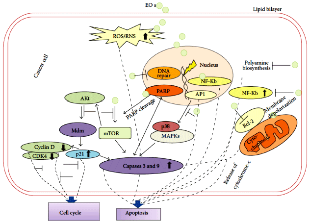 | |
| Fig. 2: | EOs and their constituents target multiple pathways in cancer cells (Reported by Gautam et al. (2014)) |
Most importantly, this molecule was very effective in the treatment of experimental metastatic melanoma reducing the number of lung tumor nodules. This is the first report on the apoptotic and antimetastatic activity of isolated α-pinene. Also, Specific induction of apoptosis by 1,8-cineole was observed in human leukemia Molt 4B and HL-60 cells (Moteki et al., 2002). The cytotoxic effect of thymol on HL-60 cells appears to be associated with induction of cell cycle arrest at sub G0/G1 phase and apoptotic cell death based on genomic DNA fragmentation pattern. Thymol also showed significant increase in production of Reactive Oxygen Species (ROS) activity, increase in mitochondrial H2O2 production and depolarization of mitochondrial membrane potential. Thymol showed increase in Bax protein level with a concomitant decrease in Bcl2 protein expression in a dose dependent manner. Also thymol showed activation of caspase -9, -8 and -3 and concomitant Poly (ADP-Ribose) polymerase (PARP) cleavage, which is the hallmark of caspase-dependent apoptosis (Deb et al., 2011). Incubation of HepG2 cells with carvacrol for 24 h induced apoptosis by the activation of caspase-3, cleavage of PARP and decreased Bcl 2 protein expression (Yin et al., 2012).
The EOs and their constituents target multiple pathways in cancer cells was reported by Gautam et al. (2014) who indicated that Essential Oils (EOs) by virtue have cell membrane permeability and act on different cellular targets involved in various pathways. The EOs increase intracellular reactive oxygen species/reactive nitrogen species (ROS/RNS) levels which results in apoptosis in cancer cells. Inhibition of protein Kinase B (Akt), mammalian target of rapamycin (Mtor) and Mitogen-Activated Protein Kinases (MAPK) pathways at different steps by EOs leads to corresponding up-/down regulation of various key biomolecules (and corresponding genes. Alteration in expression of Nuclear factor kappa-light-chain-enhancer of activated B cells (NF-KB) by EOs and further binding of NF-KB to DNA result in apoptosis in cancer cells. Dephosphorylation of Akt by the action of EOs results in overexpression of p21, which either induces apoptosis by increasing caspases level or results in cell cycle arrest by binding to cyclins. In addition, EOs-induced mitochondrial stress leads to activation of Bcl-2 gene and membrane depolarisation resulting in enhanced release of cytochrome-C to the cytoplasm which induces apoptotic cell death in cancer cells. The EOs also modulate DNA repair mechanisms by acting as DNA polymerase inhibitors and lead to poly (ADP-Ribose) polymerase (PARP) cleavage which also results in apoptosis in cancer cells (Fig. 2).
| Table 6: | Effect of administration with Myrtus communis and Origanum vulgare essential oils on serum biochemical parameters of normal male mice |
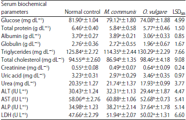 | |
Values are Mean±SE. Mean values with different small letters within a raw indicate significant differences (p<0.05), AST: Aspartate aminotransferase, ALT: Alanine aminotransferase, LDH: Lactate dehydrogenase, ALP: Alkaline phosphatase | |
Biological effects of M. communis and O. vulgare essential oils: The biological effects of M. communis and O. vulgare essential oils on normal mice were evaluated to determine the safety of using these oils as antioxidant and anti-tumor agents and results are summarized in Table 6. The oral administration of M. communis and O. vulgare essential oils to mice through 3 months showed no significant changes on serum glucose, total protein, albumin, globulin, creatinine, urea and uric acid. A significant decrease of triglycerides was observed in mice treated with M. communis essential oil (114.35 mg dL–1) compared with normal control group (125.84 mg dL–1). Also, O. vulgare essential oil showed significant increase in serum total cholesterol (98.46 mg dL–1) compared to M. communis essential oil group (86.94 mg dL–1).
On the other hand, serum ALT, AST, ALP and LDH activities (liver functions) as affected by oral administration with M. communis and O. vulgare essential oils to mice were determined (Table 6). It was noticed that no significant changes in the activities of serum ALT, AST, ALP and LDH between M. communis and O. vulgare essential oils treatments compared with normal control. Serum AST significantly decrease in mice treated with O. vulgare essential oil (52.68 IU L–1) compared with M. communis essential oil group (60.88 IU L–1). Although, all the obtained values in all experimental animals were within the normal range according to Mitruka and Rawnsley (1979).
These findings are in agreement with Sepici et al. (2004) who reported that changes in rabbit serum biochemical parameters were also investigated after M. communis administration. Myrtus communis oil induced significant decrease in triglyceride (14.3%) level but had no significant effects on the serum concentrations of other biochemical parameters (Glucose, ALT, AST, cholesterol and triglycerides) in normal animals. The supplementation of myrtle oil did not affect the ALP, ALT, AST and creatinine concentrations in laying quails (Bulbul et al., 2014). Also, Saei et al. (2013) showed that the effects of M. communis oil treatment on blood biochemical parameters of broilers at day 42 of age are reported. There was no significant difference for total protein among treatment compared with negative control (p>0.05). On the other side, the results of O. vulgare oil treatment are similar to that obtained by Corduk et al. (2013) who indicated that broilers at 21 days were treated with O. vulgare oil, then it was observed non-significantly affects on serum total protein, cholesterol, triglyceride, alanine aminotransferase enzyme, uric acid and creatinine levels, except glucose and aspartate aminotransferase enzyme (AST) levels compared with normal control. Addition of the oregano oil had no significant effects on performance, organ weights and blood parameters (Serum concentrations of total cholesterol, triglyceride, High Density Lipoprotein (HDL) and Low Density Lipoprotein (LDL)) of weaned lambs (Unal and Kocabagli, 2014).
CONCLUSION
In conclusion, the essential oils of M. communis and O. vulgare showed high scavenging activity against DPPH radicals. In addition, essential oils showed anticancer effects for the two human cell lines tested, HL-60 and NB4. On the other hand, M. communis essential oil is more effective on EACC (in vitro and in vivo) than O. vulgare essential oil. In the context, essential oils have preventive efficacies against development of tumor in transplanted animals. To conclude, M. communis and O. vulgare oils could be a promising source of natural antioxidants and anticancer agents. Nevertheless, additional in vivo studies on human patients are needed to unequivocally demonstrate this.
REFERENCES
- Wannes, W.A., B. Mhamdi, J. Sriti, M.B. Jemia and O. Ouchikh et al., 2010. Antioxidant activities of the essential oils and methanol extracts from myrtle (Myrtus communis var. italica L.) leaf, stem and flower. Food Chem. Toxicol., 48: 1362-1370.
CrossRefDirect Link - Al-Kalaldeh, J.Z., R. Abu-Dahab and F.U. Afifi, 2010. Volatile oil composition and antiproliferative activity of Laurus nobilis, Origanum syriacum, Origanum vulgare and Salvia triloba against human breast adenocarcinoma cells. Nutr. Res., 30: 271-278.
CrossRefPubMedDirect Link - Allain, C.C., L.S. Poon, C.S.G. Chan, W. Richmond and P.C. Fu, 1974. Enzymatic determination of total serum cholesterol. Clin. Chem., 20: 470-475.
CrossRefPubMedDirect Link - Asllani, U., 2000. Chemical composition of albanian myrtle oil (Myrtus communis L.). J. Essential Oil Res., 12: 140-142.
Direct Link - Bajalan, I., M. Akbarzadeh and F. Veysanlu, 2013. Chemical composition of myrtle essential oil (Myrtus communis L.) in gilane gharb from Iran. World Sci. J., 1: 59-65.
Direct Link - Baser, K.H.C., 2008. Biological and pharmacological activities of carvacrol and carvacrol bearing essential oils. Curr. Pharm. Des., 14: 3106-3119.
CrossRefPubMedDirect Link - Baydar, H., O. Sagdic, G. Ozkan and T. Karadogan, 2004. Antibacterial activity and composition of essential oils from Origanum, Thymbra and Satureja species with commercial importance in Turkey. Food Control, 15: 169-172.
CrossRefDirect Link - Ben Hsouna, A., N. Hamdi, R. Miladi and S. Abdelkafi, 2014. Myrtus communis essential oil: Chemical composition and antimicrobial activities against food spoilage pathogens. Chem. Biodivers., 11: 571-580.
CrossRefDirect Link - Bennett, J.M., D. Catovsky, M.T. Daniel, G. Flandrin, D.A.G. Galton, H.R. Gralnick and C. Sultan, 1976. Proposals for the classification of the acute leukaemias French-American-British (FAB) co-operative group. Br. J. Haematol., 33: 451-458.
CrossRefDirect Link - Berka-Zougali, B., M.A. Ferhat, A. Hassani, F. Chemat and K.S. Allaf, 2012. Comparative study of essential oils extracted from Algerian Myrtus communis L. leaves using microwaves and hydrodistillation. Int. J. Mol. Sci., 13: 4673-4695.
CrossRefDirect Link - Bouhdid, S., S.N. Skali, M. Idaomar, A. Zhiri, D. Baudoux, M. Amensour and J. Abrini, 2008. Antibacterial and antioxidant activities of Origanum compactum essential oil. Afr. J. Biotechnol., 7: 1563-1570.
Direct Link - Bradesi, P., F. Tomi, J. Casanova, J. Costa and A.F. Bernardini, 1997. Chemical composition of myrtle leaf essential oil from Corsica (France). J. Essential Oil Res., 9: 283-288.
CrossRefDirect Link - Brand-Williams, W., M.E. Cuvelier and C. Berset, 1995. Use of a free radical method to evaluate antioxidant activity. LWT-Food Sci. Technol., 28: 25-30.
CrossRefDirect Link - Bulbul, T., D. Yesilbag, E. Ulutas, H. Biricik, S.S. Gezen and A. Bulbul, 2014. Effect of myrtle (Myrtus communis L.) oil on performance, egg quality, some biochemical values and hatchability in laying quails. Revue Medecine Veterinaire, 165: 280-288.
Direct Link - Gardeli, C., P. Vassiliki, M. Athanasios, T. Kibouris and M. Komaitis, 2008. Essential oil composition of Pistacia lentiscus L. and Myrtus communis L.: Evaluation of antioxidant capacity of methanolic extracts. Food Chem., 107: 1120-1130.
CrossRefDirect Link - Da Costa, A.C., B.H.C. dos Santos, L.S. Filho and E.D.O. Lima, 2009. Antibacterial activity of the essential oil of Origanum vulgare L. (Lamiaceae) against bacterial multiresistant strains isolated from nosocomial patients. Revista Brasileira de Farmacognosia, 19: 236-241.
CrossRefDirect Link - Corduk, M., S. Sarica and G.F. Yarim, 2013. Effects of oregano or red pepper essential oil supplementation to diets for broiler chicks with delayed feeding after hatching. 1. Performance and microbial population. J. Applied Poult. Res., 24: 738-749.
CrossRefDirect Link - Cosge, B., M. Kiralan, A. Ipek, A. Bayrak and B. Gurbuz, 2011. Comparison of antiradical activities and compositions of essential oils of two Origanum spp. from Turkey. Adv. Environ. Biol., 5: 248-253.
Direct Link - Cozzi, R., R. Ricardy, T. Aglitti, V. Gatta, P. Petricone and R. De Salvia, 1997. Ascorbic acid and β-carotene as modulators of oxidative damage. Carcinogenesis, 18: 223-228.
CrossRefPubMedDirect Link - Dadalioglu, I. and G.A. Evrendilek, 2004. Chemical compositions and antibacterial effects of essential oils of Turkish oregano (Origanum minutiflorum), bay laurel (Laurus nobilis), spanish lavender (Lavandula stoechas L.) and fennel (Foeniculum vulgare) on common foodborne pathogens. J. Agric. Food Chem., 52: 8255-8260.
CrossRefPubMedDirect Link - Deb, D.D., G. Parimala, S.S. Devi and T. Chakraborty, 2011. Effect of thymol on peripheral blood mononuclear cell PBMC and acute promyelotic cancer cell line HL-60. Chem. Biol. Interact., 193: 97-106.
CrossRefDirect Link - Faleiro, L., G. Miguel, S. Gomes, L. Costa and F. Venancio et al., 2005. Antibacterial and antioxidant activities of essential oils isolated from Thymbra capitata L. (Cav.) and Origanum vulgare L. J. Agric. Food Chem., 53: 8162-8168.
CrossRefDirect Link - Fossati, P. and L. Prencipe, 1982. Serum triglycerides determined colorimetrically with an enzyme that produces hydrogen peroxide. Clin. Chem., 28: 2077-2080.
CrossRefPubMedDirect Link - Galego, L., V. Almeida, V. Goncalves, M. Costa, I. Monteiro, F. Matos and G. Miguel, 2008. Antioxidant activity of the essential oils of Thymbra capitata, Origanum vulgare, Thymus mastichina and Calamintha baetica. Acta Horticult., 765: 325-333.
Direct Link - Gautam, N., A.K. Mantha and S. Mittal, 2014. Essential oils and their constituents as anticancer agents: A mechanistic view. BioMed Res. Int.
CrossRef - Hernandez, J.M., M.H.T. Bui, K.R. Han, H. Mukouyama and D.G. Freitas et al., 2003. Novel kidney cancer immunotherapy based on the granulocyte-macrophage colony-stimulating factor and carbonic anhydrase IX fusion gene. Clin. Cancer Res., 9: 1906-1916.
PubMedDirect Link - Hung, T.M., M. Na, P.T. Thuong, N.D. Su and D. Sok et al., 2006. Antioxidant activity of caffeoyl quinic acid derivatives from the roots of Dipsacus asper Wall. J. Ethnopharmacol., 108: 188-192.
CrossRefDirect Link - Kawase, K.Y.F., C.G. Mothe, F.A. Furtado and G.L.V. Coelho, 2013. Changes in essential oil of Origanum vulgare L. affected by different extraction methods. Int. J. Res. Rev. Applied Sci., 14: 238-247.
Direct Link - Kind, P.R.N. and E.J. King, 1954. Estimation of plasma phosphatase by determination of hydrolysed phenol with amino-antipyrine. J. Clin. Pathol., 7: 322-326.
CrossRefPubMedDirect Link - Koldas, S., I. Demirtas, T. Ozen, M.A. Demirci and L. Behcet, 2015. Phytochemical screening, anticancer and antioxidant activities of Origanum vulgare L. ssp. viride (Boiss.) Hayek, a plant of traditional usage. J. Sci. Food Agric., 95: 786-798.
CrossRefDirect Link - Legrand, C., J.M. Bour, C. Jacob, J. Capiaumont and A. Martial et al., 1992. Lactate dehydrogenase (LDH) activity of the number of dead cells in the medium of cultured eukaryotic cells as marker. J. Biotechnol., 25: 231-243.
CrossRefDirect Link - Matsuo, A.L., C.R. Figueiredo, D.C. Arruda, F.V. Pereira and J.A.B. Scutti et al., 2011. α-Pinene isolated from Schinus terebinthifolius Raddi (Anacardiaceae) induces apoptosis and confers antimetastatic protection in a melanoma model. Biochem. Biophys. Res. Commun., 411: 449-454.
CrossRefDirect Link - Moteki, H., H. Hibasami, Y. Yamada, H. Katsuzaki, K. Imai and T. Komiya, 2002. Specific induction of apoptosis by 1,8-cineole in two human leukemia cell lines, but not a in human stomach cancer cell line. Oncol. Rep., 9: 757-760.
CrossRefDirect Link - Ozek, T., B. Demirci and K.H.C. Baser, 2000. Chemical composition of Turkish myrtle oil. J. Essent. Oil Res., 12: 541-544.
CrossRefDirect Link - Perera, M.G.A.N., S.S.S.B.D.P. Soysa, D.T.U. Abeytunga and R. Ramesh, 2008. Antioxidant and cytotoxic properties of three traditional decoctions used for the treatment of cancer in Sri Lanka. Pharmacogn. Mag., 4: 172-181.
Direct Link - Raina, A.P. and K.S. Negi, 2012. Essential oil composition of Origanum majorana and Origanum vulgare ssp. hirtum growing in India. Chem. Nat. Comp., 47: 1015-1017.
CrossRefDirect Link - Rajkapoor, B., B. Jayakar and N. Murugesh, 2004. Antitumor activity of Indigofera aspalathoides on Ehrlich ascites carcinoma in mice. Indian J. Pharmacol., 36: 38-40.
Direct Link - Reitman, S. and S. Frankel, 1957. A colorimetric method for the determination of serum glutamic oxalacetic and glutamic pyruvic transaminases. Am. J. Clin. Pathol., 28: 56-63.
CrossRefPubMedDirect Link - Romani, A., P. Pinelli, N. Mulinacci, F.F. Vincieri and M. Tattini, 1999. Identification and quantitation of polyphenols in leaves of Myrtus communis L. Chromatographia, 49: 17-20.
CrossRefDirect Link - Saei, M.M., A.A. Sadeghi and H. Ahmadvand, 2013. The effect of Myrtus communis oil extract on growth performance, serum biochemistry and humoral immune responses in broiler chicks fed diet containing aflatoxin B1. Archiv Tierzucht, 56: 842-850.
Direct Link - Sahin, F., M. Gulluce, D. Daferera, A. Sokmen and M. Sokmen et al., 2004. Biological activities of the essential oils and methanol extract of Origanum vulgare ssp. vulgare in the Eastern Anatolia region of Turkey. Food Control, 15: 549-557.
CrossRefDirect Link - Sarac, N., A. Ugur, M.E. Duru and O. Varol, 2009. Antimicrobial activity, antioxidant activity and chemical composition of Origanum onites L. and Origanum vulgare L. ssp. hirtum (Link) Ietswaart from Mugla (Turkey). Acta Horticulturae, 826: 397-404.
CrossRefDirect Link - Sepici, A., I. Gurbuz, C. Cevik and E. Yesilada, 2004. Hypoglycaemic effects of myrtle oil in normal and alloxan-diabetic rabbits. J. Ethnopharmacol., 93: 311-318.
CrossRefPubMedDirect Link - Snoussi, A., M.M. Chaabouni, N. Bouzouita and F. Kachouri, 2011. Chemical composition and antioxidant activity of Myrtus communis L. Floral buds essential oil. J. Essent. Oil Res., 23: 10-14.
Direct Link - Souza, E.L., T.L.M. Stamford, E.O. Lima and V.N. Trajano, 2007. Effectiveness of Origanum vulgare L. essential oil to inhibit the growth of food spoiling yeasts. Food Control., 18: 409-413.
CrossRefDirect Link - Sumbul, S., M.A. Ahmad, M. Asif and M. Akhtar, 2011. Myrtus communis Linn.-A review. Indian J. Nat. Prod. Resour., 2: 395-402.
Direct Link - Tommasi, L., C. Negro, A. Miceli and F. Mazzotta, 2009. Antimicrobial activity of essential oils from aromatic plants grown in the Mediterranean area. J. Essent. Oil Res., 21: 185-189.
CrossRefDirect Link - Trinder, P., 1969. Determination of glucose in blood using glucose oxidase with an alternative oxygen acceptor. Ann. Clin. Biochem., 6: 24-27.
CrossRefDirect Link - Unal, A. and N. Kocabagli, 2014. Effect of different dosages of oregano oil on performance and some blood parameters in lambs. Ankara Universitesi Veteriner Fakultesi Dergisi, 61: 199-204.
Direct Link - Vardar-Unlu, G., M. Unlu, E. Donmez and N. Vural, 2007. Chemical composition and in vitro antimicrobial activity of the essential oil of Origanum minutiflorum O Schwarz & PH Davis. J. Sci. Food Agric., 87: 255-259.
CrossRefDirect Link - Yadegarinia, D., L. Gachkar, M.B. Rezaei, M. Taghizadeh, S.A. Astanch and I. Rasooli, 2006. Biochemical activities of Iranian Mentha piperita L. and Myrtus communis L. essential oils. Phytochemistry, 67: 1249-1255.
CrossRefPubMedDirect Link - Yen, G.C. and P.D. Duh, 1994. Scavenging effect of methanolic extracts of peanut hulls on free-radical and active-oxygen species. J. Agric. Food Chem., 42: 629-632.
CrossRefDirect Link - Yin, Q.H., F.X. Yan, X.Y. Zu, Y.H. Wu and X.P. Wu et al., 2012. Anti-proliferative and pro-apoptotic effect of carvacrol on human hepatocellular carcinoma cell line HepG-2. Cytotechnology, 64: 43-51.
CrossRefDirect Link - Vazirian, M., M. Mohammadi, M.H. Farzaei, G. Amin and Y. Amanzadeh, 2015. Chemical composition and antioxidant activity of Origanum vulgare subsp. vulgare essential oil from Iran. Res. J. Pharmacogn., 2: 41-46.
Direct Link