
K.C. Patrick-Iwuanyanwu
Toxicological Unit, Department of Biochemistry, University of Port Harcourt, P.M.B. 5323, Choba, Port Harcourt, Rivers State, Nigeria
M.O. Wegwu
Toxicological Unit, Department of Biochemistry, University of Port Harcourt, P.M.B. 5323, Choba, Port Harcourt, Rivers State, Nigeria
Asian Journal of Biochemistry
Year: 2008 | Volume: 3 | Issue: 4 | Page No.: 213-220


ABSTRACT
The hepatoprotective effects of aqueous and alcoholic extracts of stem and leaf of Acanthus montanus pre-treatment against carbon tetrachloride (CCl4)-induced liver damage in wistar albino rats were investigated. The plant extracts were fed to the rats intragastricaly for 15 days prior to the administration of 0.5 mL CCl4 kg-1 body weight. Serum L-alanine aminotransferase (L-ALT), L-aspartate amino transferase (L-AST), lactate dehydrogenase (LDH) and alkaline phosphatase (ALP) levels, 24 h after CCl4 administration decreased significantly in rats pre-treated with aqueous and alcoholic extracts of both stem and leaf of Acanthus montanus than in CCl4-treated rats only. The aqueous extract of stem of Acanthus montanus showed a marked decrease in the levels of AST, ALT and ALP when compared with the alcoholic extract of stem including aqueous and alcoholic extract of leaf of Acanthus montanus. However, rats fed with the alcoholic extract of leaf of Acanthus montanus was lowest in the level of LDH when compared with the results obtained from the other extracts of the stem and leaf of Acanthus montanus. Determination of total serum bilirubin also showed a remarkable decrease in rats pre-treated with aqueous and alcoholic extracts of stem and leaf of Acanthus montanus when compared with those administered CCl4 alone. Aqueous leaf extract of Acanthus montanus showed the least result in total serum bilirubin when compared with the alcoholic extracts of the leaf and both aqueous and alcoholic extracts of the stem of Acanthus montanus. Lipid peroxidation expressed by malondialdehyde (MDA) concentration was significantly reduced in rats pre-treated with aqueous and alcoholic extracts of stem and leaf of Acanthus montanus than rats administered CCl4-alone. The lowest MDA concentration was observed in aqueous extracts of stem of Acanthus montanus while the highest concentration in rats pre-treated with aqueous and alcoholic extracts of stem and leaf of Acanthus montanus was observed in rats treated with alcoholic extract of stem of Acanthus montanus. Histopathological examinations in rats administered CCl4-alone showed severe hepatic damage to the liver. However, rats pre-treated with extracts of Acanthus montanus showed significant improvements in the architecture of rat liver. The results obtained in this study suggest that alcoholic and aqueous extracts of leaf and stem of Acanthus montanus may prevent liver damage induced by CCl4 in rats.
PDF Abstract XML References Citation
How to cite this article
K.C. Patrick-Iwuanyanwu and M.O. Wegwu, 2008. Prevention of Carbon Tetrachloride (CCl4)-Induced Liver Damage in Rats by Acanthus montanus. Asian Journal of Biochemistry, 3: 213-220.
DOI: 10.3923/ajb.2008.213.220
URL: https://scialert.net/abstract/?doi=ajb.2008.213.220
DOI: 10.3923/ajb.2008.213.220
URL: https://scialert.net/abstract/?doi=ajb.2008.213.220
INTRODUCTION
The use of herbal medicine in the treatment of diseases is increasing worldwide (Kaufman et al., 2002). This is traceable to the growing belief by consumers that herbal remedies are safe and effective. Indeed, the effectiveness of some medicinal herbs in the treatment of diseases has been validated through research and clinical studies (Oguntola, 2007).
The liver is known to perform a multitude of essential functions and several disease states, including hepatitis and cirrhosis alter the metabolism of this organ (Attri et al., 2001; Anonymous, 2004). In Nigeria and other developing countries, the growing incidence of liver disorder, arising from the proliferation of hepatotoxic substances in the environment has been expressed. The understanding that medicinal herbs are significant source of pharmaceutical drugs has given rise to latest trends which has shown increasing demand for phytodrugs with hepatoprotective potential (Malhorta et al., 2001).
Serum or plasma enzyme levels have been employed as markers for monitoring chemically induced tissue damages (Hukkeri et al., 2002). The enzymes L-alanine aminotransferase (L-ALT), L-aspartate amino transferase (L-AST), alkaline Phosphatase (ALP) and lactate dehydrogenase (LDH), are often used in assessing the integrity of the liver (Hukkeri et al., 2002).
Carbon tetrachloride (CCl4) is toxic to the liver and its toxicity is dose dependent and time of exposure (Junnila et al., 2000). In the liver, CCl4 is metabolized to the highly reactive trichloromethyl radical. The free radical generated would lead to auto-oxidation of the fatty acids present in the cytoplasmic membrane phospholipids and causes functional and morphological changes in the cell membrane (Pandit et al., 2004). This would lead to elevation of serum marker enzymes, namely, ALT, AST and ALP (Bhattacharryya et al., 2003a, b).
The plant Acanthus montanus belong to the family Acanthaceae and order scrophulariales. It is commonly known as Mountain Thistle or Bears Breech and is believed to have originated from Western tropical Africa (Horticopia, 2004). Traditionally, the stem and leaves are used locally to treat various illnesses. Young leaves and twigs plucked are crushed along with two cubes of sugar to make a paste which is applied on boils to induce quick suppuration. Soups made from it are also used for abdominal pains caused by indigestion. During pregnancy, the leaves are used to relax the baby. Adeyemi et al. (2004) reported that aqueous extracts of the plant showed anti-inflammatory, lack of central analgesia and antipyretic properties. They demonstrated that methanolic extracts of the plant exhibited a dose dependent inhibition of writhing and also showed a significant inhibition of both phases of the formatin pain test.
The application of Acanthus montanus in the treatment of numerous diseases by the indigenous people of Nigeria; including the works of Adeyemi et al. (2004), informed the objective of this study, which was to investigate the ability of Acanthus montanus to prevent CCl4-induced liver damage in rats. This would be achieved by the administration of both aqueous and alcoholic extracts to rats for fifteen days prior to CCl4-administration. Twenty four hour after CCl4-administration, the rats shall be anaesthetized in a chloroform-saturated chamber and blood and liver tissue shall be collected respectively for enzyme assay and histopathological examinations.
MATERIALS AND METHODS
Plant Materials
The mountain thisle plant (Acanthus montanus) was collected from Choba village, Rivers State, Nigeria. The samples were thoroughly examined to ensure that they were disease-free before they were authenticated by the Herbarium section of the Plant Science and Biotechnology Department of the University of Port Harcourt, Nigeria.
Treatment of Plant Material
After the separation of the plant into various parts, the stem and leaf were respectively cut into bits and air dried for two days. The dried plant parts were ground with a clean mortar and pestle and further milled into a fine powder using an electric blender (Moulinex) and made to pass through a 0.25 mm sieve (Ende-cotts (Test sieves) Ltd., England).
Preparation of Aqueous Extract
Four hundred milliliter of distilled water was added to 25 g of powdered stem or leaf sample in a conical flask and then shaken vigorously to allow for proper mixture. The suspension was filtered and the filtrate concentrated using a mild heat (30 °C with hot plate) for 15 min and freeze dried in a lyophilizer.
Preparation of Alcoholic Extract
The alcoholic extract was prepared as mentioned above. In this case ethanol was used instead of distilled water.
Animals
Twenty-four adult male wistar albino rats (150-160 g) used in this study were obtained from the Animal House, Department of Biochemistry, University of Port Harcourt, Nigeria. They were housed in standard cages (Griffin and George Modular Cage System) and left to acclimatize for 7 days to laboratory conditions before the commencement of the experiment. During the acclimatization, the animals were fed with pelleted rat chow and water ad libitum.
This study was conducted at the Animal House of the Department of Biochemistry, University of Port Harcourt, Nigeria in September, 2006.
Experimental Protocol
The experimental animals were divided into four groups, each group comprising six animals.
Group 1
Normal control rats (feed only)
Group 2
Received normal feed and 0.5 mL CCl4 kg-1 body weight (b.wt.)
Group 3
Orally pre-treated with 2.5 g kg-1 b.wt. of alcoholic extract of stem or leaf of Acanthus montanus prior to CCl4 induction (0.5 mL kg-1 b.wt.)
Group 4
Orally pre-treated with 2.5 g kg-1 b.wt. of aqueous extract of stem or leaf of Acanthus montanus prior to CCl4 induction (0.5 mL kg-1 b.wt.)
Induction of Hepatic Injury
Carbon tetrachloride (CCl4)-induced liver damage was achieved by injecting 0.5 mL CCl4 kg-1 b.wt. intramuscularly on the 16th day of feeding animals of groups II to IV with commercial feed, alcoholic and aqueous extracts of Acanthus montanus as stated earlier.
Preparation of Samples
Twenty-four hours after the administration of CCl4, the rats were anaesthetized in a chloroform-saturated chamber. Blood was collected from the jugular vein with the aid of a 2 mL hypodermic syringe and needle into an anti-coagulant-free bottle. The animals were dissected from anus to thorax using a surgical blade. The liver was excised immediately and fixed in 10% formalin for histological assessment of hepatic damage. Serum was separated by centrifugation and stored in a refrigerator. The levels of serum L-alanine aminotransferase (L-ALT), L-aspartate amino transferase (L-AST), lactate dehydrogenase (LDH) and alkaline phosphatase (ALP) were performed using the Humazym MUV-test kits. Bilirubin was analyzed by colorimetric method as described by Randox Bilirubin Manual (2003). Lipid peroxidation was also determined by estimating the malondialdehyde (MDA) levels using the method of Hunter et al. (1963) modified by Gutteridge and Wilkins (1980).
Analysis of Data
Statistical analysis of data was performed using analysis of variance (ANOVA) and significance was set at p≤0.05.
RESULTS
Table 1 and 2 shows group 2 rats treated with single dose of CCl4 (0.5 mL kg-1 b.wt.) with severe hepatic damage when compared with the group 1 control rats. This was evidenced by a marked elevation in the levels of serum marker enzymes, AST, ALT, ALP and LDH, bilirubin and MDA when compared to rats pre-treated with alcoholic and aqueous extracts of both stem and leaf of Acanthus montanus. The serum marker enzymes, AST, ALT and LDH recorded the lowest value in the groups pre-treated with aqueous extract of sample. However, serum ALP was observed to be lowest in rats fed alcoholic extract. A different trend was observed in the result obtained from rats fed with aqueous and alcoholic extracts of leaf of Acanthus montanus. ALT, LDH and ALP were observed to be lowest in rats fed with alcoholic extract. However, AST recorded the lowest value in rats fed with aqueous extract of leaf of Acanthus montanus (group 3) (Table 2). Table 1 and 2 shows that bilirubin was significantly reduced in the group of rats treated with aqueous extracts of stem and leaf of Acanthus montanus (group 4) when compared to their corresponding alcohol treated rats. The lowest MDA concentrations was observed in the groups fed aqueous and alcoholic extracts of Acanthus montanus (Table 1). However, the rats fed with alcoholic extract of leaf of Acanthus montanus recorded the lowest MDA concentrations (Table 2). Histopathological examinations (Fig. 1a-c) showed defects ranging from massive tissue necrosis, inflammatory cells, dilated central vacuole and fat inclusions in rats treated with CCl4 alone. However, normal with minor tissue necrosis and inflammatory cells were evident in pre-treated rats.
| Table 1: | Hepatoprotective effects of aqueous and alcoholic extracts of stem of Acanthus montanus on serum AST, ALT, LDH and ALP, bilirubin and lipid peroxidation (MDA) in CCl4-induced hepatotoxicity in rats |
 | |
| Values are means ± SEM., n = 6 rats in each group, Means with different superscript letter(s) (a, b, c, d) in the same column are significantly different at the 0.05 level | |
| Table 2: | Hepatoprotective effects of aqueous and alcoholic extracts of leaf of Acanthus montanus on serum AST, ALT, LDH and ALP, bilirubin and lipid peroxidation (MDA) in CCl4-induced hepatotoxicity in rats |
 | |
| Values are means ± SEM, n = 6 rats in each group, Means with different superscript letter(s) (a, b, c, d) in the same column are significantly different at the 0.05 level | |
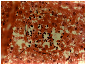 | |
| Fig. 1a: | A section of the rat liver treated with CCl4 only, showing localized area of necrosis and infiltration by inflammatory cells |
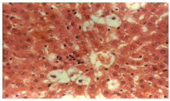 | |
| Fig. 1b: | A section of rat liver pre-treated with drugs prior to CCl4 administration showing a pattern of reduced inflammatory infiltration |
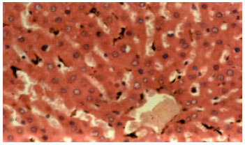 | |
| Fig. 1c: | A section of rat liver showing normal architecture (group 1) |
DISCUSSION
It has been well established that carbon tetrachloride (CCl4) is metabolized in the liver to the highly reactive trichloromethyl radical and this free radical leads to auto-oxidation of the fatty acids present in the cytoplasmic membrane phospholipids and causes functional and morphological changes in the cell membrane (Recknagel, 1967). Treatment of rats with CCl4 resulted to a significant hepatic damage as elicited by the elevated levels of serum marker enzymes: AST, ALT, LDH and ALP. These marker enzymes are cytoplasmic in origin and are released into the circulation after cellular damage (Lin et al., 2000). The rise in the enzyme AST is usually accompanied by an elevation in the levels of ALT, which plays a vital role in the conversion of amino acids to Keto acids (Salie et al., 1999).
It has been reported (Bhattacharryya et al., 2003a, b) that the antioxidant activity or inhibition of the generation of free radicals is important in the protection against CCl4-induced liver lesion.
The results in this study indicate that rats pre-treated with aqueous and alcoholic extracts of stem and leaf of Acanthus montanus showed considerable lower amounts of serum liver marker enzymes when compared with rats that received CCl4 alone. The increase in serum marker enzymes suggests that the toxicant was able to reach the liver and induce a detectable damage within 24 h. However, the significant reduction in the levels of these enzymes in rats pre-treated with stem and leaf extracts of Acanthus montanus indicate that the plant may have a considerable level of antioxidants which may be present in the tannins of the plant. The presence of the antioxidants may have protected the liver of the rats studied.
Animals treated with the alcohol extract of stem of Acanthus montanus showed variation in their liver enzymes when compared to the control group but were generally lower in comparison to animals administered CCl4 alone. This may be because alcohol has the ability of damaging the liver by causing inflammation of the cell, almost nullifying the effect of the stem extract. This is achieved by generating too much NADH, which inhibits many enzymes including those of the tricaboxylic acid cycle (TCA), gluconeogenesis and fatty acid oxidation thereby leading to accumulation of hepatic triacylglycerols (fatty liver) (Harris and Crabb, 2002). The decrease observed in the serum marker enzymes by aqueous and alcoholic extracts of stem and leaf of Acanthus montanus extracts suggest that they are able to condition the hepatocytes so as to protect the membrane integrity against CCl4-induced leakage of marker enzymes into the circulation.
The elevation of bilirubin has been reported in acetaminophen-induced hepatotoxicity (Yamaguchi et al., 1996; Ellenhorn, 1997). Bilirubin, an endogenous organic anions bind reversibly to albumin and it is transported to the liver, conjugates with glucuronic acid and excreted in the bile. Hepatobiliary disease is indicated when conjugated fraction of total bilirubin exceeds the upper limit of normal, even if the total serum bilirubin is normal or near normal (Rosen and Keefe, 1998). The lowering of bilirubin in aqueous and alcoholic extracts of stem and leaf of Acanthus montanus pre-treated rats further indicate the protective nature of Acanthus montanus on hepatic cells when compared with rats administered CCl4 alone.
Administration of 0.5 mL kg-1 b.wt. CCl4 has been reported to elevate malondialdehyde (MDA), a product of lipid peroxidation (Patrick-Iwuanyanwu et al., 2007). Therefore an increase in the liver MDA levels indicates an increase in the degree of lipid peroxidation, a well-known mechanism of liver damage (Trible et al., 1987). In addition, the extensive lipid peroxidation results in membrane disorganization by peroxidizing the highly unsaturated fatty acids. This in turn alters the ratio of polyunsaturated to other fatty acids, thus, leading to a decrease in the membrane fluidity, which may be sufficient to cause cell death (Rotruck et al., 1979). In this study, a significant elevation in the levels of end products of lipid peroxidation or MDA in the liver of rats treated with CCl4 may be attributed to hepatic damage. However, a significant decrease in the levels of lipid peroxides in aqueous and alcoholic extracts of stem and leaf of Acanthus montanus pre-treated rats suggests that the extracts may have the ability to protect the liver from free radical injury induced by CCl4. Flavonoids, tannins and microelements have been suggested to act as antioxidants and exert their antioxidant activity by scavenging the lipid peroxidation (Yuting et al., 1990).
The marked decreases in the levels of serum marker enzymes and MDA in pre-treated rats are indications of the ability of Acanthus montanus to protect the liver against CCl4 poisoning. This is corroborated by the architecture of the liver of rats that received extracts of Acanthus montanus prior to CCl4 administration (Fig. 1a-c).
This study provides conclusive evidence for the hepatoprotective effect of Acanthus montanus against carbon tetrachloride-induced hepatotoxicity. The plausible mechanism of the hepatoprotective action of Acanthus montanus might be at least partly due to its antioxidant effect.
REFERENCES
- Adeyemi, O.O., S.O. Okpo and C.C. Young-Nwafor, 1999. The relaxant activity of the methanolic extract of Acanthus montanus on intestinal smooth muscles. J. Ethnol. Pharmacol., 68: 169-173.
CrossRefDirect Link - Attri, S., S.V. Rana, K. Vaiphie, R. Katyal, C.P. Sodhi, S. Kanwar and K. Singh, 2001. Protective effect of n-acetyl-cysteine in isoniazid-induced hepatic injury in growing rats. Indian J. Exp. Biol., 39: 436-440.
PubMed - Bhattacharryya, D., R. Mukherjee, S. Pandit, N. Das and T.K. Sur, 2003. Hepatoprotective effect of Himoliv, a polyherbal formulation in rats. Indian J. Physiol. Pharmacol., 47: 435-440.
Direct Link - Gutteridge, J.M.C. and S. Wilkins, 1982. Copper-dependent hydroxyl radical damage to ascorbic acid: Formation of a thiobarbituric acid-reactive product. FEBS Lett., 137: 327-340.
CrossRefPubMedDirect Link - Hunter, F.E., J.M. Gebicki, P.E. Hoffstein, J. Weinstein and A. Scott, 1963. Swelling and lysis of rats liver mitochondria induced by ferrous ions. J. Biol. Chem., 238: 828-835.
Direct Link - Kaufman, D.W., J.P. Kelly, L. Rosenberg, T.E. Anderson and A.A. Mitchell, 2002. Recent patterns of medication use in the ambulatory adult population of the United States: The Slone survey. J. Am. Med. Assoc., 287: 337-344.
CrossRefPubMedDirect Link - Pandit, S., T.K. Sur, U. Jana, P.K. Debnath, S. Sen and D. Bhattacharyya, 2004. Prevention of carbon tetrachloride-induced hepatotoxicity in rats by Adhatoda vasica leaves. Indian J. Pharmacol., 36: 312-313.
Direct Link - Patrick-Iwuanyanwu, K.C., M.O. Wegwu and E.O. Ayalogu, 2007. Prevention of CCl4-induced liver damage by ginger, garlic and vitamin E. Pak. J. Biol. Sci., 10: 617-621.
CrossRefPubMedDirect Link - Recknagel, R.O., 1967. Carbon tetrachloride hepatotoxicity. Pharmacol. Rev., 19: 145-208.
Direct Link - Rotruck, J.T., A.L. Pope, H.E. Ganther, A.B. Swanson, D.G. Hafeman and W.G. Hoekstra, 1973. Selenium: Biochemical role as a component of glutathione peroxidase. Science, 179: 588-590.
CrossRefPubMedDirect Link - Sallie, R., J.M. Tredger and R. Williams, 1991. Drugs and the liver part 1: Testing liver function. Biopharm. Drug Dispos., 12: 251-259.
CrossRefPubMedDirect Link - Trible, D.L., T.Y. Aw and D.P. Jones, 1987. The pathophysiological significance of lipid peroxidation in oxidative cell injury. Hepatology, 7: 377-386.
Direct Link - Yuting, C., Z. Rongliang, J. Zhongjian and J. Yong, 1990. Flavonoids as superoxide scavengers and antioxidant. Free Radic. Biol. Med., 9: 19-21.
Direct Link