

P. Chattopadhyay
College of Pharmacy, IFTM, Lodhipur Rajput, Moradabad-244001, UP, India
A.K. Wahi
Delhi Institute of Pharmaceutical Sciences and Research (Formerly, College of Pharmacy), Pusph Vihar, Sector III, New Delhi II 0 017, India
S.S. Agrawal
Birla Institute of Technology and Sciences, Pilani-333031, Rajasthan, India
Asian Journal of Biochemistry
Year: 2007 | Volume: 2 | Issue: 6 | Page No.: 364-374





ABSTRACT
Genetic analysis, embryonic tissue explanation and in vivo chromatin studies have together identified the distinct regulatory steps that are necessary for the development of endoderm into a bud of liver tissue and subsequently into an organ. In this review, we discuss the acquisition of competence to express liver growth factor, which controls both stimulatory and inhibitory signals for cell proliferation. Epidermal Growth Factor (EGF), Transforming Growth Factor -α (TGF α) and Hepatocyte Growth Factors (HGF) stimulate DNA synthesis in hepatocytes in vivo and in culture but the sensitivity of cultured hepatocytes to the mitogenic effects of these factors are much higher than that of proposed that after partial hepatectomy, hepatocytes enter a state of replicative competence. Cytokine induced by Tumor Necrosis Factor -α (TNF) has main role to regulate Necrosis Factor (NF) κβ transcription factors to binding DNA. We also discuss the cytokines like EGF, TGF α and Hepatocyte Growth Factor (HGF) on liver growth. This information may contribute to the development of new targets for the treatment of liver diseases in the future.
PDF Abstract XML References
How to cite this article
P. Chattopadhyay, A.K. Wahi and S.S. Agrawal, 2007. Organogenesis and Regeneration of Liver: Mechanism and Signal Cascade. Asian Journal of Biochemistry, 2: 364-374.
DOI: 10.3923/ajb.2007.364.374
URL: https://scialert.net/abstract/?doi=ajb.2007.364.374
DOI: 10.3923/ajb.2007.364.374
URL: https://scialert.net/abstract/?doi=ajb.2007.364.374
INTRODUCTION
Little is known about the mechanism by which embryonic liver, lung and pancreas progenitor cells emerge from the endodermal epithelium to initiate organogenesis. Recent researchers elucidated a growing number of evolutionarily conserved genes and pathways that control liver development from the embryonic endoderm Adult liver tissue regeneration may recapitulate molecular events of liver organogenesis. It is increasingly clear that the genetic programs active in embryogenesis are often deregulated in several diseases. Studies have shown that within minutes following two thirds Partial Hepatotectomy (PH), the majority of cells in the remnant liver, which are normally quiescent hepatocytes rapidly re two rounds of replication and then return to a non-proliferative state. About thymidylate syntheses (TS:EC 2.1.45) and thymidine kinase (TK:EC 2.7.1.21) which catalyze the formation of thymidylate through the de novo and salvage pathways, respectively, reflected closely the total amount of DNA synthesis and that these enzymes were rate determining in DNA synthesis (Blackely, 1969; Grilli et al., 1993). Role of different signaling protein and associated growth factors has not fully elucidated. Therefore, the present review discussed role of growth factors like foxa protein HGF, TNF-α, TGF-β etc. in development and regeneration of liver.
ROLE OF foxa PROTEINS
The first genetic evidence for the importance of transcription factors in establishing the endodermal domain that gives rise to liver came from the new classical approach of identifying proteins are important for liver-specific transcription in adult hepatocytes. DNA sequences that encode these proteins are then used to trace back the expression and function of these genes in the embryo. The biochemical approach to identifying liver factors led to the cloning of three released foxa (fork head box A) formerly hepatocyte nuclear factors proteins, each of which expressed in the foetal and adult liver as well as other endoderm-derived tissues (Lai, 1990; Lai et al., 1991). The foxa proteins regulated virtually all liver-specific genes, as well as genes in the lung and the pancreas (Costa, 1994; Zarel, 1990). It is also observed that it has great role in gut organogenesis. Shortly after the discovery of foxa genes it is became clear that related genes are related gut development across the animal kingdom. The homologue to be discovered was fork head in melanogaster. It's DNA binding domain shares of 110 amino acid identified as the foxa protein (Weigel et al., 1989, 1990). The Pha-4 gene of the caenor habittis elegans, has 75% identity over foxa binding domain and Pha-4 protein required for gut development particularly anterior, pharyngel region (Axxanha et al., 1996; Horner, 1998). Interestingly, in the absence of Pha-4, pharyngel cells become ectoderm which might be analogous to the failure of epistat epiblast in foxa2 null mouse embryos to be developed into definitive endoderm during gastrulation (Dufort et al., 1998).
FIBROBLAST GROWTH FACTOR (Fgf) SIGNALING AND HEPATIC INDUCTION
There are no known mutations in any organism that block the initial induction of hepatic cells in the endoderm. To control with the possibility of redundant signaling and to more readily dissect the underlying mechanisms, an embryonic tissue explants system was developed. In this system, ventral foregut endoderm and cardiogenic mesoderm are isolated from mouse embryo and cultivated in vitro conditions that are permissive for hepatic induction (Gualdi, 1996) The system was used to show that inhibitor Fgf signaling block hepatic induction by the cardiogenic mesoderm (Jung et al., 1999). Using the same system, it was also shown that, at low concentrations, Fgf treatment alone induced early hepatic gene expression in endoderm explants that were cultured without cardiogenic mesoderm. These experiments indicate that Fgf signaling from the cardiogenic mesoderm induces the liver in the ventral foregut endoderm.
At the time of hepatic induction, the cardiogenic mesoderm express at least 3 out of the 18 known Fgf (Zhu et al., 1996; Crossley and Martin, 1995) and the ventral foregut endoderm express at least two of four tyrosine kinase receptors (Strak et al., 1991; Sugi et al., 1995).
OTHER HEPATOGENIC SIGNALING
Before hepatic induction prospective septum transverse mesenchyma cells surround the developing cardiac region near the ventral foregut endoderm and the Septum Transverse Mesenchyme (STM) explants of cardiogenic mesoderm and ventral foregut endoderm (Rossi et al., 2001).
Bone morphogenetic proteins 2 and 4 (Bmp 2 and Bmp 4) are strongly expressed in STM (Winnier et al., 2003; Hogan, 1999) before during hepatic induction and detection of Bmp 4 perturbs the development of various ventral structures in the embryos (Deutsh et al., 2001). Bmps are members of the Tgfb1. Super family of secreted signalling molecules and they together with fgfs, have many morphogenic roles in the developing embryo (Fukuda Taira, 1981) although, liver gene induction occurs normally in the ventral foregut. Induction of hepatic gene is inhibited in ventral forgut with Bmp inhibitor (Cascio and Zaret, 1991). Hestores hepatic gene are inducted by adding Bm2 or Bm4.These data are consistent with reducdant Bmp. Signalling being crucial for hepatogenesis, perhaps through enhancing the hepatic competence of the endoderm and induce a particular tissue-in such case of liver.
SECOND STAGE OF HEPATIC INDUCTION
It occurs when mesoderm-derived cells in the spectrum transverse-sum promote growth and further differentiation of the newly specific hepatic endoderm (Homas et al., 1993). The system transverse defines a region of the embryonic body cavity into which the hepatic bud grows. Locally joined mesenchyme cells in a collagens rich environment populate it (Crompton, 1992; Thomas et al., 1998).
EFFECTORS OF DEVELOP LIVER (Hex)
Two mutations that are specific to the hepatic endoderm and have the earliest known effects on liver-bud morphogenesis affect Hex (also known as Hex haemotopoietically expressed home box) (Backman et al., 2000; Martinez-Barbera, 2000) genes, both of which encode homeodomasin transcription factors. Hex in the mouse is expressed in the anterior visceral endoderm and the anterior definitive endoderm of the foregut, before hepatic and pancreatic induction (Keng, 2000). Hex expressed in the liver, thyroid and endothelial cells and in functional says Hex protein, acts as a transcriptional repressor. Homozygous in activation of Hex causes embryonic lethality in the mouse with defects in the fore-brain, which are apparently deficient signaling from the endoderm as well as abnormalities in liver and thyroid-bud development (Sladex et al., 1990). Hex seems to be required for the earliest steps of liver-bud emergence.
ROLE OF Hnf 4
Hnf is a member of the nuclear receptor super family of transcription factor and was first discovered by biochemical purification of liver nuclei (Chen, 1994; Soutolou et al., 2000). Many of yolk sac genes that tail to be activated in Hnf 4 visceral endoderm are normally expressed in differentiated hepatocytes, highlighting the common metabolic regulatory roles both tissues. Hnf 4 is crucial for hepatocytes differentiation (Beddington and Robertson, 1998). Specially, many mature hepatocytes genes fail to be activated in these mutants, including those that encode a proteins, serum factors and metabolic enzymes. Despite these extensive effects only two transcriptional regulators, Hnf α and PXR (also known as NrLi2; nuclear-receptor subfamily 1, group 1, member 2) were affected, indicating than Hnf 4 truly elicits the terminal phase of hepatocytes differentiation. Part of the regulatory cascade had been anticipated by hepatoma cell line studies, which showed that Hnf 4 activate the Hnf promoter (Lawson and Pedersen, 1987; Spath and Weiss, 1994; Fausto, 1994).
Additional insights into the role of Hnf 4 have come from cell culture genetics studies. When Hnf 4 transected in differentiated hapatoma cells that have a fibroblastic morphology, the stable transfectants now assume an epithelial morphology.
LIVER REGENERATION
The liver has a high rate of regeneration. Experimentally, liver regeneration can be induced by any acute treatment, surgical or chemical that will remove or kill a large percentage of hepatic parenchyma. The most preferred approach for inducing liver regeneration is by performing two-thirds PH in rats as described (Higgins and Anderson, 1931). A number of growth factor have been implicated as having a role, including HGF, TNF-α, TGF-β etc. But there is no established mechanism how these growth factors are regulated. In normal adult rat the annual turnover rate is of about one mitosis per year, but PH greatly stimulates the rate of mitosis (Higgins and Anderson, 1931). Liver weight doubles in 48 h reaches normal size in six days, although regeneration continues for 15 to 16 days. A bust of DNA synthesis begins 15 to 18 h after PH reaches peak in about 24 h and then declines. A second but lower maximum is reaches at about 56 h. Regenerating hepatocytes may divide more rapidly than their capacity to reestablish a sinusoidal pattern and thus maintain the live plate sinusoid plate relationship. As a result cluster of hepatocytes develop in areas of rapid replication, particularly in periportal areas where regeneration seems be more active. Rapidly generating hepatocytes are swollen and hydrophic compared with normal hepatocytes, their nuclei are enlarged with more crisply defined chromatin and nucleoli are prominent. The functional capacity of effectively regenerating hepatocytes is diminished compared with that of the normal hepatocytes. The regenerative response involves hypertrophy and hyperplasia. Studies with hepatic resections in larger animals (dogs and primates) and human have been established that the regenerative response to proportional to the amount of liver removed.
Even small resections (<10%) are followed by eventual restoration of the liver to full size when liver from large dogs is transplanted into small dogs, large size gradually decrease until the size of the organ becomes proportional to the new body size. In cases of baboon liver transplanted to humans, the transplanted in fact liver of the baboon rapidly grew in size (with in a week) until it reached the size of human liver. Liver regeneration after PH is carried out by proliferation of all the existing mature cellular populations composing the intact organ. These include hepatocytes (the main functional cells of the organ), billary epithelial cells (using bilary ducts; fenestrated endothelial cells (a unique type of endothelial cell with large cytoplasmic gaps that allow maximal contact between the circulating blood and hepatocytes); kuffer cells (macrophages in hepatic sinusoids) and cells of D-lto (Satellite cells unique to the liver and located under the sinusoids; they surround hepatocytes with long processes, store vitamin A, synthesize connective tissue proteins and secrete several growth factors). All of these cells proliferate to rebuild the most hepatic tissue. We observed in our previous study that mitotic division with karyomegali and anisocytosis observed in liver regeneration (Chattopadhyay et al., 2006a and b).
EFFECT OF PARTIAL HEPATECTOMY ON LIVER CELL FUNCTION
Liver regeneration is controlled by multiple signaling pathways induced by a variety of hormones, growth factor and cytokines (Michalopoulus and Zarnegar, 1992). Recent evidence from knockout mice deficient in interleukin-6 (IL-6) suggests that the following signaling sequences are critical for initiating liver regeneration. It also been shown that various growth factors and hormones, as well as PH can activate p42/44 mitogen activity of protein kinase (p42/44 MAPK), p38 mitogen activated protein kinase (p38 MAPK), c-Jun NH2-terminal kinase (JNK also termed p46/54 stress activated protein kinase) in the rat liver. Previous study suggested that tri-iodothyroxine (T3) regulate liver regeneration after 70% partial hepatectomised albino rat (Chattopadhyay, 2006b). Also we observed that insulin activity increased in liver regeneration after 70% partial hepatectomised albino rats (Chattopadhyay et al., 2007). The role of different kinase and event of cell condition are briefly described.
INDUCTION OF PHF/NF-kB AFTER PARTIAL HEPATECTOMY
Some transcription factor that pre-exist in hepatic cell are activated within minutes of post hepatectomy and one of genes expressed in the regenerating liver encodes 1Q I κB-α, a specific inhibitor of P65/RelA and other Rel family members during liver regeneration. It has found that PHF/NF-κB (post hepatectomy factors), a κB site binding complex induced immediately after the partial hepatectomy (Cressman et al., 1994a and b)
In comparing with normal liver cells κB complex has some difference in mobility and cross linking nature, so NF-κB are can be called PHF (post hepatectomy factors)/NF-κB. After3h of hepatectomy induction of mRNA, 1 κB-α takes place. The activation of PHF/NF-κB is one of major mechanism by which hepatocytes regulate their mitogenic programme during liver regeneration. High molecular weight PHF/NF-κB complexes rapidly disappear after 1 h post hepatectomy and only lower-molecular weight complexes persist. This mechanism occurs through physiological nuclear proteolysis which appears to involve proteases (Cressman and Taub, 1994).
A p50/NF-κB-P35/Rel a heterodimer that contains a proteolyzed amino terminal DNA binding fragment of p65/RelA is present in the nuclei of hepatic cells and express within minutes of PH. It appears in the absence of I κB-α resynthesis increases turnover of PHF/NF-κB via conversion of p50/NF-κBI-P65/RelA to p50/NF-κBI-p35/RelA (DNA binding complex). This proteolytic conversion of p65/RelA into p35/RelA and subsequent degradation of p35/RelA is a part of the rapid turnover of PHF/NF-κB in the cell nuclei. Nuclear proteolysis provides a potential mechanism for tightly regulating the level of active NF-κB and also synthesis of I κB-α via transactivation of the IκB-α gene by NF-κB. This could in part account for the down regulation of NF/κB binding. The target of PHF/NF-κB during liver regeneration are not known.
ROLE OF STAT 3
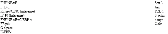
With the treatment of growth factor or interferon treatment of cells, it is observed that cells are activates of transcription (SIF/Stat), which binds to a serum inducible element first identified in the C-fos promoter (Stain and Baldwin, 1993). EGF treatment of animal cells induces Stat 3 activity in the liver. It is reported that IL-6 also activate Stat 3 (Ruff-Jamison et al., 1993). It was revealed that Stat3 DNA binding activity increased in the remnant liver within 30 min of PH and peaks at more than 30 fold after 3 h (Table 1).
The induction of Stat 3 appears to be part of the initial response of the remmant liver of PH, because it occurs in the presence of cyclo heximide-mediated protein synthesis blockade. Activation of Stat 3 is unusual because it extends beyond the immediately early time period and remains near peak level at 5 h post hepatectomy. Stat 3 contributes to the transcriptional activation of the immediate early genes that are induced over a prolonged time in the G1 phase of hepatic cell after PH. The identification of Stat 3 as an early factor for liver regeneration provides clues as to the signal transduction pathways that are activated in the remnant liver within the first minute to several hours after PH.
| Table 1: | Potential target genes of PHF/NF-κB and Stat 3 Potential target genes of |
 | |
EPIDERMAL GROWTH FACTOR (EGF) ACTIVITY DURING LIVER GROWTH
In the mouse, EGF functions as an endocrine factor that has profound effect on liver regeneration. EGF is produced mostly in salivary glands and is abundant in male animals. Removal of salivary glands delays the peak of DNA synthesis after PH by 24 h (Noguechi et al., 1991). In mice, with intact salivary glands, EGFR, mRNA and receptor binding activity increases during the first 8 h PH and decrease there after (Noguechi et al., 1992). Although, EGF is present in blood of normal mice, it apparently acts hepatocyte mitogens for hepatocytes after PH, a change the receptor is required to permit ligand binding and activation of signal transduction. Lack of circulating EGF in salaoadenectomized mice alters the timing of DNA synthesis after PH but does not decreases the proportion of hepatocytes that replicate during the process. Synthesis of EGF mRNA and peptide have been detected in rat liver shortly after PH, indicating that in these animals EGF may act by autocrine as well as endocrine mechanisms (Mead and Fausto, 1980; Fausto and Webber, 1983).
TGF-α EXPRESSION DURING LIVER DEVELOPMENT AND REGENERATION
Expression of TGF-α in the liver is associated with hepatocyte proliferation (Farts et al., 1982; Webber et al., 1993). TGF-α is produced by hepatocytes which can respond to the factor because they contain the specific receptor (EGFR). The autocrine loop of TGF-α synthesis is stimulated in liver cell cultures as well as in vivo by TGF-α itself or ECF providing an amplification mechanism for TGF-α synthesis (Wu et al.,1991; Russel et al., 1993). TGF-α is synthesized as a 160 amino acid precursor that is anchored in the cell membrane. The extra cellular domain of the precursor contains the 50 amino acid bounded in alanine residues at each end which are sites for cleavage by elastases. The precursor also has a transmembrane domain and a 35 amino acid cytoplasmic domain. The carboxy-terminal valine residue of the intercellular domain may serve as a signal site for cleavage of processed TGF-α peptide from the precursor molecule. TGF-α has 35 homology with EGF and binds to the same receptor (EGFR). During liver regeneration after partial hepatectomy in rats, TGF-α mRNA stat increase at about 4 h after PH and reaches a maximum before the peak of DNA synthesis. Peptide levels are increased at 24 and 48 h after the operation. The 50 amino acids diffusible from TGF-α is detected only at 48 h when the major wave of hepatocyte replication has taken place. These observations imply that membrane anchored, non diffusible forms of TGF-α may be active in hepatocytes and may account for a significant proportion of total TGF-α activity. One possibility that needs to be evaluated experimentally is that diffusible TGF-α becomes detectable only after its cell membrane receptors are completely accepted by the legend.
HEPATOCYTE GROWTH FACTOR (HGF) EXPRESSION DURING LIVER REGENERATION
HGF is a heterodimeric glycoprotein consisting of a heavy (α) and a light (β) chain of approximate molecular weight of 64000 and 32000, respectively. The heterodimeric form is generated from a single chain precursor peptide with a molecular weight of 87000-92000. The α-chain has four kringle domains (double loop structure with three disulfide bridges) with 40% homology with plasminogen. The β chain has homology with serine proteases but has no proteolytic activity of its own because of amino acid substitutions in the catalytic site residues.
On a molar basis HGF is the most potent of the liver mitogen (Strain, 1993; Schimacher et al., 1992). The factor are not produced by hepatocytes or other epithelial cells but is made by mesenchymal cells throughout the body. In the liver it is made by Kuffer cells and endothelial cells (Crilli et al., 1993; Eion and Baltimore, 1993). After PH HGF levels in blood increase sharply during the first 4-6 h. In addition, HGF mRNA produced by non parenchymal cells increases and reaches n maximum 18-24 h after the operation. Thus HGF could act on hepatocytes during liver regeneration endocrine or paracrine mechanism. Although the rapid increase in circulating levels of HGF shortly after PH indicates that the factor plays a role in the early events of the process. However, more precise experiments need to be done to determine whether the rise in circulating HGF triggers liver regeneration.
ROLE OF CYTOKINES AND TRANSCRIPTION FACTOR DURING LIVER REGENERATION TNF-KB
NF-KB comprises a family of protein that are related to bomo and heterodimers and also related to the rel oncogene and the drosophila gene dorsal. Originally described in lymphocytes NF-KB proteins have been detected in many cell types and found to participate in gene activation related to defense mechanisms and cell proliferation process. NF-KB (Eion and Baltimore, 1993) activation is a rapid process that involves post translation modification of protein which is triggered by many different types of stimulate such as endotoxins (LPS), TNF α, IL-I, IL-2 UV light and oxidants (Tewari et al., 1992). Its target genes include, among others, surface immunoglobulin, adhesion molecules, cytokines, acute-phase response genes, the c-myc protoncogeneis and several viruses including HIV. NF-κB activation induced by most if not all, agents converges into an intracellular pathway that involves oxidant molecules. Anderson et al. (1994) proposed that there is a common pathway of NF-κB activation involving the generation of mitochondrial oxidant, followed by protein phosphorylation and degradation of IKBS (inhibitor of KB) leading to the production of active NF-κB (composed of two subunits designated (p50 and p65), which migrates into the nuclease. It has recently been found that knockout mice that lack p65 die during embryonic development with massive apoptotic liver cell death.
Tewari et al. (1992) reported that a protein complex named PHF (post hepatectomy factor) was activated and bound to DNA within minutes after PH. PHF was found to be protein related to the transcription factor NF-κB whereas RL-F1 was identified as IKB α, a specific inhibitor of NF-κB activities.The authors proposed that PHF may act by competing with NF-κB for the same DNA binding sites because PHF binding was insensitive to RL-IF1 (IKBα) inhibition. Tewari et al. (1992) concluded that at the strat of liver regeneration (IκBα) gene activities would suppresses NF-κB binding and enhance PHF activation. Increased NF-κB binding was detected in nuclear extract from hepatocytes isolated 30 min after PH (Cressman et al., 1994b). Extracts from these cells also contained p50 homodimers (which bind to DNA but lack transcriptional activities) and the minor complex, which correspond to PHF. These three complexes could also be detected in nuclear extracts from normal lvier nonparenchymal cells but so increase in NF-κB binding (that is p50/p65 heterodimer) was detected in nonparenchymal cell extracts after PH. Increased NF-κB binding to DNA was also detected after 30% hepatectomy, suggest that NF-κB activation may be involved in the initial steps of liver regeneration. However, NF-κB acitivation by itself is obviously not sufficient to cause DNA synthesis because animals with 30% hepatectomy, although capable of responding to growth factors, do not undergo steps of liver regenertion. However, NF-κB activation by itself is obviously not sufficient to cause DNA synthesis because animals with 30% hepatectomy, although capable of responding to growth factor, do not undergo DNA synthesis.
ROLE OF TNF AND NF-kB AFTER PARTIAL HEPATECTOMY
The rapid activation of NF-κB after PH indicates that a signal for gene activation has been received in the hepatocyte nucleus almost immediately after the operation. It then becomes essential to identify the mechanisms and agents responsible for NF-κB activation at the start of liver regeneration and to determine the target genes of the NF-κB response. Intraperitoneal injection of 5 μg of TNF to intact liver of rats caused strong induction of NF-κB 30 min after the injection, similar to what was found after PH (Diehl et al., 1994). Diehl and colleagues have concluded from the experiments using TNF antibodies that TNF may cause elevation of c-jun, jun-kinase and AP1 after PH and blockage of TNF activity inhibits liver regeneration. It is thus, conceivable that TNF may contribute to the initiation of liver regeneration by being responsible for both NF-κB and AP1. On the other hand, NF-κB activation depends on many other factors and might be regulated at the start of liver regeneration by phosphorylation and proteolytic steps causing the degradation of the inhibitor (IκB).The phosphorylation step is modulated by the red-ox state of the cell whereas the proteolytic cleavage of (IκB) depends on proteosome activity. Thus, NF-κB activation could be initiated by extra cellular stimuli such as TNF or by intracellular signal involving the generation of reactive oxygen intermediates and proteolytic activity.
REFERENCES
- Axxanha, M., B. Goszczynski, M.A. Chung, M. Kalb and J.D. Meanee, 1996. A fork head/HNF-3 homolog expressed in hepharynx and intestine of the caenorhabditis, elegans embryo. Dev. Biol., 178: 289-303.
Direct Link - Beddington, R.S. and E.J. Roberson, 1998. Anterior patterning in mouse. Gene, 14: 277-284.
Direct Link - Cascio, S. and K.S. Zaret, 1991. Hepatocyte differentiation initiates during endoermal mesenchymal interactions prior to liver formation. Development, 113: 217-225.
Direct Link - Chattopadhyay, P., S.S. Agrawal and A. Garg, 2006. Liver regeneration effects of Phyllanthus amarus Linn. Against alcohol induced cell injury in partially hepatectomised albino rats. Int. J. Pharmacol., 2: 426-430.
Direct Link - Chattopadhyay, P., S.S. Agrawal and A. Garg, 2006. Tridothyronone (T3) regulation on liver cell regeneration in alcohol induced liver cell injury in partial hepatectomised albino rats. Ind. J. Gastroenterol., 25: 165-166.
Direct Link - Chattopadhyay, P., A. Garg, V.P. Varshey, A.K. Sharma and S.S. Agrawal, 2007. Increase insulin activity by Phyllanthus amarus linn on liver cell regeneration in partially hepatectomised albino rats. Res. J. Medicinal Plant, 1: 17-20.
CrossRefDirect Link - Chen, W.S., 1994. Disruption of the HNF-4 gene, expressed in visceral endoderm leads to cell death in embryonic ectoderm and impaired gastrulation of mouse embryo. Genes Dev., 8: 2466-2477.
Direct Link - Cressman, D.E. and R. Taub, 1994. Physiologic tumor of NF-κB by nuclear proteolysis. J. Biol. Chem., 269: 26594-26597.
Direct Link - Cressman, D.E., L. Gereenbalum and B.A. Haber, 1994. Rapid post hepatectomy fator/nuclear factor κB in hepatocytes a primary responded in the regenerating liver. J. Biol. Chem., 269: 30129-30435.
Direct Link - Crompton, M.R., 1992. Identification of a novel vertebrate homeobox gene expressed in haematopoietic cells. Nucl. Acids. Res., 20: 5661-5667.
Direct Link - Crossley, P.H. and G.R. Martin, 1995. The mouse Fgf8 gene encodes a family of polypeptide and is expressed in regions that direct outgrowth and patterning in the developing embryo. Development, 121: 439-451.
Direct Link - Deutsch, G., J. Jung, M. Zheng, J. Lara and K.S. Zaret, 2001. A bi potential precursor population for population for pancreas and liver within the embryonic endoderm. Development, 128: 871-881.
Direct Link - Grilli, M., J.J. Chiu and M.J. Lenardo, 1993. NF-κB and Rel: Participants in a multiform transcriptional regulatory system. Int. Rev. Cytol., 143: 1-62.
Direct Link - Gualdi, R., 1996. Hepatic specification of the gut endoderm in vitro: Cell signalling and transcriptional control. Genes. Dev., 10: 1670-1682.
Direct Link - Homas, R., J. Radich and S. Collins, 1993. PCR cloning of an orphan homo box gene (PRH) preferentially expressed in myeloid and liver cells. Biochem. Biophys. Res. Commun., 195: 976-983.
Direct Link - Horner, M.A., 1998. Pha-4, a HNF-3 homolog, specific pharyngeal organ identity in caenorhabditis elegans. Gene Dev., 12: 1947-1952.
Direct Link - Jung, J., M. Zheng, L. Goldfarb and K.S. Zaret, 1999. Initiation of mammalian liver development from endoderm by fibroblast growth factor. Science, 284: 1998-2003.
CrossRefDirect Link - Keng, V.W., 2000. Homeobox gene hex is essential for onset of mouse embryonic liver development and differentiation of monocyte lineage. Biochem. Biophys. Res. Commun., 276: 1155-1161.
Direct Link - Lai, E., V.R. Prezioso, W. Taa, W.S. Chen and J.E. Darnell, 1991. Hepatocyte nuclear factor 3A belongs to a gene family in mammals that is homologous to the Drosophila homeotic gene fork head. Genes Dev., 5: 416-427.
Direct Link - Michalopoulos, G. and R. Zarnegar, 1992. Hepatocyte growth factor. Hepatology, 15: 119-155.
Direct Link - Noguechi, S., Y. Ohba and T. Oka, 1991. Influence of epidermal growth factor on liver regenertion after partial hepatectomy in mice. J. Endocrinol., 128: 125-131.
Direct Link - Noguechi, S., Y. Ohba and T. Oka, 1992. The role of transcription and messenger RNA stability in the regulator ion of epidermal growth factor receptor gene expression in regeneration mouse liver. Hepatology, 15: 88-96.
Direct Link - Ruff-Jamison, S., K. Chen and S. Cohen, 1993. Induction by EGF and interferon-γ of tyrosine phosphorylated DNA-binding proteins in mouse liver nuclei. Science, 261: 1733-1736.
Direct Link - Rusell, W., P. Dempsay, S. Siraric, A. Peck and R. Cottey, Jr., 1993. Transforming growth factor-alpha (TGF-alpha) concentration increases in regenerating rat liver: Evidence for a delayed accumulation of mature TGF alpha. Endocrinology, 133: 1731-1738.
Direct Link - Sladex, F.M., W. Zhong, E. Lai and J.E. Darnell Jr., 1990. Liver enriched transcription factor HNF-4 is a novel member of the steroid hormone receptor super family. Genes Dev., 4: 2353-2365.
Direct Link - Soutolou, E., N. Katrakilim and I. Talianidis, 2000. Acetylating regulates transcription factor activity at multiple levels. Mol. Cell, 5: 745-751.
Direct Link - Spath, G.F. and M.C. Weiss, 1994. Hepatocytes nuclear factor 4 provokes expression of epithelial marker genes, acting as a morphogen in differentiated hepatoma cells. J. Cell Biol., 140: 935-946.
Direct Link - Stain, B. and A.S. Baldwin, Jr., 1993. Distinct mechanisms for regulation of the interleukin-8 gene involve synergism and cooperativity between C/EBP and NF-κB. Mol. Cell. Biol., 13: 7191-7198.
Direct Link - Strain, A., 1993. Hepatocyte growth factor. Another ubiquitous cytokine. J. Endocrinol., 137: 1-5.
Direct Link - Strak, K.L., J.A. McMohan and A.P. McMohan, 1991. FGFR-4, a new member of fibroblast growth factor receptor family, expressed in definite endoderm and skeletal muscle uneages of the mosue. Development, 113: 641-651.
Direct Link - Sugi, Y., J. Sasse, M. Barron and J. Lough, 1995. Developmental expression of fibroblast growth factor receptor-1 (cck-1; Ffg) during hear development. Dev. Dynam., 202: 115-125.
Direct Link - Thomas, P.Q., A. Brown and R.S.P. Beddington, 1998. Hex: A homeobox gene revealing peri-implantation asymmetry in the mouse embryo and an early transient marker of endothelial cell precursors. Development, 125: 85-94.
Direct Link - Winnier, G., M. Blessin, P.A. Labosky and B.L.M. Hogan, 2003. Bone morphogenetic protein-4 is required for mesodern formationand patterning in the mouse. Genes Dev., 9: 2105-2216.
Direct Link - Wu, J.C., G. Merlino, K. Cbeklova, B. Mosinger, Jr. and N. Fausto, 1991. Autonomous growth in serum-free medium and production of hepatocellular carcinomas by differentiated hepatocyte liver that express ransforming growth factor alpha 1. Cancer Res., 54: 5964-5973.
Direct Link - Zarel, K., 1990. Developmental competence of the gut endoderm: Genetic potentiation by GATA and HNF 3/fork head proteins. Dev. Biol., 209: 1-10.
Direct Link - Zhu, X., J. Sasse, S. McAllister and J. Lough, 1996. Evidence that fibroblast growth factors 1 and 4 participate in regulation of cardiogenesis. Dev. Dynam., 287: 429-438.
Direct Link