
M. Kumar
Veterinary Polyclinic, Bharatpur, Rajasthan, 321001, India
B. Sharma
Department of Epidemiology and Veterinary Preventive Medicine, Pandit Deen Dayal Upadyay Veterinary and Animal Science University (DUVASU) Mathura, Uttar Paradesh, 281001, India
A. Kumar
Division of Animal Health, Central Institute for Research on Goats, Makhdoom P.O. Farah, Mathura, Uttar Paradesh, India
H.P. Lal
Department of Veterinary Medicine,
V. Kumar
Extension Education and Socio Economic Section, Central Institute for Research on Goats, Makhdoom P O Farah, Mathura, Uttar Paradesh, India
M.K. Tripathi
Devision of Veterinary Physiology and Biochemistry, F.V.Sc and A.H., Sher-e-Kashmir University of Agricultural Sciences and Technology of Jammu, Ranbir Sing, Pura, Jammu, India
Asian Journal of Animal and Veterinary Advances
Year: 2014 | Volume: 9 | Issue: 10 | Page No.: 653-663


ABSTRACT
Toxocara canis is a very important gastrointestinal nematode affecting canines with considerable public health importance. This study was conducted to find out the prevalence and corresponding haemato-biochemical changes in dogs infested with T. canis and to determine its zoonotic implication to dog owners. A total of 121 dogs were screened from October 2008 to May 2009 by direct smear and Mc-master technique, to determine correlation between overall prevalence of T. canis infestation with respect to sex, age, breed, size and season-wise infestation. Haemato-biochemical profile was performed in 24 infested dogs, randomly selected to evaluate changes in Hb, PCV, TEC, TLC, DLC count, serum protein, serum glucose and serum enzymes. The overall prevalence was found to be 28.93%. The prevalence was not influenced by sex but non-descript breeds had significantly higher rates. Pups were more infested than adults and the disease was more prevalent in winters. Dogs having active infection with T. canis infestation showed anemia, leucocytosis and significant eosinophilia (p<0.05). A significant decrease (p<0.05) was observed in serum protein and glucose whereas highly significant increase (p<0.01) was found for both serum enzymes (SGOT and SGPT). Very few dog owners (4.13%) were aware about potential public health significance of the parasite. Survey revealed that unaware owners who belonged to lower/middle/upper middle class, did not maintain hygiene and scheduled deworming and always remain at high zoonotic risk. Being zoonotic, the parasite poses a significant danger to humans mainly children who remain in their vicinity. Thus immediate action needs to be taken to control this parasite and to increase awareness among the dog-owners about the zoonoses being spread by the companion animals.
PDF Abstract XML References Citation
Received: July 19, 2014;
Accepted: September 15, 2014;
Published: October 01, 2014
How to cite this article
M. Kumar, B. Sharma, A. Kumar, H.P. Lal, V. Kumar and M.K. Tripathi, 2014. Prevalence and Haemato-Biochemical Studies of Toxocara canis Infestation in Dogs and Risk Perception of Zooneses by Dog Owners in Mathura, India. Asian Journal of Animal and Veterinary Advances, 9: 653-663.
DOI: 10.3923/ajava.2014.653.663
URL: https://scialert.net/abstract/?doi=ajava.2014.653.663
DOI: 10.3923/ajava.2014.653.663
URL: https://scialert.net/abstract/?doi=ajava.2014.653.663
INTRODUCTION
Toxocara canis is one of the most common parasites found in the small intestine of dogs, producing eggs passing out with the faeces and becoming a source of infection to other susceptible dogs (Roberts and Janovy, 1996). Dogs can also acquire the infection by ingesting infective larvae in tissues of paratenic hosts or in the vomitus of infected pups (Sprent, 1961), transplacental migration and transmammary passage (Stone and Giardeau, 1968). Heavy infestations of parasites occur most commonly in kennels with poor hygienic conditions (Jacobs et al., 1977). On ingestion, eggs hatch in small intestine and second stage larva penetrate into intestinal wall and reach liver and lungs via bloodstream from where they are coughed up, swallowed and finally enter the small intestine where they mature (Urquhart et al., 1996). Heavy infestations in young pups may cause nervous signs and death (Urquhart et al., 1996). In adult dogs, T. canis infestation causes emaciation, anaemia, constipation, intermittent diarrhea, a pot-bellied appearance and even death due to acute intestinal obstruction.
A dog excretes thousands of T. canis eggs into the environment that are potential source for humans who acquire the infection directly by ingesting unwashed vegetables and fruits contaminated with Toxocara eggs or indirectly through contact with animal secretions and excreta (Daryani et al., 2008; Overgaauw and Nederland, 1997; Magnaval et al., 2001). Now a days, dogs are living with humans as companion animals all over the world. Children are at special risk as compared to adults because of their closer contact with dogs. Two syndromes of toxocariasis have been described in humans, i.e., Visceral Larva Migrans (VLM) and Ocular Larva Migrans (OLM) (Bugg et al., 1999; Soulsby, 1986).
Epidemiological pattern of the parasitic diseases in the different agro-climatic zones of the country usually provides a basis for developing strategic and tactical control systems against them (Traub et al., 2007). Keeping in view the importance of T. canis infection in relation to human health, the present study was designed to determine the prevalence of T. canis and various epidemiological factors affecting its occurrence as well as its effect on different haemato-biochemical parameters in dogs along with an attempt to determine the awareness regarding potential risk of zoonotic infection to dog owners in and around Mathura district.
MATERIALS AND METHODS
Prevalence of toxocariasis: A total of 121 dogs (both stray and pet) under one year age and of either sex were screened from October 2008 to May 2009 to find out the prevalence of Toxocariasis in and around Mathura district, on the basis of qualitative (direct smear method) and quantitative (will’s floatation method) faecal examination. Individual faecal samples without any preservatives were collected directly from rectum and examined immediately. To obtain accurate information with regard to severity of infection, the fecal samples have been screened using Mc-Master technique (Fig. 1) which uses saturated salt solution (Maff, 1977). To determine different epidemiological factors influencing the prevalence of T. canis infestation, animals were grouped according to breed, sex, age and size. To study the influence of age, three groups were made as 0-1, 1-3 and >3 months. Dogs were grouped according to their size as large breeds (Doberman, German shepherd, Labrador, Boxer and Great Dane) and small breeds (Dachshund, Samoyed and Pomeranian).
Questionnaire survey: A structured questionnaire was prepared in local (Hindi) language to survey the dog owners regarding their awareness about the zoonotic risks to human (dog owners) from dogs.
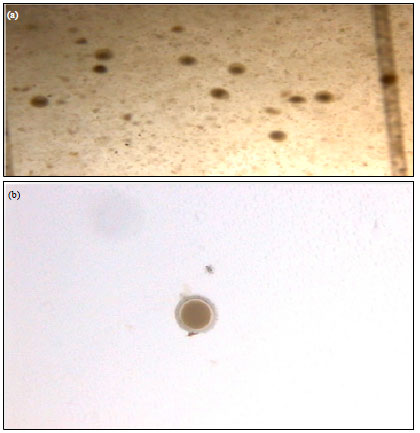 | |
| Fig. 1(a-b): | Photograph showing (a) Toxocara canis eggs at phase contrast microscope 10x and (b) Eggs in the Mc-master slide 10x |
The questionnaire was designed to gather information on deworming, housing, feeding schedule and defecation pattern of dogs, hygienic measures employed, use of neck collar in dogs, pet clinic visits, the extent of awareness on dog parasites, activities with dog (recreational purposes), economic and social status of the dog owners, attitude towards zoonoses and other related factors.
Haemato-biochemical analysis: To compare haemato-biochemical parameters in healthy and infected dogs, a case group A consisting of randomly selected 24 infected dogs was created. The parameters of these dogs (group A) were compared to those of healthy dogs (group B).
Blood sample collection and processing: For haematological studies, About 5 mL blood, was collected from cephalic vein in clean dry test tubes containing anticoagulant (EDTA at 1.5 mg dL-1 of blood) from each dog. For biochemical analysis, 6 mL blood was taken in another test tube without anticoagulant and serum was collected and stored at -20°C.
Estimation of blood parameters
Hb, PCV, TEC, TLC, DLC: Hemoglobin (Hb) (g dL-1) was estimated by Sahli’s method as recommended by Benjamin (1986). Packed Cell Volume (PCV) was determined using Wintrobe’s method as described by Coles (1986). Erythrocyte count (TEC) and total leukocyte (TLC) was determined according to the method described by Benjamin (1986). Differential Leukocyte Count (DLC) was done by hematocrit tube method and blood films stained by Giemsa staining method (Schalm et al., 1986).
Estimation of serum biochemical parameters
TSP, SG, SGOT, SGPT: Biochemical analysis of serum samples was done to estimate total serum protein by Modified Biuret method (Koller, 1984), Serum glucose by GOD-POD method (Tietz, 1976), Serum Glutamate Oxaloacetate Transaminase (SGOT) and Serum Glutamate Pyruvate Transaminase (SGPT) (Reitman and Frankel, 1957). All the estimations were carried out by using commercial biochemical kits (Span Diagnostic Ltd., Surat, India) with the help of auto serum analyser (Mindray GS 120).
Statistical analysis: The data analysis has been done with the aid of Excel spreadsheet to arrive at the descriptive statistics for summarizing the data. Chi-square test was used to assess the association of risk factors on the prevalence of parasites. All statistical analyses were performed using the SPSS statistical software.
RESULTS
Questionnaire survey: Table 1 shows the detailed questionnaire survey. In most of the cases, dogs were maintained unhygienically (90.08%), defecating haphazardly within the house premises (70.24%). Around 80.16% pet dogs shared common space with owners and were fed on mostly home food or leftover food (80.99%) with no considerations to special nutritional requirements of the canines. Very few owners (4.13%) knew that canine parasites could be transmitted to humans with 69.49% dog owners with majority of their children having close contact with their pups without taking any precautions. Only 15.70% of the dog owners had followed a standard deworming schedule in their lifetime. This study showed that 60.33% dog owners used neck collars for their dogs and only 12.39% visited pet clinic regularly.
Prevalence of toxocariasis: Table 2 shows the factors associated with prevalence of Toxocara canis. An overall prevalence of T. canis infestation was found to be 28.93% in this study. The classical clinical symptoms observed in the affected dogs were pot bellied appearance, stunted growth, intermittent diarrhoea and constipation, dull hair coat, pseudo-rickets with pale mucous membrane and abdominal bloating after nursing. The prevalence revealed an increasing trend from October 2008 to February 2009 and a decreasing trend from March to May 2009, with maximum prevalence (41.66%) in February and minimum prevalence (9.09%) in May (Fig. 2). Autumn or spring season was found to be the most conducive for the spread of toxocariasis.
 | |
| Fig. 2: | Month wise prevalence of percentage infection Toxocara canis in dogs |
The sex wise prevalence was found to be 29.23 and 28.57% in males and females, respectively. Among descript breeds, the prevalence rate was found to be 14.74% as compared to 40.68% in local/non-descript breeds. Prevalence rate in large and small sized dogs of descript breeds was 17.14 and 18.52%, respectively. In different age groups (0-1, 1-3 and >3 month age), the prevalence was 31.82, 55.56 and 18.06%, respectively. The prevalence rate during winter season was 37.33% whereas, in other seasons, it was 15.22% only.
| Table 1: | Knowledge of dog owners regarding zoonotic risk from Toxocara canis infestation in dogs in Mathura district |
 | |
| NSp>0.05, ** p<0.01, *p<0.05 | |
| Table 2: | Factors associated with prevalence of Toxocara canis in dogs |
 | |
| NSp>0.05, **p<0.01, @(March, April, May, October), large size breed (Doberman, German shepherd, Labrador, Boxer and Great Dane), small size breed (Dachshund, Samoyed and Pomeranian). Inference: Toxocara canis are equally prevalent in male and female dogs (χ2 0.018, p>0.05). Non-descript (local) breeds had significantly higher prevalence than descript breed, it might be due to descript breed get good management and health care prophylactic under their owners. Descript breeds of different size i.e., large and small were equally prevalent. Different age groups of dogs differ significantly (p<0.01) on prevalence. In winter season prevalence of disease is significantly more (p<0.01) than other season of the year | |
| Table 3: | Values of blood and serum parameters of healthy and Toxocara canis infested dogs |
 | |
Blood parameters: The values of PCV, Hb and TEC were found to be lower in infected group than in healthy dogs. There was a significant increase in TLC values in infected group as compared to healthy group. The lymphocytes slightly decreased whereas, neutrophil and eosinophil counts showed an increasing trend in the infected dogs. The results of packed cell volume, Haemoglobin, total erythrocyte count, Total leucocyte count and DLC Mean±SE values for infected (group A) and healthy dogs (group B) are presented in Table 3.
Serum biochemical parameters: Total protein (TSP) value for infected group was found to be significantly lower than in the healthy group. The same trend was followed by Serum Glucose (SG) value. Both Serum Glutamate Oxaloacetate Transaminase (SGOT) and Serum Glutamate Pyruvate Transaminase (SGPT) values were markedly increased for infected group. The results of TSP, SG, SGOT and SGPT Mean±SE values for infected (group A) and healthy dogs (group B) have been presented in Table 3.
DISCUSSION
Based on the screening of fecal samples, overall prevalence of Toxocara canis infestation in dogs was found to be 28.93%. Similar findings were also obtained by several workers in their respective studies across the world including India with the prevalence ranging from 8.7-36.6% (Coman et al., 2009; Panigrahi et al., 2014; Bahrami et al., 2011; Katagiri and Oliveira-Sequeira, 2008).
Sex of dogs did not influence the likelihood of acquiring Toxocara canis infestation as prevalence in male and female dogs was 29.23 and 28.57%, respectively, the difference being non significant (p>0.05). This observation is supported by the findings of Swai et al. (2010) and Anene et al. (1996).
Non-descript local dogs were more prone to infestation of Toxocara canis with prevalence being 40.68% whereas, in descript breeds dogs, prevalence was only 14.74%, the difference being highly significant (p<0.01). Similar findings are reported by Panigrahi et al. (2014) and Sahu et al. (2014). The reasons of significantly higher prevalence in non descript dogs may be due to the factors like climatic conditions of semiarid zone, lack of awareness, care and hygiene and poor management practices adopted by the owners, who were mostly from socio-economically poor classes. The owners of foreign breeds generally belonged to middle to upper classes and were more aware about proper management of pets including regular deworming which lead to low rate of prevalence in their pets as compared to non-descripts (Anene et al., 1996). Similar observations were also recorded by other co workers who described the prevalence ranging between 6.5-93% in non-descript dogs (Doganay and Oge, 1993; Jani et al., 1995). Reports with a little lower prevalence in descript dogs were published by various workers (Jani et al., 1995; O’Lorcain, 1994; Saito et al., 1995).
Descript breeds of different size i.e., large and small were equally predisposed for infestation as prevalence rate in large and small size dogs were 17.14 and 18.52%, respectively. This difference was not significant (p>0.05).
Age of animal played a very significant role (p<0.01) in prevalence of Toxocara canis infestation. Among the different age groups studied (0-1, 1-3 and >3 month age), the prevalence of Toxocara canis infestation was 31.82, 55.56 and 18.06%, respectively, the difference being highly significant (p<0.01). The highest prevalence was in pups of 1-3 month age group. Findings similar to our study have been reported by Khante et al. (2009) whereas, much higher prevalence upto 81% has been reported in pups by Coman et al. (2009). Higher prevalence in pups have also been reported by various workers (Panigrahi et al., 2014; Swai et al., 2010; Senlik et al., 2006; Luty, 2001). This may be due to premature immune development in pups that are susceptible to the infection through prenatal, colostral or lactogenic transmission (Stone and Giardeau, 1968).
Season of area of study also played a very significant role (p<0.01) in prevalence of infestation of Toxocara canis. In winters, the prevalence was 37.33% whereas, in other season it was 15.22% only. This difference was highly significant (p<0.01). This low rate of infection in summers could possibly be due to the hot, arid climatic conditions which might render eggs non-infective or by the sandy soil in the area that might cover faeces containing T. canis ova making eggs unavailable to other dogs (Gross et al., 1984). These findings are in close resemblance with the earlier reports (Das et al., 2009; Radwan et al., 2009; Avcioglu and Burgu, 2008; Andresiuk et al., 2007).
In the present study, a significant decrease (p<0.05) was observed in the values of Hb, PCV and TEC in the infected group, in comparison to healthy group. These findings tally with the results of several other workers (Qadir et al., 2011; Sharma et al., 2010; Chattha et al., 2009; Ogunkoya et al., 2006). Total Leucocyte Count (TLC) was significantly higher (p<0.05) in infected dogs indicating leucocytosis (Sharma et al., 2010; Chattha et al., 2009). This increase in TLC might be due to liberation of histamine and other histamine like substances from the damaged tissues. These values were also in close agreement with the findings of (Karadam et al., 2008; Lai and Chen, 2007). No significant change (p>0.05) was observed in the values of lymphocyte, neutrophil and monocyte count in the infected group whereas, significant eosinophilia was observed (p<0.05) (Sharma et al., 2010; Ogunkoya et al., 2006; Lai and Chen, 2007). Eosinophilia is induced by changes in local and circulating histamine concentration caused by tissue injury (Schalm et al., 1986). Rising Blood histamine concentration may provide the stimulus for increased release of eosinophils from bone marrow, producing a subsequent eosinophilia (Ettinger, 1975).
A significant drop (p<0.05) in the value of Total Serum Protein (TSP) in infected dogs was recorded. Similar trend has already been reported earlier by Hayden and Kruiningen (1975), Kaymaz et al. (1999) and Nwoha et al. (2013). The reduction in TSP may be due to chronic internal haemorrhage during infection and loss of serum, via exudation or leakage in the lumen of gut causing enteropathy. The altered rate of intestinal absorption of nutrients especially protein from the injured gut during infection might also have contributed to the drop in the TSP (Dargie and Allonby, 1975). Hypoproteinaemia may also be attributed to the interference with efficacy of digestion and absorption by damaged intestinal mucosa and diarrhoea due to mechanical irritation. A significant drop (p<0.05) in values of Serum Glucose (SG) was also recorded in infected dogs. These findings tally with the result of Lloyd et al. (1981). In infected dogs, the increase in the values of Serum Glutamic Oxalo-acetic Transaminase (SGOT) and Serum Glutamic Pyruvic Transaminase (SGPT) values were highly significant (p<0.01) (Nwoha et al., 2013; Hayden and Kruiningen, 1975). Such an increase in transaminase activity supports greater liberation of enzymes from damaged and necrosed intestinal tissues. In advanced cases of infection, excessive liberation of enzymes may be correlated with the increased permeability of liver cells and intestinal tissue allowing more diffusion of GOT and GPT enzymes in to blood stream and their elevation in serum. The necrosis of cells during intestinal and extra-intestinal phases of Toxocara canis in dogs was directly responsible for the increase in enzymes.
The role of dogs as companion animals and the close relationship between humans and dogs although significantly beneficial, also represents a potential public health risk as natural transmission of parasitic infections from dogs to humans which may occur directly or indirectly via environmental factors. All kinds of dogs (owned and stray dogs) are involved in transmission, even if the particular implication of each population is not clearly established (Dogan et al., 2007). Survey revealed that unaware owners belonging either to lower/middle/upper middle class, who did not maintain hygiene and scheduled deworming, shared home or played with their dogs, fed their dog with home food cooked or human leftover food and did not visit pet clinic usually, always remain at high zoonotic risk with T. canis infection. The difference was highly significant (p<0.01) in this group. Similar findings were also recorded in respective studies by Panigrahi et al. (2014) and Ahmed et al. (2014). Question regarding the use of neck collar to dogs also revealed a significant difference (p<0.05) with the group not using neck color. Neck collar prevented dogs becoming exposed to toxocara eggs.
CONCLUSION
It could be concluded that stray dogs and pups infected with T. canis pose high zoonotic risk to humans especially who live in vicinity of pets like dog trainers, veterinarians and children. Most of dog owners are unaware about zoonotic risk from this parasite, whilst they maintain their pets unhygienically without regular deworming or visit to pet clinics. The prevalence of T. canis infestation in dogs was found to be 28.93% and it was found to be influenced by age, breed and season. The infestation caused significant haemato-biochemical derangement in the affected dogs. Interventions needed should focus on health education provided to dog owners, strategic deworming of dogs using broad spectrum anthelmintic and proper sanitation and hygiene.
ACKNOWLEDGMENT
The authors are highly thankful to Dean, College of Veterinary Science and Animal Husbandry and Director, Teaching Veterinary Clinical Complex for their cooperation and provision of research facilities.
REFERENCES
- Ahmed, W.M., W.M. Mousa, S.M. Aboelhadid and M.M. Tawfik, 2014. Prevalence of zoonotic and other gastrointestinal parasites in police and house dogs in Alexandria, Egypt. Vet. World, 7: 275-280.
Direct Link - Andresiuk, V., N. Sardella and G. Denegri, 2007. Seasonal fluctuations in prevalence of dog intestinal parasites in public squares of Mar del Plata city, Argentina and its risk for humans. Rev. Argent. Microbiol., 39: 221-224.
PubMed - Anene, B.M., T.O. Nnaji and A.B. Chime, 1996. Intestinal parasitic infections of dogs in the Nsukka area of Enugu State, Nigeria. Prev. Vet. Med., 27: 89-94.
CrossRef - Avcioglu, H. and A. Burgu, 2008. Seasonal prevalence of Toxocara ova in soil samples from public parks in Ankara, Turkey. Vector-Borne Zoonot. Dis., 8: 345-350.
CrossRefDirect Link - Bahrami, A.M., A.Z. Doosti, H. Nahrevanian, A.M. Noorian and S.A. Asbchin, 2011. Epidemiological survey of gastro-intestinal parasites in stray dogs and cats. Aust. J. Basic Applied Sci., 5: 1944-1948.
Direct Link - Bugg, R.J., I.D. Robertson, A.D. Eliot and R.C.A. Thompson, 1999. Gastrointestinal parasites of urban dogs in Perth, Western Australia. Vet. J., 157: 295-301.
CrossRef - Chattha, M.A., A. Aslam, Z.U. Rehman, J.A. Khan and M. Avais, 2009. Prevalence of Toxocara canis infection in dogs and its effects on various blood parameters in Lahore (Pakistan). J. Anim. Plant Sci., 19: 71-73.
Direct Link - Coman, S., I.C. Dida and B. Bacescu, 2009. Incidence and treatment of the diarrhoeic syndrome with parasite aetiology in dogs and cats. Revista Scientia Parasitologica, 10: 106-111.
Direct Link - Daryani, A., M. Sharif, A. Amouei, F. Askarian, H. Ziaei, S.H. Gohardehi and R. Bastani, 2008. Prevalence of Toxocara canis in stray dogs in Northern Iran. Int. J. Infect. Dis., 12: e379-e379.
CrossRefDirect Link - Das, S.S., D. Kumar, R. Sreekrishnan and R. Ganesan, 2009. Gastrointestinal parasitic infections in dogs in Puducherry. J. Vet. Parasitol., 23: 77-79.
Direct Link - Dogan, N., E.C. Dinleyici, O. Bor, S.O. Toz and Y. Ozbel, 2007. Seroepidemiological survey for Toxocara canis infection in the Northwestern part of Turkey. Turkiye Parazitoloji Dergisi, 31: 288-291.
PubMedDirect Link - Dargie, J.D. and E.W. Allonby, 1975. Pathophysiology of single and challenge infections of Haemonchus contortus in Merino sheep: Studies on red cell kinetics and the self-cure phenomenon. Int. J. Parasitol., 5: 147-157.
CrossRefPubMedDirect Link - Gross, E.M., R. Zeitan and V. Torok, 1984. Toxocara canis infection in dogs in Beersheba, Israel. J. Helminthol., 58: 139-141.
PubMed - Hayden, D.W. and H.J. Kruiningen, 1975. Experimentally induced canine toxocariasis: laboratory examinations and pathologic changes, with emphasis on the gastrointestinal tract. Am. J. Vet. Res., 36: 1605-1614.
PubMed - Jacobs, D.E., E.J. Pegg and P. Stevenson, 1977. Helminths of British dogs: Toxocara canis: A veterinary perspective. J. Small Anim. Pract., 18: 79-92.
CrossRef - Jani, R.G., B.M. Jani and M.R. Dave, 1995. Prevelance of intestinal parasites in dogs at Anand (Gujarat). J. Vet. Parasitol., 9: 51-53.
Direct Link - Karadam, S.Y., S. Ertug, H. Ertabaklar and P. Okyay, 2008. The comparision of IgG antibodies specific to Toxocara spp. among eosinophilic and non-eosinophilic groups. Microbiol. Q. J. Microbiol. Sci., 31: 113-116.
Direct Link - Katagiri, S. and T.C.G. Oliveira-Sequeira, 2008. Prevalence of dog intestinal parasites and risk perception of zoonotic infection by dog owners in Sao Paulo State, Brazil. Zoonoses Public Health, 55: 406-413.
CrossRefPubMedDirect Link - Kaymaz, A.A., U. Bakirel, R. Gonul and H. Tan, 1999. Serum protein electrophoresis in dogs with intestinal parasites. Truk. J. Vet. Anim. Sci., 23: 457-459.
Direct Link - Khante, G.S., L.A. Khan, A.M. Bodkhe, P.R. Suryawanshi, M.A. Majed, U.S. Suradkar and S.S. Gaikwad, 2009. Epidemiological survey of gastro-intestinal parasites of non-descript dogs in Nagpur city. Vet. World, 2: 22-23.
Direct Link - Sharma, S.K., S.R.P. Sinha, S. Sinha, C. Jayachandran and M. Kumar, 2010. Post-treatment haematological studies on toxocariosis in dogs. J. Vet. Parasitol., 24: 63-65.
Direct Link - Lai, S.C. and K.M. Chen, 2007. Changes to plasminogen activators and matrix metalloproteinase-9 in dogs with toxocarosis. Vet. Parasitol., 150: 122-127.
PubMed - Lloyd, S., S. Kristensen and E.J.L. Soulsby, 1981. The effect of corticosteroid therapy on infection with Toxocara canis in dogs. Zeitschrift Parasitenkunde, 66: 57-61.
CrossRefPubMedDirect Link - Luty, T., 2001. Prevalence of species of Toxocara in dogs, cats and red foxes from the Poznan region, Poland. J. Helminthol., 75: 153-156.
PubMed - Magnaval, J.F., L.T. Glickman, P. Dorchies and B. Morassin, 2001. Highlights of human toxocariasis. Korean J. Parasitol., 39: 1-11.
CrossRefPubMedDirect Link - Nwoha, R.I.O., I.O. Eze and B.M. Anene, 2013. Serum biochemical and liver enzymes changes in dogs with single and conjunct experimental infections of Trypanosoma brucei and Ancylostoma caninum. Afr. J. Biotechnol., 12: 618-624.
Direct Link - Ogunkoya, A.B., K.A.N. Esievo and N.M. Useh, 2006. The haemogram of dogs with gastrointestinal parasites in Zaria, Nigeria. J. Anim. Vet. Adv., 5: 782-785.
Direct Link - O'Lorcain, P., 1994. Epidemiology of Toxocara spp. in stray dogs and cats in Dublin, Ireland. J. Helminthol., 68: 331-336.
PubMed - Overgaauw, P.A.M. and V. Nederland, 1997. Aspects of Toxocara epidemiology: Human toxocarosis. Crit. Rev. Microbiol., 23: 215-231.
CrossRef - Qadir, S., A.K. Dixit, P. Dixit and R.L. Sharma, 2011. Intestinal helminths induce haematological changes in dogs from Jabalpur, India. J. Helminthol., 85: 401-403.
CrossRef - Radwan, N.A., A.I. Khalil and R.A. El Mahi, 2009. Morphology and occurrence of species of Toxocara in wild mammal populations from Egypt. Comp. Parasitol., 76: 273-282.
CrossRefDirect Link - Reitman, S. and S. Frankel, 1957. A colorimetric method for the determination of serum glutamic oxalacetic and glutamic pyruvic transaminases. Am. J. Clin. Pathol., 28: 56-63.
CrossRefPubMedDirect Link - Sahu, S., S. Samanta, N.R. Sudhakar, O.K. Raina and S.C. Gupta et al., 2014. Prevalence of canine toxocariasis in Bareilly, Uttar Pradesh, India. J. Parasit. Dis., 38: 111-115.
CrossRef - Senlik, B., V.Y. Cirak and A. Karabacak, 2006. Intestinal nematode infections in Turkish military dogs with special reference to Toxocara canis. J. Helminthol., 80: 299-303.
PubMedDirect Link - Stone, W.M. and M. Girardeau, 1968. Transmammary passage of Ancylostoma caninum larvae in dogs. J. Parasitol., 54: 426-429.
Direct Link - Swai, E.S., E.J. Kaaya, D.A. Mshanga and E.W. Mbise, 2010. A survey on gastro-intestinal parasites of non-descript dogs in and around arusha municipality, Tanzania. Int. J. Anim. Vet. Adv., 3: 63-67.
Direct Link