
Z.B. Seyed
Department of Clinical Sciences, Faculty of Veterinary Medicine, University of Tehran, P.O. Box 14155-6453, Tehran, Iran
S.S. Tabrizi
Department of Clinical Sciences, Faculty of Veterinary Medicine, University of Tehran, P.O. Box 14155-6453, Tehran, Iran
G.R. Abdollahpour
Department of Clinical Sciences, Faculty of Veterinary Medicine, University of Tehran, P.O. Box 14155-6453, Tehran, Iran
Asian Journal of Animal and Veterinary Advances
Year: 2011 | Volume: 6 | Issue: 11 | Page No.: 1089-1093


ABSTRACT
Stomatocytosis, cup-shaped cell with a slit-like indentation, is an autosomal recessive trait which was reported from some dog breeds. A 4-day-old Holstein-Friesian female calf, which had been suffered tremendously from incomplete passive transfer of immunoglobulin, was referred to a veterinary hospital in a state of shock and lateral recumbency. The calf had been drenched with 150 mL of sulfadimidine by an inexperienced labor. Hematological findings demonstrated 50% stomatocytes in complete blood count and mild macrocytic normochromic anemia. Moreover, Wright's staining of RBCs shows 0.4% reticulocytosis. Despite supportive treatment, the animal didn't recover and died after 1 h. On microbiologic investigation, Escherichia coli were isolated from all tissues, body fluids and bone marrow. This case study has described in detail the first report of ruminant stomatocytosis in a Holstein-Friesian calf that was administrated by toxic dose of sulfadimidine (sulfamethazine) and suffered enormously from coli septicemia and meningitis.
PDF Abstract XML References Citation
Received: March 30, 2011;
Accepted: August 15, 2011;
Published: September 28, 2011
How to cite this article
Z.B. Seyed, S.S. Tabrizi and G.R. Abdollahpour, 2011. Stomatocytosis in the Ruminant: A First Report. Asian Journal of Animal and Veterinary Advances, 6: 1089-1093.
DOI: 10.3923/ajava.2011.1089.1093
URL: https://scialert.net/abstract/?doi=ajava.2011.1089.1093
DOI: 10.3923/ajava.2011.1089.1093
URL: https://scialert.net/abstract/?doi=ajava.2011.1089.1093
INTRODUCTION
Stomatocytes, rounded and cup-shaped cells are recognized as linear areas of central pallor and slit-like indentations on blood smears (Feldman et al., 2000; Greer and Wintrobe, 2008; Lichtman et al., 2005). Although, in humen, stomatocytes are seen in a variety of acquired and inherited disorders, it is an autosomal recessive trait in some dog breeds. The etiologies are undefined and didn’t represent in other species of animals (Bonfanti et al., 2004; Feldman et al., 2000; Pinkerton et al., 1974). Acquired disorders and medical conditions associated with stomatocytosis include malignant neoplasms, alcoholism, cardiovascular and hepatobiliary disease. Therapy with several amphipathic and cationic drugs can transform discocytes into stomatocytes (Greer and Wintrobe, 2008; Lichtman et al., 2005; Suwalsky et al., 2006). This brief communication described in detail the first report of ruminant stomatocytosis in a Holstein-Friesian calf that suffered enormously from coli septicemia and meningitis. Animal was administrated by toxic dose of sulfadimidine (sulfamethazine).
CASE PRESENTATION
A 4-day-old Holstein-Friesian female calf was referred to the animal veterinary hospital on April 5, 2009. She had been born with no administration in severe cold weather, in a traditional dairy open shed, located at a high-altitude region. Her dam had been culled after parturition due to non-treated, dry period mastitis and udder gangrene; therefore, the calf had been suffered tremendously from incomplete passive transfer of immunoglobulin.
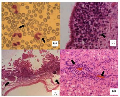 | |
| Fig. 1(a-d): | Microscopic findings of hematological and histopathological study. (a): Blood smear. Several stomatocytes and toxic band neutrophils are present (Giemsa stainx x1000). (b): Brain. Periventricular severe gliosis (H and E x600). (c): Small intestine. Exudation in mucous and sub mucosal tissues (H and E x100). (d): Liver parenchyma. Severe cholestasis with the bile casts in the portal bile duct, numbers of mononuclear cells infiltrate into the vicinity of the cholestasis (H and E x450) |
At the third day of life, the calf became severely depressed, so had been drenched with 150 mL of sulfamethazine (sulfadimidine, 33.3%-PO, Tolide-Darou®), by an inexperienced labor. Consequently, after 26 h, she had been carried to the hospital in a state of shock and lateral recumbency. Clinical examination revealed hypothermia (37.5°C rectal temperature), tachypnea (70 min-1) and tachycardia (160 min-1); the other signs included marked colic on abdominal palpation, tremor, convulsion, paddling, opisthotonos, hyperesthesia and cystic impaction.
Blood was drawn by jugular venipuncture and transferred into a tube that contained citrate. Hematological findings demonstrated 50% stomatocytes in complete blood count (Fig. 1a). Sampling repeated whereas a blood sample was taken simultaneously from a 2-day-old calf as control. However, a result was same as previous. She had mild macrocytic normochromic anemia [27.4% Packed cell volume (PCV); 8.3 g dL-1 Hemoglobin (Hb); 4.23x10⁶ μL-1 Red Blood Cell (RBC); 63 fl Mean Corpuscular Volume (MCV); 31.4 g dL-1 Mean Corpuscular Hemoglobin Concentration (MCHC)]. Wright's staining of RBCs demonstrated 0.4% reticulocytosis. The Calf had thrombocytopenia with 42x03 μL-1 platelet count. Differential count of white blood cells (17200 μL-1) indicated 58% neutrophils (53% segmented cells and 5% toxic band cells), 40% lymphocytes and 2% monocytes. Plasma protein to the fibrinogen ratio was 12.5 (5 g dL-1 plasma protein and 0.4 g dL-1 fibrinogen). Despite supportive treatment, the animal didn't recover and died after one hour. Postmortem examination revealed the meninges and mucous membranes were hyperemic or hemorrhagic, especially on the abomasal and intestinal mucosa in a form of numerous petechia (Fig. 2). The stomach was full of milk but the small intestine was relatively empty.
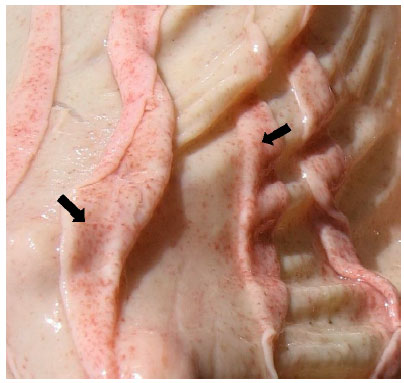 | |
| Fig. 2: | Multiple erosions and petechiae of the abomasal mucosa |
Congested, blue-red slightly rubbery lungs and hyperemia kidney were seen. On microbiologic investigation, Escherichia coli was isolated from all tissues, body fluids and bone marrow. Tissue samples from formalin-fixed, paraffin-embedded sections were processed for histopathology studies. Microscopically, brain edema with distended Virchow-Robin spaces and severe gliosis were present (Fig. 1b). The kidneys showed acute tubular necrosis. Interstitial pneumonia without involving of alveolar spaces was observed in the lungs. Exudation in mucous and sub mucosal tissues of an intestine accompanied by infiltration of mononuclear cells and neutrophils were evidence of acute enteritis (Fig. 1c). The liver demonstrated cholestasis, with bile casts in the bile ducts and the canaliculi. Small numbers of mononuclear cells and neutrophils infiltrated into the vicinity of the cholestasis and fatty change was obvious. The liver architecture was severely necrotic multifocally as a massive necrosis (Fig. 1d).
Due to normal RBC's smears of all fifteen dairy cows and their calves that were kept in mentioned farm, results from the hematological investigation could not confirm inherited disorder.
DISCUSSION
In animal, stomatocytosis is an autosomal recessive trait with undefined etiologies and has been described in Alaskan Malemutes (3.7% of RBCs), Miniature Schnauzers (5-40% of RBCs), Drentse-Prtrijshond (14-38% of RBCs) and recently in 1-15% of standard schnauzers RBCs (Bonfanti et al., 2004; Feldman et al., 2000; Pinkerton et al., 1974). Stomatocytosis was not reported in other species of animals. Therefore, this case study represented the first report of stomatocytosis in the ruminant. Approximately, 50% of RBCs were involved in the present report. On the other hand, human stomatocytes are seen in a variety of acquired and inherited disorders, whereas few stomatocytes are commonly found on blood films of normal subjects (Greer and Wintrobe, 2008; Lichtman et al., 2005).
The calf RBC was macrocytic normochromic, presumably, 0.4% reticulocytosis was normal finding in this case because at birth, about 9% of the RBCs are reticulocytes. Due to neonatal calves had the highest leukocyte counts at birth (5.7 to 20.5x03 cells μL-1) that decline in the few days of life, it seems, there was leukocytosis in this calf (Feldman et al., 2000).
In addition, the ruminant neonate begins life with fewer lymphocytes than neutrophils, at five days of age the lymphocyte has become the dominant WBC with a neutrophil to the lymphocyte ratio of 0.51 (Feldman et al., 2000). To sum up, about 10x103 neutrophils μL-1 and 860 band cells μL-1, in this colostrum deprived calf, on the 4th-day of life, showed neutrophilia. Moreover, total serum protein concentration in pre suckled calves is about 4 g dL-1 and handsome fibrinogen is low (160±130 mg dL-1); all of these data accompanied by microbiologic investigation, confirmed coli septicemia (Radostits et al., 2007).
Last but not least, Sulfadimidine (33.3% PO, Tolide-Darou®) has not been recommended, by producer, for calves under 30 days old and its dosage is 30 mL 50 kg body weight in daily water intake. It is noteworthy that the calf received a toxic dose of drug, but it is not clear, the oral intake of toxic dose of sulfonamide in 26 h period, could make this appearance in RBCs morphology and tissue lesions. Therapy with several amphipathic and cationic drugs can transform discocytes into stomatocytes (Greer and Wintrobe, 2008; Lichtman et al., 2005; Suwalsky et al., 2006). Not only continuing treatment of sulfonamide induced aplastic anemia, crystalluria, hepatic necrosis and cholestasis, but also cholestasis, hepatic injury, edema and hyperemia are common effects of sepsis (Maxie et al., 2007; Poirier et al., 1999; Radostits et al., 2007). This case study has described in detail the stomatocytosis in a Holstein-Friesian calf that was administrated by toxic dose of sulfadimidine (sulfamethazine) and suffered enormously from coli septicemia and meningitis. Although this is a preliminary work, it shows the first report of occurrence of stomatocytosis in ruminant.
ACKNOWLEDGMENT
The authors would like to thank Dr. Hesaraki, Dr. Khaki and Dr. Garagozloo for their praiseworthy support.
REFERENCES
- Pinkerton, P.H., S.M. Fletch, P.J. Brueckner and D.R. Miller, 1974. Hereditary stomatocytosis with hemolytic anemia in the dog. Blood, 44: 557-567.
PubMedDirect Link - Poirier, L.A., D.R. Doerge, D.W. Gaylor, M.A. Miller and R.J. Lorentzen et al., 1999. An FDA review of sulfamethazine toxicity. Reg. Toxicol. Pharmacol., 30: 217-222.
CrossRefDirect Link - Suwalsky, M., S. Mennickent, B. Norris, F. Villena and C.P. Sotomayor, 2006. Effects of the antiepileptic drug carbamazepine on human erythrocytes. Toxicol. In vitro, 20: 1363-1369.
CrossRefPubMedDirect Link