
A. Esfandiari
Department of Anatomical Sciences, Islamic Azad University, Kazerun Branch, Iran
A. Yousof
Department of Pathological Sciences, Islamic Azad University, Kazerun Branch, Iran
A. Aliabadi
Department of Clinical Sciences, School of Veterinary Medicine, Islamic Azad University, Kazerun Branch, Iran
Asian Journal of Animal and Veterinary Advances
Year: 2010 | Volume: 5 | Issue: 4 | Page No.: 246-252


ABSTRACT
The aim of this study was to determine what histological changes occur in the photoreceptor layer of male dogs after unilateral ligation of common carotid artery of different times. Twenty adult male dogs were randomly divided into 4 groups: control group, experimental group I, experimental group II and experimental group III. All of experimental groups (I, II, III) were done ligation of common carotid artery for 12 h , 24 h and 1 week, respectively. The retinas of eye were removed and studied by transmission electron microscopy. The experimental group 1 showed the least photoreceptor damage, such as some loss of mitochondrial cristae in the inner segment, but other layers appeared normal. The experimental II animals showed sublethal injury in the inner segment such as a few vacuolization and loss of mitochondrial cristae, but normal appearance in other layers. In the experimental III animals, disorganization and distention in the outer segments, cell swelling in the inner segment and pyknosis and karyolysis in the outer nuclear layer were present. Present findings have confirmed that photoreceptor cells damage occurs in dog retina after ligation of common carotid artery for 1 week.
PDF Abstract XML References Citation
How to cite this article
A. Esfandiari, A. Yousof and A. Aliabadi, 2010. The Survey of Effects of Unilateral Ligation of Common Carotid Artery on Ultra Structural of Photoreceptor Cells of Retina in Dog. Asian Journal of Animal and Veterinary Advances, 5: 246-252.
DOI: 10.3923/ajava.2010.246.252
URL: https://scialert.net/abstract/?doi=ajava.2010.246.252
DOI: 10.3923/ajava.2010.246.252
URL: https://scialert.net/abstract/?doi=ajava.2010.246.252
INTRODUCTION
The eye is a complex and highly developed photosensitive organ that permits an accurate analysis of the form light intensity and color reflected from objects. Each eye is composed of 3 concentric layers: an external layer (sclera and cornea), middle layer (choroid, ciliary body and iris) and an inner layer of nervous tissue (retina). The optical retina consists of an outer layer of photosensitive cells, the rods and cones. They are sensory retinal receptors and play an important role in visual system.
The effects of chronic retinal ischemia in an animal model involving permanent carotid occlusion investigated by Stevens et al. (2002). They indicated that ischemic damage to the optic nerve caused loss of pupillary reflex and death of retinal ganglion cells in a subset of rats. Block et al. (1997) suggested that the intraluminal suture method leads to retinal ischemia and immunohistochemistry of the retinas revealed that the vascular occlusion induced the expression of glial fibrillary acidic protein in retinal Müller cells. Barnett and Osborne (1995) showed that reduction of the retinal blood flow by occlusion of both common carotid arteries suppressed the b-wave of the rat's electroretinogram. Optic nerve atrophy in rats has been shown following bilateral ligation of common carotid artery (Takamatsu, 1988). The permeability of retinal blood vessels to intravenously injected horseradish peroxidase was examined two to seven days after bilateral common carotid ligation in rats by Korte and Henkind (1986).
Various histological studies were undertaken on photoreceptor structures of the retina in different animals using light and electron microscopy (Braeckevelt, 1983, 1987, 1990, 1992, 1993, 1998; Braeckevelt et al., 1996, 1998; Garcia and De Juan, 1999; Haacke et al., 2001).
No ultrastructural evaluation of the effect of unilateral ligation of common carotid artery on the photoreceptor layer has yet been conducted. The objective of the present study, therefore, was to determine effects of unilateral ligation of common carotid artery on photoreceptor cells of retina.
MATERIALS AND METHODS
For this study the eyes of 20 adult healthy male dogs were investigated by light and electron microscopy. The animals were obtained from the animal house of Islamic Azad University Kazerun Branch. This study was conducted from Dec. 2008 to Nov. 2009. The animals had free access to food and water. The same environmental conditions (temperature, light history and diet) were used before and after ligation of common carotid artery.
The dogs were randomly divided into 4 groups (n = 5 in each group): control group (CON), experimental group I (EXP-I), experimental group II (EXP-II) and experimental group III (EXP-III). The dogs in the CON group were kept in normal environmental conditions. Unilateral ligation of common carotid artery was done in the animals of EXP-I, EXP-II and EXP-III for 12 h, 24 h and 1week, respectively.
The histological effects of ligation of common carotid artery were evaluated using a transmission electron microscope.
The dogs were anesthetized with xylazine-ketamine and their eyeballs quickly removed. The cornea, lens and vitreous body were removed and opened at the equator and thereafter fixed for 4 h in 4% glutaraldehyde buffer (pH 7.3) with sodium cacodylate at 4°C. The posterior half of the eyeball was removed and the retina was then separated near the optic nerve of the superior hemisphere and fixed for an additional hour. Thereafter, the retinal tissue was washed in sodium cacodylate and cut into less than 1 mm2 pieces. The tissue was postfixed for 1.5 h in 1% osmium tetroxide, washed briefly in distilled water, dehydrated through a graded ethanol series and then cleared in propylene oxide and embedded in agar resin.
Semithin sections (0.5 μm) were obtained using an ultramicrotome (Omu 3; Reichert, Vienna, Austria), stained with toluidine blue and examined first under a light microscope. Ultrathin sections (60 nm) were also obtained and collected on copper grids. These sections were stained in aqueous uranyl acetate and lead citrate and were examined under a Philips CM-10 transmission electron microscope (Philips, Eindhoven, The Netherlands).
The laboratory care, anesthesia and euthanasia of animals used in this study were performed in accordance with the Guide for the Care and Use of Laboratory Animals (Institute of Laboratory Animal Resources, 1996).
RESULTS AND DISCUSSION
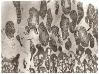
The rod outer segments were slender, whilst the cone outer segments were slightly less slender. The rod and cone outer segments were composed of bimembraneous discs (Fig. 1, 2). The inner segments of the photoreceptor layer had few mitochondria (Fig. 1).
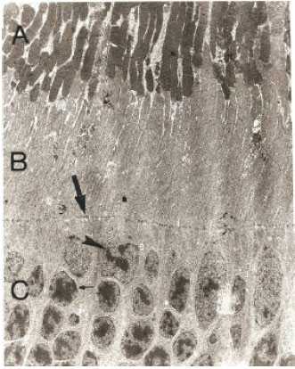 | |
| Fig. 1: | Electromicrograph of photoreceptor layer from the control group. A: Outer segment; B: Inner segment; C: Outer nuclear layer; Arrow: Outer limiting membrane; Arrowhead: Cone nucleus; Thin arrow: Rod nucleus (x1650) |
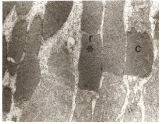 | |
| Fig. 2: | Electromicrograph of outer segment of photoreceptor cells from the control group. r: Outer segment of rod cell; c: Outer segment of cone cell; Asterisk: Disc membrane of outer segment (x8900) |
The outer limiting membrane was composed of a series of zonulae adherents between the rods and cones (Fig. 1). The cone nuclei are located nearest the outer limiting membrane. They are larger than rod nuclei (Fig. 1).
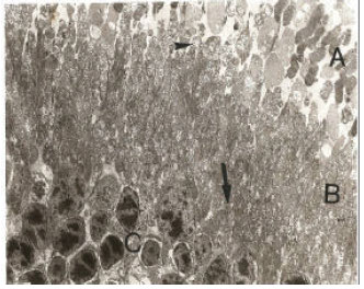 | |
| Fig. 3: | Electromicrograph of photoreceptor layer from the EXP-1 group. A: Outer segment; B: Inner segment; C: Outer nuclear layer; Arrow: Outer limiting membrane; Arrowhead: Loss of mitochondrial cristae (x1650) |
The unilateral ligation of common carotid artery produced damage in the dog retina. The ultrastructure of the dog retina was examined after unilateral ligation of common carotid artery of different times. The photoreceptor layers in EXP-I group showed the least damage, such as only some loss of mitochondrial cristae (Fig. 3). In addition, in the EXP-I group, the outer segment, mitochondria, outer limiting membrane and outer nuclear layer appeared normal (Fig. 3).
The dogs in EXP-II group showed that a few vacuolization and loss of mitochondrial cristae in inner segments (Fig. 4). But outer segment, outer limiting membrane and outer nuclear layer had normal. The retina in EXP-III group had major signs of damage (Fig. 5, 6). Distention and disorganization of the outer segment disc membrane appeared in this group (Fig. 5, 6). Extensive vacuolization, more loss of mitochondrial cristae and cell swelling of the inner segment were obvious (Fig. 5). Pyknotic and karyolysis nuclei were apparent in the outer nuclear layer (Fig. 5).
The retina is the eye tissue layer that converts light into visual signals transmitted to the brain. This process is carried out by two major types of photoreceptors, rods and cones that are distinguished by their shape, type of photopigment, retinal distribution and pattern of synaptic connections. These properties reflect the fact that rod and cone systems are specialized for different aspects of vision. The photoreceptor layer of the dog retina is very similar to that of other animals (Braeckevelt, 1983, 1987, 1990, 1992, 1993, 1998; Braeckevelt et al., 1996, 1998; Garcia and De Juan, 1999; Haacke et al., 2001). In the present study, the photoreceptor damage produced by EXP-I group, EXP-II group and EXP-III group was evaluated. At the electron microscopic level, the dogs in EXP-II and EXP-III showed disorganization and distention of the outer segment disc membrane, similar to the findings of Esfandiari et al. (2008). By comparison, the outer segment of dogs in EXP-I appeared normal. However, it is equally plausible that diurnal changes in RPE function unrelated to outer segment phagocytosis and renewal underlie the effect of unilateral ligation of common carotid artery for different times.
In the histopathology results presented in our study, ligation of common carotid artery for 1 week appeared to cause more damage to the inner segment, outer limiting membrane and outer nuclear layer of the dog retina than did ligation of common carotid artery for the 12 and 24 h.
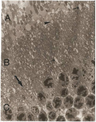 | |
| Fig. 4: | Electromicrograph of photoreceptor layer from the EXP-2 group. A: Outer segment; B: Inner segment; C: Outer nuclear layer; Arrow: Outer limiting membrane; Arrowhead: Loss of mitochondrial cristae; Thin arrows: Vacoules (x1650) |
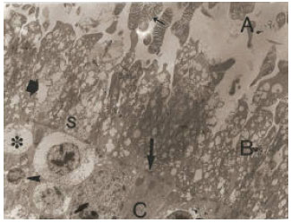 | |
| Fig. 5: | Electromicrograph of photoreceptor layer from the EXP-3 group. A: Outer segment; B: Inner segment; C: Outer nuclear layer; Thin arrow: Disorganization of outer segment; Thick arrow: Vacoule; S: Cell swelling; Arrow: Outer limiting membrane; Arrowhead: Pyknotic nucleus; Asterisk: Karyolysis (x1650) |
Present results suggested that the mitochondrial cristae in the inner segment of dog retinas were more loss in EXP-III than EXP-II. Cell swelling and vacuoles observed in the inner segment of retina in the EXP-III group. Vacuoles are a common response to injury in sublethally damaged cells.
 | |
| Fig. 6: | Electromicrograph of outer segment of photoreceptor cells from the EXP-3 group. Arrows indicates disorganization and distension of outer segment (x2950) |
In the EXP-3 group, mitochondrial cristae were lost, aerobic oxidative phosphorylation stopped and ATP levels fell. Ultimately, this deficiency of ATP lead to a failure of Na+-K+ pumps and loss of cell volume control (Na+ and H20 moved into cells), maybe also causing cell swelling. In addition, lethal injury to photoreceptor nuclei resulted in pyknotic and karyolysis nuclei.
We concluded that unilateral ligation of common carotid artery for 1 week caused more photoreceptor cells damage than did short time ligation. Furthermore, ligation of common carotid artery for 12 h did not cause photoreceptor cells damage.
ACKNOWLEDGMENT
This study was conducted under the sponsorship of the Islamic Azad University-Kazerun Branch.
REFERENCES
- Barnett, N.L. and N.N. Osborne, 1995. Prolonged bilateral carotid artery occlusion induces electrophysiological and immunohistochemical changes to the rat retina without causing histological damage. Exp. Eye Res., 61: 83-90.
CrossRef - Block, F., C. Grommes, C. Kosinski, W. Schmidt and M. Schwarz, 1997. Retinal ischemia induced by the intraluminal suture method in rats. Neurosci. Lett., 22: 45-48.
CrossRefDirect Link - Braeckevelt, C.R., 1983. Retinal fine structure in the domestic sheep. Acta Anat., 116: 265-275.
CrossRefDirect Link - Braeckevelt, C.R., 1987. Photoreceptor fine structure in the vervet monkey (Cercopithecus aethiops). Histol. Histopathol., 2: 433-439.
PubMedDirect Link - Braeckevelt, C.R., 1990. Photoreceptor fine structure in light-and dark-adaptation in the butterfly fish (Pantodon buchholzi). Anat. Anz., 171: 351-358.
PubMedDirect Link - Braeckevelt, C.R., 1992. Retinal photoreceptor fine structure in the red-backed salamander (Plethodon cinereus). Histol. Histopathol., 7: 463-470.
PubMedDirect Link - Braeckevelt, C.R., 1993. Retinal photoreceptor fine structure in the red-tailed hawk (Buteo jamaicensis). Anat. Histol. Embryol., 22: 222-232.
PubMedDirect Link - Braeckevelt, C.R., 1998. Fine structure of the retinal photoreceptors of the emu (Dromaius novaehollandiae). Tissue Cell., 30: 137-148.
CrossRefDirect Link - Braeckevelt, C.R., S.A. Smith and B.J. Smith 1996. Fine structure of the retinal photoreceptors of the barred owl (Strix varia). Histol. Histopathol., 11: 79-88.
PubMedDirect Link - Braeckevelt, C.R., S.A. Smith and B.J. Smith, 1998. Photoreceptor fine structure in Oreochromis niloticus in light-and dark-adaptation. Anat. Rec., 252: 453-461.
PubMedDirect Link - Esfandiari, A., S. Gholami and A. Safavi, 2008. Morphology of retinal photoreceptor layer in continuous light exposed and dark adapted male cats. Iranian J. Vet. Res., 9: 36-41.
Direct Link - Garcia, M. and J. de Juan, 1999. Fine structure of the retina of black bass, Micropterus salmoides (Centrarchidae, Teleostei). Histol. Histopathol., 14: 1053-1065.
PubMedDirect Link - Haacke, C., M. Hess, R.R. Melzer, H. Gebhart and U. Smola, 2001. Fine structure and development of the retina of the grenadier anchovy Coilia nasus (Engraulididae, Clupeiformes). J. Morphol., 248: 41-55.
CrossRef - NRC, 1996. Guide for the Care and Use of Laboratory Animals. National Academy Press, Washington, DC., USA., ISBN-10: 0309053773, Pages: 140.
Direct Link - Korte, G.E. and P. Henkind, 1986. Vascular permeability and the optic disc. Changes after bilateral common carotid ligation in the rat. Arch. Ophthalmol., 104: 273-276.
PubMedDirect Link - Stevens, W.D., T. Fortin, A. Bruce and B. Pappas, 2002. Retinal and optic nerve degeneration after chronic carotid ligation. Stroke, 33: 1107-1112.
CrossRefDirect Link - Takamatsu, J., 1988. Bilateral carotid artery ligation in rat serial pathology of the optic nerve. No To Shinkei., 40: 739-746.
PubMedDirect Link