
Nadia M.S. Arafa
Department of Physiology, National Organization for Drug Control and Research, Egypt
Pakistan Journal of Biological Sciences
Year: 2010 | Volume: 13 | Issue: 20 | Page No.: 966-976


ABSTRACT
The study aimed to evaluate the effect Echinacea extract (E) on the testicular antioxidants function in normal rats or that subjected to anti-androgenic compound, cyproterone acetate (CA). Rats were divided into 5 groups treated daily via an oral tube for two intervals 2 and 4 weeks, 1st control, 2nd E (Echinacea treated group in dose 63 mg kg-1), 3rd CA (cyproterone acetate treated group in dose 25 mg kg-1), 4th E+CA and 5th E as prophylactic one week before E+CA treatment with the same aforementioned E or CA doses. The body, testes, epididymis and vas deferens weights were recorded. Sperm count, Nitric Oxide (NO), calcium ion (Ca2+) and malondialdhyde (MDA) contents in addition to superoxide dismutase (SOD), glutathione S-transferase (GST) activities were determined in testicular tissues. CA exhibited direct negative effect on reproductive organs weight and significant reducing effect on sperm count and Ca2+ contents. SOD and GST activities significantly decreased in addition to significant increase in NO, MDA contents reflecting the oxidative status of testis in CA treated rats. The prophylactic effect of E treatment, in time related manner, showed significant improvement in the antioxidant status of the testicular tissue which is more pronounced as compared to E+CA treatment.
PDF Abstract XML References Citation
Received: June 19, 2010;
Accepted: August 13, 2010;
Published: September 24, 2010
How to cite this article
Nadia M.S. Arafa, 2010. Efficacy of Echinacea on the Action of Cyproterone Acetate in Male Rats. Pakistan Journal of Biological Sciences, 13: 966-976.
DOI: 10.3923/pjbs.2010.966.976
URL: https://scialert.net/abstract/?doi=pjbs.2010.966.976
DOI: 10.3923/pjbs.2010.966.976
URL: https://scialert.net/abstract/?doi=pjbs.2010.966.976
INTRODUCTION
The antioxidant system plays an effective role in protecting testes and other biological tissues below a critical threshold of Reactive Oxygen Species (ROS), preventing testicular dysfunction (Ochsendorf, 1999). ROS mediated oxidative stress is one of crucial reasons of infertility, decreased sperm viability and increased level of free radicals which may cause degeneration of testicular tissue (Koksal et al., 2002; Turner and Lysiak, 2008). The administration of antioxidants to normal animals, not suffering from induced oxidative stress, appears to improve testicular function, signifying oxidative stress a consistent feature of testicular physiology (Orozco et al., 2003; Juan et al., 2005). Antioxidants administered to infertile men advocate promising their use in male infertility treatment (Suleiman et al., 1996; Keskes-Ammar et al., 2003).
Cyproterone acetate is a synthetic steroidal drug which competes with testosterone and dihydrotestosterone for the androgen receptor. It is used to suppress male fertility in oral contraceptives formulations and also in the treatment of various sexual and metabolic disorders (Meriggiola et al., 1996). Cyproterone also has progesterone like activity and reduces pituitary luteinizing hormone and plasma testosterone (McLeod, 1993) and suppresses the secretion of gonadotropins thus, interferes with testosterone production (Barradell and Faulds, 1994; Neumann, 1994; Honer et al., 2003). Due to the direct interaction with the cellular receptors CA inhibits the action of the androgens and due to this property it is used for the prostatic carcinoma treatment (Torri and Floriani, 2005).
Cyproterone is genotoxic and tumor initiating agent (Kasper, 2001) and the genotoxic effects postulated to be reduced by the use of antioxidants (Siddique et al., 2008).
Echinacea (E) is herbaceous plant genus, consisting of nine species and the current study used one of them Echinacea purpurea or purple coneflower. Echinacea (E) preparations have been marketed as possible immune stimulators or enhancers worldwide. Echinacea became popular as remedies for the common cold and rhinovirus infection (Barrett et al., 2006; Shah et al., 2007; Simasek and Blandino, 2007). It has anti-tumor effect (Block et al., 2002). The therapeutic effect of Echinacea has been assigned to the presence of caffeic acid derivatives such as cichoric acid, echinacoside, chlorogenic acid and lipophilic polyacetylene-derived compounds, such as alkylamides, constituting isobutylamides are parts of the active principles and suitable for standardization of E. purpurea row material and finished products (Letchamo et al., 1999). Pellati et al. (2005) reported that Echinacea roots are a good source of natural antioxidants possessing radical scavenging activity.
The objective of this study is to evaluate the physiological and antioxidant effect of Echinacea extract consumption on normal male rat reproductive system or that subjected to cyproterone acetate in addition to the prophylactic effect of Echinacea extract supplementation by subjecting it to normal rat a week before cyproterone acetate treatment to explore its impact in reducing the postulated cyproterone related side effects.
MATERIALS AND METHODS
Experimental animals: This study was carried out on adult male albino rats (Wistar strain). Sixty animals were obtained from the farm of National Organization for Drug Control and Research (NODCAR) in 2008. Their weights were (130.2 ± 18.5 g). Male rats were housed in iron mesh cages, each cage contained six rats. Animals were kept under controlled temperature of 21 ± 2°C and 12 h light/12 h dark cycle throughout the experiment. A commercial pelleted diet was used during the experiment and allowed with water ad libitum. The animals were allowed to adapt to the laboratory conditions for two weeks before the beginning of the experiment.
Drugs: Cyproterone acetate (C24H29ClO4) was obtained from Schering, Germany as ANDROCURE tablets each containing 50 mg active ingredient Cyproterone acetate (6-chloro-17-hydroxy-1alpha,2alpha-methylene-pregna-4,6-diene-3,20-dione-acetate) (LOT. No. 481A). Echinacea purpurea dry extract was supplied by MEPACO (Arab Company for Pharmaceutical and Medicinal Plants), Egypt as capsules each containing 175 mg from dray extract.
Experimental design: Animals divided into five groups using random selection each of twelve rats treated oral daily as 1st Normal control (C) received 0.1 mL of 0.5 g/100 mL carboxymethyl cellulose, (CMC) for each 100 g b.wt. 2nd Echinacea treated group (E) received 63 mg kg 1, 3rd Cyproterone acetate treated group (CA) received 25 mg kg 1, 4th Echinacea+Cyproterone acetate treated group (E+CA) received 63 mg kg 1 (E) + 25 mg kg 1 (CA) and 5th Echinacea (week) + Cyproterone acetate treated group (E w + CA) received 63 mg kg 1 Echinacea for 1 week before 63 mg kg 1 (E) + 25 mg kg 1 (CA) treatment. All groups were treated daily for either two or four consecutive weeks from the beginning of the experiment. These doses used calculated equivalent to the human therapeutic dose (Reagan-Shaw et al., 2008). The groups treated with E+CA received cyproterone acetate first followed by E. after 3.5 h as the peak plasma concentration of CA is being achieved in 3 to 4 h from the gastrointestinal tract (Sweetman, 2005). The animal experiments in relation to maintaining and handling animals were strictly conducted in accordance with the internationally agreed guidelines.
Fingerprints of Echinacea extract: Chromatographic identification of Echinacea caffeic acid derivatives: one capsule was extracted with methanol: water (80: 20; v/v) with 15 min ultra-sonication at room temperature. Caftaric, chlorogenic and chichoric acid in addition to echinacoside active ingredients of Echinacea extract (Fig. 1) was determined by HPLC, UV detector at 330 nm wavelength according to the method of Liu and Murphy (2007).
Echinacea extract by GC-MS analysis according to the method described in Song et al. (2000). Phytosterols identified by their fragmentation pattern while scanning the mass range m/z 50-700 and retention times and the representation of the analysis included in Fig. 2 (B1, B2 and B3).
Biochemical analysis: Body weight of the rats recorded weekly throughout the duration of the experiment. After two and four weeks of treatment, rats sacrificed 12 h following the last dose by rapid decapitation. Testis, epididymis and vas deferens dissected out, cleaned and weighed.
 | |
| Fig. 1: | HPLC chromatogram of Echinacea purpurea extracts active ingredients separation |
Testis tissues were used for the measurement of enzyme activities of superoxide dismutase (SOD) according to Minami and Yoshikawa (1979) and Glutathione transferases (GST) according to the method of Habig et al. (1974) and Asmah et al. (1993). In addition to Malondialdehyde (MDA) was determined by HPLC according to the procedure of Karatas et al. (2002), nitric oxide (NO) (nitrites/nitrate) was determined according to the method of Papadoyannis et al. (1999) by HPLC and calcium ion was determined by colorimetric assay kit according to Ladenson (1980), Bradley and Schumann (1984) and Woo and Cannon (1984). Testicular sperm count was performed according to Blazak et al. (1993).
Statistical analysis: Statistical analysis was evaluated by one-way ANOVA. Once a significant F test was obtained, LSD comparisons were performed to assess the significance of differences among various treatment groups. Statistical Processor System Support SPSS for Windows software, Release 10.0 (SPSS, Chicago, IL) was used.
RESULTS
Figure 1 represents the HPLC analysis of the Echinacea purpurea extract and identified the active ingredients of the root extract which possesses the antioxidant potential. Figures 2a-c represent the gas chromatographic separation using the mass spectrum detector to identify the persistence of the vegetative sterols in the extract.
Testes weights in Table 1 showed significant decrement at p<0.05 in CA, E+CA and E w+CA groups treated rats after two or four weeks in comparison with the corresponding control weights, while rats treated with E exhibited not statistically different decrease and increase in testes weights as compared to the control values throughout the experimental periods.
Testes/body weight ratio decreased significantly after 4 weeks of treatments in CA and E w+CA groups as compared to the control or the 2 weeks of treatment values. The ratio in E+CA groups decreased significantly throughout the 2 periods as compared to that of the control.
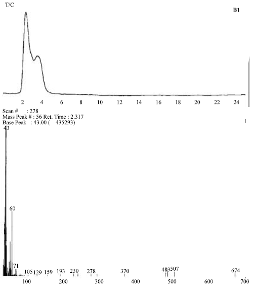 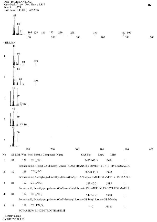 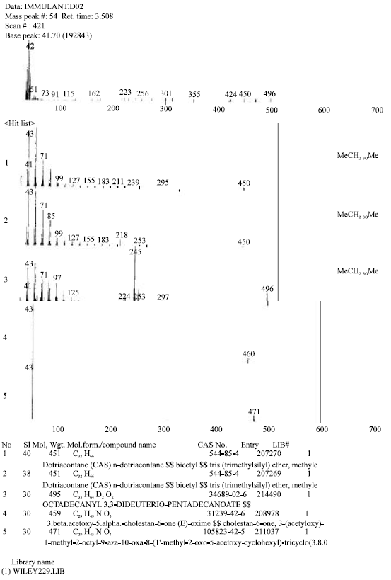 | |
| Fig. 2: | (a-c): GC mass spectrums of Echinacea purpurea extract showing ingredients and identification of some library names |
While in E treated groups in 4 weeks of treatment the ratio significantly increased in comparison with that of the 2 weeks of treatment without statistical difference comparing to control values.
The data depicted in Table 1 showed that epididymis weights decreased significantly at p<0.05 in CA, E+CA and E w+CA treated rats after two or four weeks in comparison with the corresponding control weights. In contrast, not statistically different decrease in epididymis weight in E treated rats after two weeks of treatment, followed by a significant decrease after the four weeks as compared with the corresponding control value.
A significant reduction in vas deferens weight reported in Table 1 in CA, E+CA and E w+CA treated groups of rats after two and four weeks of treatments in comparison with the corresponding control weights at the two periods. However rats treated with E showed a significant decrease after two weeks followed by not statistically different decrease in vas deferens weight after four weeks of treatment as compared with that of control.
The data in Table 2 revealed that CA treatment throughout the two experimental periods exhibited significant decrease in the testicular SOD and GST enzyme activities and sperm counts and calcium ions contents. In addition to the significant increase in the MDA and nitric oxide (nitrites/nitrates) concentrations as compared with the corresponding control or E treated groups’ values.
Rats treated for two weeks in E groups showed significant increase in GST activity and nitric oxide values and significant decrease in sperm counts and calcium ions contents in comparison with the control values of the two week duration. While treatment with E for four weeks significantly affected the decrease in SOD and GST activities and the MDA, sperm counts and calcium ions contents comparing to four weeks control values.
E+CA treated group for two weeks showed significant decrease in the testicular SOD enzyme activity and sperm counts and calcium ions contents, moreover, significant increase in the MDA and nitric oxide levels as compared with their control values. E+CA treated group for four weeks exhibited significant decrease in the testicular GST enzyme activity and calcium ions contents and significant increase in the nitric oxide level comparing with the four weeks control values. E+CA treatment for four weeks significantly increased the testis SOD activity and the sperm count as compared to the corresponding CA treated group values. While the MDA and nitric oxide levels decreased in non-statistical difference as compared to that of the CA treated group values.
The groups of E w+CA treated rats showed no statistical difference in SOD activities and MDA and sperm count contents but the calcium contents decreased and nitric oxide increased significantly throughout the experimental periods comparing to the control values. Data showed significant increase in the SOD and GST activities and sperm counts values, while the MDA and nitric oxide contents decreased significantly as compared to the CA treated groups throughout the two experimental periods.
| Table 1: | Effect of Echinacea (E) and/or cyproterone acetate (CA) supplementation on the organs weights (g) of male rats |
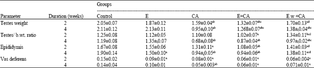 | |
The results are presented as Means ± SE of 6 rats. a: Significant change from the corresponding control value, b: Significant change from the E group, c: Significant change from the CA group, d: Significant change from the E + CA treated group, t: Significant change between two and four weeks with the same treatment. Significance at 0.05 level | |
| Table 2: | Effect of Echinacea (E) and/or cyproterone acetate (CA) supplementation on testicular SOD and GST activities, MDA, calcium ions and nitric oxide contents and sperm count |
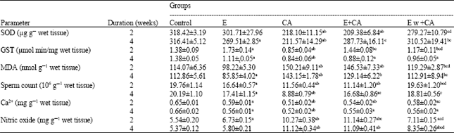 | |
The results are presented as Means ± SE of 6 rats. a: Significant change from the corresponding control value, b: Significant change from the E group, c: Significant change from the CA group, d: Significant change from the E + CA treated group, t: Significant change between two and four weeks with the same treatment. Significance at 0.05 level | |
DISCUSSION
In the present study treatment of male rats with cyproterone acetate reduced the testes, testes-body weight ratio, epididymis and vas deferens weights during the experimental periods as expected according to the anti-androgenic action of this drug in rat (O’ Connor et al., 2002; Campos et al., 2003) and in line with Aleem et al. (2005) results about CA treatment in a dose of 20 mg kg 1 for 15 days which reduced the fertility and weights of accessory sex glands of the male rats.
The oxidative stress in testis was discernible in terms of perturbations in the activities of antioxidant enzymes SOD and GST and nitric oxide level. SOD protects against spontaneous O2 toxicity and lipid peroxidation. The reduction of SOD suggests that it is involved in antioxidant defense and it has been shown to act as an alternate regulatory switch in testicular steroidogenesis (Pradeep et al., 1990). It is evident, that SOD plays an important role in scavenging of reactive oxygen species (ROS) in testes, because in comparison to rat liver, the activity of catalase and glutathione peroxidase is much lower in the testicular tissue (Peltola et al., 1992). The GST conjugates GSH with compounds containing an electrophilic center and thereby provides critical protection against products of oxidative stress. Since the GSH-conjugate is transported out of the cell, intracellular GSH is consumed irreversibly in the conjugation and thus the maintenance of intracellular GSH levels is essential for the optimal function of GST (Zhang et al., 1998). On the other hand GST significant reduced activity in CA treated groups may be explained through the tissue degeneracy, as the enzyme activities in the animal systems upon exposure to carcinogenic chemical have evolved various defense mechanisms to protect themselves from the oxidative damage. As GSTs form a part of adaptive response of germ cells to oxidative stress and are important constituents in detoxifying the products of lipid peroxidation that inhibition of GST leads to the augmentation of lipid peroxidation and resulting in germ cell apoptosis (Rao and Shaha, 2000; Yang et al., 2001).
The testicular tissue nitrate/nitrite, as an index of NO production and MDA levels were significantly elevated in rat testes in CA treated groups. The tissue NO increase in CA group may be attributed to the injury of testicular tissue in relation to abundant ROS production and consequent migration of macrophages and polymorphonuclear leucocytes to the region (Sikka, 2001). The significant increase in lipid peroxidation marker (MDA) enhanced ROS generation in testis after two and four weeks of treatment suggesting cyproterone acetate potential to induce significant oxidative stress in the reproductive milieu of rats. The results are consistent with the possible role of reactive oxygen species for the genotoxicity previously suggested by Siddique and Afzal (2005) NO radicals have been found to regulate multiple biological functions in inflammation and in mediating many cytotoxic and pathological events. NO has a bimodal effect on sperm motility whereby low concentrations of NO enhance sperm motility, whereas high concentrations of NO decrease it. This effect might be due to the dual nature of NO, which is a signal transduction molecule at low concentrations, while being cytotoxic at higher concentrations (Sikka, 2001; Sheweita et al., 2005). In general imbalance in peroxidant and antioxidant status could produce oxidative stress (Ong et al., 2002).
Also, the formation of free radicals and oxidative stress by CA was involved in the destruction of Sertoli cells and the azospermia occurred and this result occurred indirectly via the androgen deprivation effect of CA causing diminution of antioxidant detoxification (Tam et al., 2003).
As regard to Echinacea treatment the data in this study showed not statistically different decrease in testes and vas deferens weights but significant decrease in epididymis weight after four weeks of treatment with Echinacea. Echinacea treatment for 2 to 4 weeks showed gradual antiandrogenic activity through the effect on male sexual hormone testosterone producing organ which may be associated with the vegetative sterols which are from the constituents of Echinacea extract (Skaudickas et al., 2003; 2004). The chemical structure of these compounds is very similar to cholesterol. In the digestive duct vegetative sterols reduce the absorption of cholesterol, creating a certain competition between sterols and cholesterols thus dietary phytosterols may reduce the risk of prostate cancer by lowering the activities of the enzymes of testosterone metabolism (Awad and Fink, 2000; Trautwein and Demonty, 2007).
In Mishima et al. (2004) study, they assumed that SOD activity in peripheral blood was increased because of antioxidants such as echinacocide and caffeine acid in Echinacea purpurea (identified by HPLC) which eliminate superoxide (O 2) by a free radical scavenging effect. Pellati et al. (2004) indicate that E roots are a good source of natural antioxidants and could be used to prevent free-radical-induced deleterious effects, capable of scavenging hydroxyl radicals, nitration inhibition and to suppress the oxidation of human low-density lipoprotein, particularly at the molecular level and also transition metal chelating (Hu and Kitts, 2000; Weiss and Landauer, 2003; Masteikova et al., 2007).
These results are agreed with the results in this study as treatment with E caused a significant reduction in MDA content in testes and not statistically different change in SOD in the first two weeks. Weiss and Landauer (2003) documented a protective effect of polyphenols from E against free radical damage and a class of specific antioxidants known as caffeoyl derivatives in appreciable amounts. SOD remove (O 2) generated by NADPH-oxidase in neutrophils and can play an important role in protecting spermatozoa during genitourinary inflammation (Baker et al., 1996).
In the present results CA induced a significant decrease in sperm count and this reduction was improved by using treatment with E+CA. this improvement may be due to improvement of hemoglobin levels and number of erythrocytes by Echinacea treatment (Abouelella et al., 2007).
Cyproterone acetate treatment alone or with E induced a significant reduction in testicular No. Lue et al. (2003) suggested NO physiological role in regulation of germ cell number and in determining testicular size. Concomitant treatment with CA + E improves but did not inhibit the significant reduction of testicular No. Endothelial production of NO is at least partially regulated by the inositol trisphosphate (IP3)-dependent calcium (Ca2+) signalling pathway, which may be activated by ligand-receptor binding and/or wall shear stress. The elevated levels of free Ca2+ in the cytosol reversibly bind with calmodulin; the resulting Ca2+-calmodulin complexes activate endothelial nitric oxide synthase enzyme, thus causing an increase in synthesis and release of No. In addition, NO leads to increased levels of cyclic guanosine monophosphate (cGMP), this activates protein kinase. This is thought to inhibit Ca2+ influx, thus providing a negative feedback mechanism and limiting the concentration of Ca2+ in the cytosol. (Shen et al., 1992; Lin et al., 2000; Plank et al., 2007). Ca2+ can directly activate antioxidant enzymes, increase the level of SOD in animal cells (Gordeeva et al., 2003) and induce mitochondrial GSH release (Brookes et al., 2004). The result in this study in the same trend, there is a significant reduction in testicular SOD and intra cellular testicular Ca ions. Yan et al. (2006) studies indicated that Ca2+ plays dual roles in regulating ROS homeostasis.
The net Ca2+ effects on ROS generation and annihilation appear to be tissue-specific and context-sensitive, and, within a given cell, are differentially regulated in local subcellular compartments. These results give an explanation for intracellular testicular calcium ion reduction. Also, Lyng et al. (2000) suggested that the calcium signals are probably coupled to the regulation of gap junctional efficiency between Sertoli cells.
The low-affinity receptors may convey complementary androgen signals at elevated local levels such as in the testis, when nuclear receptors are (over) saturated. Androgens can also induce rapid calcium fluxes in a variety of cell types, including human prostatic cancer cells and rat sertoli cells (Steinsapir et al., 1991; Gorczynska and Handelsman, 1995; Gorczynska-Fjalling, 2004). This suggestion explains the effect of CA treatment on decreasing the testicular Ca ion content.
The study suggests that Echinacea supplementation especially a week before cyproterone acetate shows improvement in the oxidative stress induced by cyproterone acetate treatment. That may be due to the Echinacea antioxidant activity. The anti-androgenic effect of E may in addition to the anti-inflammatory and immune enhancing activity add valuable for using it as a co-treatment for male contraceptives or in treatment of prostate cancer which needs more investigations.
ACKNOWLEDGMENTS
The author gratefully acknowledge the help of NODCAR (The national organization for drug control and research), Department of Physiology, for facilities in the HPLC analysis. As regard to conflicts of interest, the author declares that there is no conflict of interest associated with this study. And the Author contributed to the design of the study, elaboration of bench work and analytical procedures in addition to interpretation of the data, writing the manuscript.
REFERENCES
- Abouelella, A.M.K., Y.E. Shahein, S.S. Tawfik and A.M. Zahran, 2007. Phytotherapeutic effects of Echinacea purpurea in gamma-irradiated mice. J. Vet. Sci., 8: 341-351.
Direct Link - Aleem, M., V. Padwal, J. Choudhari, N. Balasinor, P. Parte and M. Gill-Sharma, 2005. Cyproterone acetate affects protamine gene expression in the testis of adult male rat. Contraception, 71: 379-391.
PubMed - Awad, A.B. and C.S. Fink, 2000. Phytosterols as anticancer dietary components: Evidence and mechanism of action. J. Nutr., 130: 2127-2130.
PubMed - Baker, H.W.G., J. Brindle, D.S. Irvine and R.J. Aitken, 1996. Protective effect of antioxidants on the impairment of sperm motility by activated polymorphonuclear leukocytes Fertil. Steril., 65: 411-419.
CrossRefPubMedDirect Link - Barradell, L.B. and D. Faulds, 1994. Cyproterone. A review of its pharmacology and therapeutic efficacy in prostate cancer. Drugs Aging, 5: 59-80.
PubMed - Barrett, B., R. Brown, R. Voland, R. Maberry and R. Turner, 2006. Relations among questionnaire and laboratory measures of rhinovirus infection. Eur. Respir. J., 28: 358-363.
PubMed - Block, K.I., D.B. Boyd, N. Gonzalez and A. Vojdani, 2002. Point-counterpoint: The immune system in cancer. Integr. Cancer Ther., 1: 294-316.
PubMed - Brookes, P.S., Y. Yoon, J.L. Robotham, M.W. Anders and S.S. Sheu, 2004. Calcium, ATP and ROS: A mitochondrial love-hate triangle. Am. J. Physiol. Cell Physiol., 87: 817-833.
CrossRefDirect Link - Campos, M., P.L. Morais and A.S. Pupo, 2003. Effect of cyproterone acetate on alpha1-adrenoceptor subtypes in rat vas deferens. Braz. J. Med. Biol. Res., 36: 1571-1581.
Direct Link - Trautwein, E.A. and I. Demonty, 2007. Phytosterols: Natural compounds with established and emerging health benefits. Oleagineux Corps Gras Lipides, 14: 259-266.
CrossRef - Gorczynska, E. and D.J. Handelsman, 1995. Androgens rapidly increase the cystolic calcium concentration in Sertoli cells. Endocrinology, 136: 2052-2059.
Direct Link - Gorczynska-Fjalling, E., 2004. The role of calcium in signal transduction processes in Sertoli cells. Reprod. Biol., 4: 219-241.
PubMed - Gordeeva, A.V., R.A. Zvyagilskaya and Y.A. Labas, 2003. Cross-talk between reactive oxygen species and calcium in living cells. Biochem. (Moscow), 68: 1077-1080.
CrossRef - Habig, W.H., M.J. Pabst and W.B. Jakoby, 1974. Glutathione S-transferases: The first enzymatic step in mercapturic acid formation. J. Biol. Chem., 249: 7130-7139.
CrossRefPubMedDirect Link - Honer, C., K. Nam, C. Fink, P. Marshall and G. Ksander et al., 2003. Glucocorticoid receptor antagonism by cyproterone acetate and RU486. Moll. Pharmacol., 63: 1012-1020.
PubMed - Hu, C. and D.D. Kitts, 2000. Studies on the antioxidant activity of Echinacea root extract. J. Agric. Food Chem., 48: 1466-1472.
CrossRefPubMedDirect Link - Juan, M.E., E. Gonzalez-Pons, T. Munuera, J. Ballester, J.E. Rodriguez-Gil and J.M. Planas, 2005. Trans-Resveratrol, a natural antioxidant from grapes, increases sperm output in healthy rats. J. Nutr., 135: 757-760.
Direct Link - Kasper, P., 2001. Cyproterone acetate: A genotoxic carcinogen. Pharmacol. Toxicol., 88: 223-231.
PubMed - Keskes-Ammar, L., N. Feki-Chakroun, T. Rebai, Z. Sahnoun and H. Ghozzi et al., 2003. Sperm oxidative stress and the effect of an oral vitamin E and selenium supplement on semen quality in infertile men. Arch. Androl., 49: 83-94.
PubMed - Koksal, T., T. Erdogru, B. Toptas, K.H. Gulkesen, M. Usta, A. Baykal and M. Baykara, 2002. Effect of experimental varicocele in rats on testicular oxidative stress status. Andrologia, 34: 242-247.
PubMed - Lin, S., K.A. Fagan, K.X. Li, P.W. Shaul, D.M.F. Cooper and D.M. Rodman, 2000. Sustained endothelial nitric-oxide synthase activation requires capacitative Ca2+ entry. J. Biol. Chem., 275: 17979-17985.
PubMed - Liu, Y. and P.A. Murphy, 2007. Alkamide stability in echinacea purpurea extracts with and without phenolic acids in dry films and in solution. J. Agric. Food Chem., 55: 120-126.
PubMed - Lue, Y., A.P. Sinha Hikim, C. Wang, A. Leung, and R.S. Swerdloff, 2003. Functional role of inducible nitric oxide synthase in the induction of male germ cell apoptosis, regulation of sperm number and determination of testes size: Evidence from null mutant mice. Endocrinology, 144: 3092-3100.
PubMed - Lyng, F.M., G.R. Jones and F.F. Rommerts, 2000. Rapid androgen actions on calcium signaling in rat sertoli cells and two human prostatic cell lines: Similar biphasic responses between 1 picomolar and 100 nanomolar concentrations. Biol. Reprod., 63: 736-747.
PubMed - Masteikova, R., J. Muselik, J. Bernatoniene and R. Bernatoniene, 2007. Antioxidative activity of Ginkgo, Echinacea and Ginseng tinctures. Medicina (Kaunas), 43: 306-309.
PubMed - Meriggiola, M.C., W.J. Bremner, C.A. Paulsen, A. Valdiserri and L. Incorvaia et al., 1996. A combined regimen of cyproterone acetate and testosterone enanthate as a potentially highly effective male contraceptive. J. Clin. Endocrinol. Metab., 81: 3018-3023.
PubMed - Masayasu, M. and Y. Hiroshi, 1979. A simplified assay method of superoxide dismutase activity for clinical use. Clin. Chim. Acta, 92: 337-342.
CrossRefPubMedDirect Link - Mishima, S., K. Saito, H. Maruyama, M. Inoue, T. Yamashita, T. Ishida and Y. Gu, 2004. Antioxidant and immuno-enhancing effects of Echinacea purpurea. Biol. Pharm. Bull., 27: 1004-1009.
CrossRefPubMedDirect Link - Neumann, F., 1994. The antiandrogen cyproterone acetate: Discovery, chemistry, basic pharmacology, clinical use and tool in basic research. Exp. Clin. Endocrinol., 102: 1-32.
PubMedDirect Link - O`Connor, J.C., S.R.Frame and G.S. Ladics, 2002. Evaluation of a 15-day screening assay using intact male rats for identifying antiandrogens. Toxicol. Sci., 69: 92-108.
PubMed - Ong, C.N., H.M. Shen and S.E. Chia, 2002. Biomarkers for male reproductive health hazards: Are they available?. Toxicol. Lett., 134: 17-30.
PubMed - Orozco, T.J., J.F. Wang and C.L. Keen, 2003. Chronic consumption of a flavanol- and procyanindin-rich diet is associated with reduced levels of 8-hydroxy-2-deoxyguanosine in rat testes. J. Nutr. Biochem., 14: 104-110.
PubMed - Ochsendorf, F.R., 1999. Infections in the male genital tract and reactive oxygen species. Human Reprod. Update, 5: 399-420.
PubMed - Papadoyannis, L.N., V.F. Samanidou and C.C. Nitsos, 1999. Simultaneous determination of nitrite and nitrate in drinking water and human serum by high performance anion-exchange chromatography and UV detection. J. Liquid Chromatogr. Related Technol., 22: 2023-2041.
CrossRefDirect Link - Pellati, F., S. Benvenuti, L. Magro and T. Lasseigne, 2005. Variability in the composition of antioxidant compounds in Echinacea species by HPLC. Phytochem. Anal., 16: 77-85.
PubMed - Pellati, F., S. Benvenuti, L. Magro, M. Melegari and F. Soragni, 2004. Analysis of phenolic compounds and radical scavenging activity of Echinacea spp. J. Pharmaceut. Biomed. Anal., 35: 289-301.
CrossRefPubMedDirect Link - Peltola, V., I. Huhtaniemi and M. Ahotupa, 1992. Antioxidant enzyme activity in the maturing rat testis. J. Androl., 13: 450-455.
Direct Link - Plank, M.J., D.J.N. Wall and T. David, 2007. The role of endothelial calcium and nitric oxide in the localisation of atherosclerosis. Mathematical Biosci., 207: 26-39.
Direct Link - Pradeep, K.G., N. Seerwani, M. Laloraya, M. Nivsarkar, S. Verma and A. Singh, 1990. Superoxide dismutase as a regulatory switch in mammalian testicular steroidogenesis. Biochem. Biophys. Res. Commun., 173: 302-308.
PubMed - Asmah, R., W.Z. Wan-Ngah, A. Gapor and B.A.K. Khalid, 1993. Long-term tocotrienol supplementation and glutathione-dependent enzymes during hepatocarcinogenesis in the rat. Asia Pacific. J. Clin. Nutr., 2: 129-134.
Direct Link - Reagan-Shaw, S., M. Nihal and N. Ahmad, 2008. Dose translation from animal to human studies revisited. FASEB J., 22: 659-661.
CrossRefDirect Link - Shah, S.A., S. Sander, C.M. White, M. Rinaldi and C.I. Coleman, 2007. Evaluation of echinacea for the prevention and treatment of the common cold: A meta-analysis. Lancet Infectious Dis., 7: 473-480.
PubMed - Shen, J., F.W. Luscinskas, A. Connolly, C.F. Dewey and M.A. Gimbrone, 1992. Fluid shear stress modulates cytosolic free calcium in vascular endothelial cells. Am. J. Physiol., 262: 384-390.
Direct Link - Sheweita, S.A., A.M. Tilmisany and H. Al-Sawaf, 2005. Mechanisms of male infertility: Role of antioxidants. Curr. Drug Metab., 6: 495-501.
Direct Link - Siddique, Y.H. and M. Afzal, 2005. Genotoxic potential of cyproterone acetate: A possible role of reactive oxygen species. Toxicol. In vitro, 19: 63-68.
PubMedDirect Link - Siddique, Y.H., G. Ara, T. Beg, M. Faisal, M. Ahmad and M. Afzal, 2008. Antigenotoxic role of Centella asiatica L. extract against cyproterone acetate induced genotoxic damage in cultured human lymphocytes. Toxicol. In vitro, 22: 10-17.
PubMedDirect Link - Sikka, S.C., 2001. Relative impact of oxidative stress on male reproductive function. Curr. Med. Chem., 8: 851-862.
Direct Link - Simasek, M. and D.A. Blandino, 2007. Treatment of the common cold. Am. Fam. Physician., 75: 515-520.
PubMedDirect Link - Skaudickas, D., A. Kondrotas and K. Baltrusaitis, 2004. The effect of Echinacea purpurea extract on sexual glands of male rats. Medicina (Kaunas), 40: 1211-1218.
PubMed - Skaudickas, D., A.J. Kondrotas, K. Baltrusaitis and G. Vaitiekaitis, 2003. Effect of Echinacea (Echinacea Purpurea L. Moench) preparations on experimental prostate gland. Medicina, 39: 761-766.
PubMed - Song, Y.S., C. Jin and E.H. Park, 2000. Identification of metabolites of phytosterols in rat feces using GC/MS. Arch. Pharmacal Res., 23: 599-604.
CrossRefDirect Link - Steinsapir, J., R. Socci and P. Reinach, 1991. Effects of androgen on intercellular calcium of LNCaP cells. Biochem. Biophys. Res. Commun., 179: 90-96.
PubMed - Suleiman, S.A., M.E. Ali, Z.M. Zaki, E.M. El-Malik and M.A. Nasr, 1996. Lipid peroxidation and human sperm motility: Protective role of vitamin E. J. Androl., 17: 530-537.
PubMedDirect Link - Sweetman, S.C., 2005. Martindale, The Complete Drug Reference. 34th Edn., Pharmaceutical Press, Chicago, London, pp: 1544.
CrossRef - Tam, N.N., Y. Gao, Y.K. Leung and S.M. Ho, 2003. Androgenic regulation of oxidative stress in the rat prostate: Involvement of NAD(P)H oxidases and antioxidant defense machinery during prostatic involution and regrowth. Am. J. Pathol., 163: 2513-2522.
PubMed - Torri, V. and I. Floriani, 2005. Cyproterone acetate in the therapy of prostatic carcinoma. Arch. Ital. Urol. Androl., 77: 157-163.
PubMed - Turner, T.T. and J.J. Lysiak, 2008. Oxidative stress: A common factor in testicular dysfunction. J. Androl., 29: 488-498.
CrossRefPubMedDirect Link - Weiss, J.F. and M.R. Landauer, 2003. Protection against ionizing radiation by antioxidant nutrients and phytochemicals. Toxicology, 189: 1-20.
CrossRefDirect Link - Yan, Y., C.L. Wei, W.R. Zhang, H.P. Cheng and J. Liu, 2006. Cross-talk between calcium and reactive oxygen species signaling. Acta Pharmacol. Sin., 27: 821-826.
CrossRef - Yang, Y., J.Z. Cheng, S.S. Singhal, M. Saini, U. Pandya, S. Awasthi and Y.C. Awasthi, 2001. Role of glutathione S-transferases in protection against lipid peroxidation: Overexpression of hGSTA2-2 IN K562 cells protects against hydrogen peroxide-induced apoptosis and inhibits Jnk and caspase 3 activation. J. Biol. Chem., 276: 19220-19230.
CrossRefDirect Link - Zhang, K., P. Mack and K.P. Wong, 1998. Glutathione-related mechanisms in cellular resistance to anticancer drugs. Int. J. Oncol., 12: 871-882.
PubMed - Rao, A.V.S.K. and C. Shaha, 2000. Role of glutathione S-transferases in oxidative stress-induced male germ cell apoptosis. Free Radic. Biol. Med., 29: 1015-1027.
CrossRefPubMedDirect Link