
Hashem Al Sheikh
Department of Biology, College of Science King, Faisal University, Al-Hassa, Saudi Arabia
Research Journal of Microbiology
Year: 2009 | Volume: 4 | Issue: 6 | Page No.: 229-234


ABSTRACT
This study was conducted for one year period during March 2008 to February 2009 in the Eastern Province of Saudi Arabia. Out of a total 250 samples collected during this period 178 (71.54%) were found positive. The dermatophytes causing different types Tinea were Epidermatophyton floccossum, Microsporum canis, M. gypseum, Trihophyton mentagrophytes, T. rubrum, T. schoelneinii, T. soudanense, T. violaceum and T. verrucossum. Besides these non-dermatophytes fungi Candida albicans, C. krusei, C. tropicalis and Fusarium solani were also isolated causing infection at different sites of human body. Samples from females yielded higher percentage of dermatophytes as compared to males. The percentage of infection of T. capitis and T. corporis were found to be higher in the age group of 0-15 years, while, T. pedis and T. cruris dominates in the age group of 16-30 years. Orychomycosis was dominated among the age group of 31-45 followed by 46-60 years. While, above 60 years yielded very low percentage of dermatophytes. Present study showed that more females were affected by dermatophytes (almost double in number) than males. Result of present study clearly indicates that the epidemiology of dermatophytes significantly differs from other regions of Saudi Arabia.
PDF Abstract XML References Citation
How to cite this article
Hashem Al Sheikh, 2009. Epidemiology of Dermatophytes in the Eastern Province of Saudi Arabia. Research Journal of Microbiology, 4: 229-234.
URL: https://scialert.net/abstract/?doi=jm.2009.229.234
URL: https://scialert.net/abstract/?doi=jm.2009.229.234
INTRODUCTION
Dermatophytes with an estimated life time risk 10-20% affects millions of people worldwide (Sahin et al., 2004). Dermatophytosis usually caused by true dermatophytes (Epidermatophyton, Microsporum, Trichophyton), yeasts (mostly Candida species) and moulds (Aspergillus, Alternaria, Fusarium etc.) and usually classified as different types of tinea depending upon the site of human body they cause infection. The characteristic and epidermiology of these dermatophytes significantly affected by cultural back ground, geographic location and population migration pattern. A very significant variations in the pattern of dermatophytosis different countries is clearly evident from studies performed by Kasai (1997), Weitzman et al. (1998), Gupta and Sammerbell (1998), Costa et al. (1999), Pawardhan and Dave (1999) and Coloe and Baird (1999). The heterogenicity in the distribution pattern of dermatophytes in different parts of the word has been attributed to factors of climate, life style, prevalence of immunodeficiency disease in the community and also the reluctance of patient to seek treatment because of embarrassment or minor nature of disease unless the condition becomes sufficiently serious to affect the quality of life (Ungpakron, 2005). Studies aimed at determining the intensity and nature of dermatophytosis in different regions of the world are important for the prevention and management of disease (Sahin et al., 2004; Singh et al., 2003; Falahati et al., 2003; Vella Zahra et al., 2003; Lari et al., 2005). Although, dermatophytic infections are quite common in Saudi Arabia (Abanmi et al., 2008), but very little record are available from this part of world (Al-Sogair et al., 1991; Venugopal and Venugopal, 1992). The aim of this study was to determine the causative agents of dermatophytosis among different age, group, gender and different sites of body,
among the patients seeking medical care for dermatophytosis at different hospitals and clinics in the Eastern Province of Saudi Arabia including cities like Hofuf, Al Mubarraz and other cities and villages of Al-Hassa Region.
MATERIALS AND METHODS
This study was conducted for a period of one year during March 2008 to February 2009. The sample which were clinically diagnosed for dermatophytic infection were collected from different hospitals and clinics of Ministry of Health from Eastern region of Saudi Arabia (Hofuf, Al Mubarraz and other cities and villages of AlHassa’s region). All necessary precautions were taken to avoid any contamination during collection, transport and identification of pathogen (Gupta et al., 2003). Although, KOH test (20% potassium hydroxide + 4% dimethyl sulfoxide was done for all clinically diagnosed specimen but only those which were both KOH and culture positive for dermatophytes were included in the study. A specimen of mycotic lesion (clinically diagnosed) was collected carefully by scraping the skin, after disinfection with a 70% alcohol solution using a sterile scalpel. Samples of clinically abnormal nails were collected by vigorously scraping the distal portion of the nail, the underside area and the nail bed. Specimen were cultured on dermatophyte test medium (Becton Dickinson, Sporsk, MD, USA) and Sabouraud dextrose agar (Oxoid Limited, London, UK). The inoculated plates were incubated at 30°C for two week (Eran and Richardson, 1989). After the colonies had developed, the fungi were identified based on colony and microscopical morphology (Ellis, 1994; Eran and Richardson, 1989). A total number of 250 samples were collected including skin scrapings and nail clippings.
RESULTS
Out of 250 samples collected, 125 samples were from male patients and 125 from female patients. Tinea capitis (hair and scalp) and T. corporis (body) showed higher number of cases among the age group of 0-15 with 4/7 (male/female cases) in the case of T. capitis and 5/9 cases in the case of T. corporis while in Tinea pedis (4/9) and T. cruris (5/8) showed highest number of cases among the age group of 16-30 years. Onychomycosis showed the highest number of cases 9/16 (male/female) in the age group of 31-45 years. Age group of 61 years and above showed very few cases of dermatophytic infections. The dermatophytes infection in females were found to be almost double compared to males. Altogether onychomycosis showed the highest number of cases followed by T. corporis, T. crunis, T. capitis and T. pedis in descending order. The percentage positive cases for males was 26% out of 250 samples collected while percent positive cases for females was 49.6% out of total 250 samples (Table 1).
| Table 1: | Distribution of dermatophytes according to age group and infected sites (n = 250; 125 sample from males and 125 samples from females) |
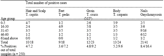 | |
| % Positive out of 250 samples, Male = 26% and Female = 49.6%, M: Male; F: Female | |
| Table 2: | Distribution of dermatophytes causing Tinea at different human body sites |
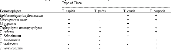 | |
| +: Present, -: Absent | |
| Table 3: | Onychomycoses caused by dermatophytes |
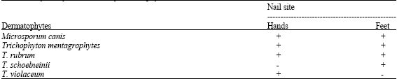 | |
| +: Present, -: Absent | |
| Table 4: | Non-dermatophytes (opportunistic fungi) isolated from different body site samples |
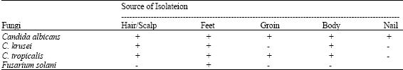 | |
| +: Present, -: Absent | |
Trichophyton mentagrophytes and T. rubrum were the most common species causing all types of tinea, while Microsporum canis causes T. capitis, T. cruris and T. corporis. Microsporum canis was not isolated in the case of T. pedis. Trichophyton schoelneinii and T. soudanense were also not isolated from foot (T. pedis). Epidermatophyton floccussum caused T. cruris and T. corporis, Microsporum gypseum caused T. capitis and T. cruris, Trichophyton violaeum caused only T. corporis, while, T. verrucossum caused T. cruris and T. corporis (Table 2).
Microsporium canis, T. memtagrophytes and T. rubrum caused infection both in the nails of hand and foot, while T. schoelneinii caused infection in foot nails and T. violaceum isolated only from nails of hand (Table 3).
Candida albicans, C. krusei, C. tropicalis and Fusarium solani were the non-dermatophytes (Table 4) isolated from different sites of the body. Aspergillus species isolated from dermatophytic samples are not included here. Candida albicans was isolated from samples from all sites followed by C. tropicalis isolated from 4 types of samples except nails. Fusarium solani restricted to feet and nails samples. Candida krusei was not isolated from groin and nail samples.
DISCUSSION
Among Tinea, T. corporis and T. cruris were found to be most common in the Eastern Province of Saudi Arabia, while in contrast Abanmi et al. (2008) found Tinea capitis and T. pedis as the most common and T. corporis as the least common from central (Riyadh) region of Saudi Arabia. This might be due to different in environmental condition. Eastern region stretched along the Arab Gulf, while Central region has very dry climate.
Children under 15 years of age appeared to be more susceptible to T. capitis and T. corporis which are in agreement with earlier finding from Saudi Arabia (Abanmi et al., 2008; Al-Sogair et al., 1991; Venugopal and Venugopal, 1992) and from other regions of the world (Lari et al., 2005; Brajac et al., 2004). This may be due to low level of fungi static fatty acids at an early stage and large family size may cause some neglect (in terms of hygiene standard), the sharing of towels, clothing and hair accessories may lead to spread of dermatophytes (Ansarin et al., 2001). The humidity and temperature are well known factors affecting fungal penetration through the stratum corneum of the skin (Morishita et al., 2003). Exposure to high temperature with high humidity is common in the Arabian Gulf, moreover, traditional and religious habit may affect the prevalence of dermatophytes (Sahin et al., 2004). Trichophyton Mentagrophytes was the most common isolates in this study which was also reported as a most common caused organism of Tinea from different countries (Coloe and Baird, 1999; Ungpakorn, 2005). Earlier studies indicate that dog and cat play an important role in the spreading of tinea (Dolenc-Voljc, 2005; Zdovc, 1998).
Present study showed that more female were affected by dermatophytes (almost double in number) than male, Ealier reports also indicated a higher prevalence of dermatophytes in females as compared to males (Singh et al., 2003; Ellabib et al., 2002; Brilhante et al., 2004). Although, some studies recorded higher prevalence of dermatophyte, in males than females (Falahati et al., 2003; Lari et al., 2005).
In the present study, Onyhomycosis was most prevalent disease and more than any type of tinea specially in the adults of age group between 16-45 years. Which is in agreement with the earlier finding is in agreement with the earlier finding from Saudi Arabia (Abanmi et al., 2008), neighbouring countries (Sahin et al., 2004; Falahati et al., 2003) and other parts of the world (Gupta et al., 2003; Pierard and Pierard-Franchimont, 2005; Garg et al., 2004; Lange et al., 2006). Candida albicans was the most common isolates among the Candia species causing Onychomycosis (Garg et al., 2004). Candida species may colonies skin, hair, nails and may become pathogenic in case of any immunodeficiency, trauma and loss of epidermal barrier function. Fusariumj solani is well known to cause onychomycosis (Romano et al., 2005; Garccia-Matos, 2000).
In Saudi Arabia, nearly 25% of the population suffers from diabetes and other immunodeficiencies and these population are at a high risk of dermatophytic infection which may leads to deep mycosis specially if caused by molds. Therefore it is suggested, that a routine check ups of these immunocompromised patient should be done for any short of dermatophytes and also samples collected from these patient should be send to diagnostic laboratory with clinically details, with changing a population density, migration of rural population to urban areas and influence of exports worker from different countries affect the epidemiology of dermatophytes, therefore, a regular survey should be conducted for spreading of dermatophytes. Result of present study clearly indicates that the epidemiology of dermatophytes significantly differs from other regions of Saudi Arabia.
ACKNOWLEDGMENTS
The author is thankful to King Faisal University for financial support of this project (Project No. 10104). The author would like to acknowledge all specialists in different governmental hospitals of AlHassa’s region especially, Dr. Manal Sayed Mohamed a Dermatology Specialist, Dr. Eman Elmasry Eldamarany, Consultant Biochemistry, Dr. Manar AlTablawy Consultant Haematology and Mr. Hashem Almusalam a Microbiology specialist.
REFERENCES
- Abanmi, A., S. Bakheshwain, N. El-Khizzi, A.R. Zouman and S. Hantirah et al., 2008. Characteristics of superficial infection in the Riyadh region of Saudi Arabia. Int. J. Dermatol., 47: 229-235.
PubMedDirect Link - Al-Sogair, S.M., M.K. Moawad and Y.M. AlHomaidan, 1991. Fungal infections as a cause of skin disease in the eastern province of Saudi Arabia: Tinea corporis and T. capitis. Mycoses, 34: 423-427.
PubMedDirect Link - Brajac, I., L. Stojnic-Sosa, L. Parpic, K. Loncarek and F. Gruber, 2004. The epidemiology of Microsporum canis infections in Rijeka area, Croatia. Mycoses, 47: 222-226.
Direct Link - Brilhante, R.S.N., R.A. Cordeiro, M.F.G. Rocha, A.J. Monteiro, T.E.F. Meireles and J.J.C. Sidrim, 2004. Tinea capitis in a dermatology center in the city of Fortaleza, Brazil: The role of Trichophyton tonsurans. Int. J. Dermatol., 43: 575-579.
CrossRefDirect Link - Coloe, S.V. and R.W. Baird, 1999. Dermatophyte infectioin in Melbourne trends from 1961/64 to 1995/96. Pathology, 31: 395-397.
PubMed - Costa, T.R., M.R. Costa M.V. Da-Silva, A.B. Rodrigues, F. Fernandes-Ode, A.J. Soares and R. Silva-Mdo, 1999. The etiology and epidemiology of dermatophytosis in Goiania G.O. Brazil. Rev. Soc. Bras. Med. Trop., 32: 367-371.
PubMedDirect Link - Dolenc-Volje, M., 2005. Dermatophyte infections in the Ljubljana regioin, Slovenia, 1995-2002. Mycoses, 48: 181-186.
PubMedDirect Link - Ellabib, M.S., M. Agaj, Z. Khalifa and K. Kavanagh, 2002. Trichophyton violaceum is the dominant cause of tinea captitis in children in Tripoli, Libya: Result of two years survey. Mycopathologia, 153: 145-147.
CrossRefDirect Link - Falahati, M., L. AKhlag, A.R. Lari and R. Alaghehbandan, 2003. Epidemiology of dermatophytosis in an area South of Tehran. Iran. Mycopathologia, 156: 279-287.
CrossRefDirect Link - Garcia-Matos, P., T. Domingaez, P. Martin, M. Linares, J. Mira and J. Calap, 2000. Onchomycosis caused by non-dermatophytic filamentous fungi in Cadiz. Enform. Infec. Microbiol. Clin. 18: 319-324.
PubMed - Gupta, A.K., J.E. Ryder and R.C. Summerbell, 2003. The diagnosis of non-dermatophyte mold Onyhomycosis. Int. J. Dermatol., 42: 272-273.
Direct Link - Gupta, A.K. and R.C. Summerbell, 1998. Increased incidence of Trichophyton tonsurans tinea capitis in Ontario, Candia between 1985 and 1996. Med. Mycol., 36: 55-60.
PubMed - Kasai, T. and Epidemiological Investigation Committee for Human Mycoses in the Japanese Society for Medical Mycology, 2001. 1997 epidemiological survey of dermatophytoses in Japan. Nippon. Ishinkin. Gakkal. Zasshi, 42: 11-18.
PubMedDirect Link - Lange, M., J. Roszkiewicz, A. Szczerkowska-Dobosz, E. Jasiel-Walikowska and B. Bykowska, 2006. Onychomycoses is no longer a rare finding in children. Mycoses, 49: 55-59.
PubMedDirect Link - Lari, A.R., L. Akhalagi, M. Falahati and R. Alaghehbandan, 2005. Characteristics of dermatophytoses among children in an area South of Tehran Iran. Mycoses, 48: 32-37.
CrossRefDirect Link - Morishita, J. Ninomiya, Y. Sei and I. Takiuchi, 2003. Effect of temperature, humidity, minor injury and washing on penetration of dermatophytes into human stratum corneum. Nippon. Ishinkin. Gakkai. Zahsshi, 44: 269-271.
PubMedDirect Link - Pawardhan, N. and R. Dave, 1999. Dermatomycosis in and around Aurangabad. Indian J. Pathol. Mirobial., 42: 455-462.
PubMedDirect Link - Pierard, G.E. and C. Pierard-Franchimont, 2005. The nail under fungal siege in patient with type II diabetes mellitus. Mycoses, 48: 339-342.
CrossRefDirect Link - Romano, C., C. Gianni and E.M. Difonzo, 2005. Petrospective study of Onyhomycosis in Italy: 1985-2000. Mycoses, 48: 42-44.
CrossRefDirect Link - Sahin, I., S. Oksuz, D. Kaya, I. Sencan and R. Cetinkaya, 2004. Dermatophytes in the rural area of Duzce, Turkey. Mycoses, 47: 470-474.
PubMedDirect Link - Singh, D., D.C. Patel, K. Rogers, N. Wood, D. Riley and A.J. Morris, 2003. Epidemiology of dermatophyte infections in Auckland New Zealand. Aust. J. Dermatol., 44: 263-266.
PubMedDirect Link - Ungpakron, R., 2005. Mycoses in Thailand: Current concerns. Jap. J. Med. Mycol., 46: 81-86.
Direct Link - Vella-Zahra L., P. Gatt, M.J. Boffa, E. Borg and E. Mifsud et al., 2003. Characteristics of superficial mycoses in Malta. Int. J. Dermatol., 42: 265-271.
ASCIDirect Link - Venugopal, P.V. and T.V. Venogopal, 1992. Superficial mycoses in Saudi Arabia. Aust. J. Dermatol., 33: 45-48.
CrossRefDirect Link - Weitzman, I., N.X. Chin, N. Kunjukunju and P. Della-Latta, 1998. A survey of dermatophytes isolated from human patients in the United States from 1993 to 1995. J. Am. Acad. Dermatol., 39: 255-261.
PubMedDirect Link - Zdovc, I., 1998. Epidemiological and diagnostic features of animal dermatophytosis. Acta Dermatol. Venereol., 7: 113-119.
Direct Link