
N.S. AL-Sowyan
Department of Biology, Faculty of Science and Arts, AL-Qassim University, P.O. Box 30230, Buraydah 51744, Saudi Arabia
International Journal of Pharmacology
Year: 2009 | Volume: 5 | Issue: 3 | Page No.: 208-214


ABSTRACT
Paracetamol, a major cause of acute liver failure represents a significant clinical problem. Intake of a large dose of paracetamol (APAP) may result in severe hepatic necrosis. Oxidative stress mediated by oxidative capacities of the APAP metabolite (N-acetyl-P-benzoquinon-imine (NAPQI)) is considered as the main cause of hepatotoxicity of APAP. This labor therefore seeks to induce liver damage in rats using single dose of APAP and to evaluate the possible protective effects of administration of folic acid on APAP induced liver damage in rats. Serum transaminases and lactic dehydrogenase levels were assessed as markers of hepatic damage. Also, bilirubin, total protein, albumin, globulin and A/G ratio were analyzed. Equally, comparative effects of folic acid on the markers were also evaluated. Paracetamol caused liver damage as evident by statistically significant increased in the activities of alanine aminotransferase, aspartate aminotransferase and alkaline phosphates. There were general statistically significant losses in the activities of lactic dehydrogenase and an increase in total bilirubin and protein with significant decrease in A/G ratio in paracetamol treated group compared with the control group. Also, the histopathological examination of liver showed marked degeneration of hepatic cells and necrosis with congested portal vein and dilated hepatic sinusoid. However, folic acid was able to counteract the effect of APAP. The present results suggest that folic acid can act as hepatoprotective against paracetamol toxicity and that the mechanism by which they do this is by acting as antioxidants.
PDF Abstract XML References Citation
How to cite this article
N.S. AL-Sowyan, 2009. Efficacy and Safety of Folic Acid During Toxic Hepatitis Induced by Acute Overdose of Paracetamol. International Journal of Pharmacology, 5: 208-214.
DOI: 10.3923/ijp.2009.208.214
URL: https://scialert.net/abstract/?doi=ijp.2009.208.214
DOI: 10.3923/ijp.2009.208.214
URL: https://scialert.net/abstract/?doi=ijp.2009.208.214
INTRODUCTION
Liver dysfunction is one of the major public health problems encountered both in developing and developed countries. Since, liver function affects almost every system in the body, liver diseases usually result in a profound change in patient life profile. Owing to its ability to concentrate, biotransform and excrete a wide array of xenobiotics and to its anatomical proximity to the blood supply of the gastrointestinal tract, the liver is markedly vulnerable to damage. Liver injury induced chemically can be experimentally used to mimic many types of liver diseases (John-Cullen, 2005).
Paracetamol (APAP) is a commonly used analgesic antipyretic drug. It is generally considered a safe drug. However, in large single dose ingestion (usually as an intentional overdose) it causes massive centrilobular necrosis that may be fatal in both humans and experimental animals (Shayiq et al., 1999; Olaleye and Rocha, 2008). There is also increasing awareness that combining therapy doses of APAP with prolonged fasting might lead to hepatotoxicity (Bonkovsky et al., 1994).
Availability of paracetamol as an over-the-counter medication alone and in combination with other prescription and over-the-counter drugs therefore, creates a situation that may lead to exposure to excessive quantities of the drug (Andringa et al., 2008). The factors that influence hepatotoxicity after an acute dose of paracetamol are relatively well understood. However, the mechanisms that affect susceptibility after exposure to paracetamol are more complex. Understanding these mechanisms is important because of the extensive use of paracetamol as an over-the-counter analgesic drug (Anker and Smilkstein, 1994). Oxidative stress mediated by oxidative capacities of APAP metabolite, is considered as the main cause of hepatotoxicity. Folate is being utilized to treat a wide variety of diseases like hepatotoxicity via., selective cellular markers can reduce the toxicity of therapeutic agents (Antelava et al., 2007). Because oxidative stress plays a major role in several liver diseases, it is of interest to evaluate the role of folic acid in protection against liver injury. Therefore, the aim of the present study is to shed more light on the possible hepatoprotective effect of folic acid on acute paracetamol intoxication. This study therefore seeks to induce liver damage in rats using single dose of paracetamol and the chosen parameter will be examined both in treated and untreated rats. Also, the hepatoprotective potential of folic acid will be evaluated regarding its effect on liver function parameters including, serum alanine aminotransferase (ALT), aspartate aminotransferase (AST), alkaline phosphates (ALP), total bilirubin, total protein, albumin, globulin and A/G ratio. The study design includes evaluation of oxidative stress biomarkers as lactate dehydrogenase (LDH). In addition, histopathological examination of liver sections from all studied groups will be performed.
MATERIALS AND METHODS
The present study was planned to investigate the hepatoprotective effect of folic acid intake on the prevention of acute hepatotoxicity induced by overdose of paracetamol.
The study was conducted on 30 adult male albino rats, weighing 120-200 g. All rats included were fed on normal balanced diet (rat chow) and allowed to adapt the prevailing environment for one week prior to the beginning of the experiment in the laboratory of physiology.
The experiment was carried out through January 2008-January 2009. The rats were divided into 3 equal groups:
| • | Control group consisted of rats maintained on the commercial rat chow diet all over the experimental period and served as a control |
| • | Paracetamol treated group consisted of rats maintained on the commercial rat chow diet and received a single oral dose of paracetamol 640 mg kg-1 b.wt. according to Devi et al. (2005) to induce acute hepatotoxicity. Both blood and tissue samples were collected 3 days after paracetamol administration to APAP treated rats caused insignificant changes in these parameter when compared to |
| • | Folic acid and acute paracetamol treated group consisted of rats maintained on the commercial rat chow diet, they were given a single oral dose of paracetamol 640 mg kg-1 b.wt. and folic acid in a dose of 8 mg kgGo diet (Endoh et al., 2007) mixed with diet for 3 weeks |
Liver and blood samples were collected at the end of experimental period for biochemical and histological analysis.
Blood samples were obtained from the orbital sinus of an over night fasted rats under light ether anesthesia (Simmons and Brick, 1987), then centrifuged. The separated sera were analyzed for estimation of alanine aminotransferase (ALT), aspartate aminotransferase (AST), alkaline phosphates (ALP), lactate dehydrogenase (LDH), total bilirubin, total protein, albumin, globulin and albumin/globulin ratio (A/G ratio). Also, liver sample were taken for liver histopathology.
RESULTS AND DISCUSSION
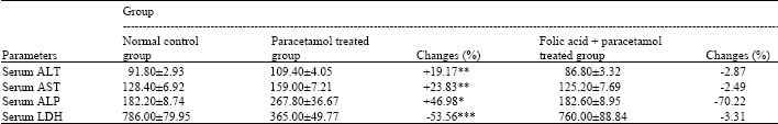
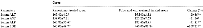
Paracetamol treated group showed a significant increase in serum activity of ALT, AST and ALP with significant decrease in the activity of LDH in comparison to normal control rats. Folic acid when given to APAP treated rats caused insignificant decreases in all parameter. But, when compared to APAP treated group it caused significant decrease serum liver transaminases with elevation of LDH activity (Table 1-2).
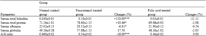
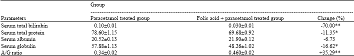
As regarding to serum level of total bilirubin, total protein, albumin, globulin and A/G ratio administration of APAP led to significant increase in bilirubin and total protein with significant decrease in serum albumin and A/G ratio. Folic acid supplementation to APAP treated rats caused insignificant changes in these parameter when compared to normal control group. But when compared to APAP treated group it showed significant decrease in serum bilirubin, total protein and globulin with increased A/G ratio (Table 3-4).
Histological results: In the normal group, histological examination of the liver revealed normal architecture of the hepatic lobules and hepatocytes (Fig. 1a).
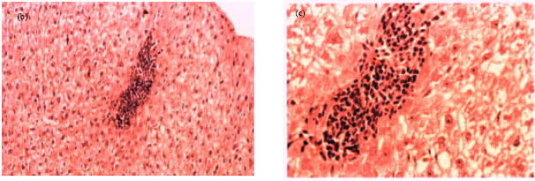
On the other hand, the hepatocytes of paracetamol treated rats showed severe degeneration and necrosis with periportal inflammation and congested portal vein (Fig. 1b, c).
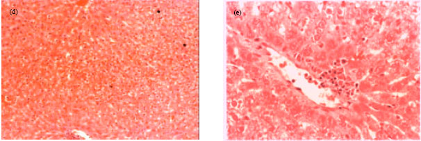
Folic acid administration produced less degenerative changes and reduction of inflammatory cell number at the periportal area to reach nearly to the normal hepatic morphology (Fig. 1d, e).
Paracetamol is a widely used analgesic and antipyretic since its production in 1950 (Graham and Scott, 2005). It is considered safe at the therapeutic doses. However, intake of a large dose of APAP may result in severe hepatic necrosis (Olaleye and Rocha, 2008). It also causes liver damage in therapeutic doses at certain circumstances e.g., fasting (Willis, 2005). In the present study, paracetamol caused liver damage as evident by statistically significant increased in serum activities of ALT, AST and ALP, with significant losses in the activity of LDH compared with the control group. The marked significant increase in serum ALT and AST were consistent with the finding of Helieh et al. (2004), Janbaz et al. (2004) and Omar et al. (2005). They reported that, administration of APAP causes severe centrilobuler necrosis with marked increase in serum ALT and AST activity.
This findings were explained by Kirchain and Gill (1997), on the basis that the presence of these enzymes in high concentration and their liberation from the hepatocytes cytoplasm make them a very sensitive indicator lesions within the liver.
| Table 1: | The changes in serum ALT (μ L-1), AST (μ L-1), ALP (μ L-1) and LDH (μ L-1) of normal and treated groups |
 | |
| Values are expressed as Mean±SE; *Significant at p<0.05, **Highly significant at p<0.01, ***Most significant at p<0.005 | |
| Table 2: | The changes in serum ALT (μ L-1), AST (μ L-1), ALP (μ L-1) and LDH (μ L-1) of treated groups. |
 | |
| Values are expressed as Mean±SE; *Significant at p<0.05, **Highly significant at p<0.01 | |
| Table 3: | The changes in serum total bilirubin (mg dL-1), total protein (g L-1), albumin (g L-1), globulin (g L-1) and A/G ratio of normal and treated groups |
 | |
| Values are expressed as Mean±SE; *Significant at p<0.05, ***Most significant at p<0.005 | |
| Table 4: | The changes in serum total bilirubin (mg dL-1), total protein (g L-1), albumin (g L-1), globulin ( g L-1 ) and A/G ratio of treated groups |
 | |
| Values are expressed as Mean±SE; *Significant at p<0.05, **Highly significant at p<0.01 | |
On the other hand, Olaleye and Rocha (2008) reported that oxidative stress mediated by oxidative capacities of APAP metabolite N-acetyl-P-benzoquinon-imine (NAPQI) is considered as the main cause of hepatotoxicity. Also, the increase in serum ALP activities is supported by the findings of Shivashangari et al. (2004) and Omar et al. (2005) that, paracetamol overdose is usually accompanied by the increase in ALP activity due to hepatocellular injury. On the other hand, significant reduction in the activity of serum LDH in the present study after administration of APAP is in the contrary with the findings of Bernal et al. (2002) and Shivashangeri et al. (2004) that, APAP toxicity was associated with increased release of LDH. The decreased activity of LDH in the present study may be due to ischemic hepatitis (Martin et al., 2004). The fall in LDH concentration could explain one of the mechanisms of APAP induced toxicity by stimulating the release of reactive intermediate species from macrophages (Hinson et al., 2000).
The marked increase in serum globulin, bilirubin and total protein with significant decrease in albumin and A/G ratio after paracetamol administration were consistent with the findings of Bourdi et al. (2002), Ju et al. (2002) Ridruejo et al. (2007) and Terneus et al. (2008). They reported that oxidative stress is a process that account for increased globulin level by APAP administration through the release of inflammatory mediators. Also, several studies argued that, oxidative stress is an additional or alternation process that accounts for hepatocellular damage (Manov et al., 2002). It has been shown that, non parenchymal cells, such as Kupffer and endothelial cells, located adjacent to the damaged areas undergo a series of morphological and functional changes and once activated, these cells are capable of releasing inflammatory mediators (anti-inflammatory cytokines ) and reactive oxygen species that may cause additional hepatic damage (Blazka et al., 1995) .
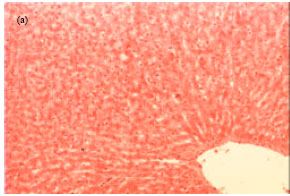 | |
| Fig. 1: | (a) Photomicrograph of a transverse section of the live. The normal hepatic architecture of the liver and the hepatocytes having dense round nuclei and darkly stained granular cytoplasm. The central veins and the hepatocytes arranged in cords radiating from the central vein. The hepatic sinusoids are present in between the hepatic cords of the liver (Hx. and E. x250), (b) The hepatocytes lose their normal architecture. Some of them are swollen and other cells showed marked degeneration and necrosis with dilated hepatic sinusoids and congested portal vein surrounded by inflammatory cells. The portal vein markedly congested and some inflammatory cells with rounded deeply stained nuclei can be seen around the portal vein (Hx. and E. x250), © The earlier picture is magnified to show degeneration and necrosis of hepatocytes and hepatic sinusoids are markedly dilated . Notice the presence of more cavities (Hx. and E. x400), (d) The most of hepatocytes appear normal with less focal degeneration. Most of hepatic sinusoids appear normal and less of them are dilated (Hx. and E. x250) and (e) The earlier picture photomicrograph of a transverse section of the liver. The periportal area show some inflammatory cells. Most of hepatocytes appear normal and less of cells show signs of degeneration. Most of hepatic sinusoids appear normal and less of them are dilated (Hx. and E. x400) |
As regard to the marked decrease in albumin level and A/G ratio in rats subjected to acute overdose of paracetamol, this findings is in agreement with Guyton and Hall (2006) that, A/G ratio is decreased in advanced liver disease due to decreased synthesis of albumin and increased synthesis of gamma globulin.
The histopathological examination of liver sections of the rats subjected to the acute APAP overdose, confirms the changes observed in the levels of liver biochemical markers. The histopathological examination of the liver revealed, degeneration of hepatic cells and necrosis of their nuclei with periportal infiltration, in addition to irregular blood sinusoids. This result is in agreement with Bushel et al. (2007) and Ramaiah and Jaeschkel (2007) that hepatic infiltration of neutrophils is an acute response to liver injury and hepatic stress induced by drug toxicity. These changes correlated positively with the increase in serum transaminases activity, such as AST and ALT were considered as a necrotic event by Ray et al. (1996). The pathological hepatic lesion produced by APAP with the concomitant increase in ALT, AST, ALP activities were generally though to be initiated by a high reactive metabolic product N-acetyl-P-benzoquinonemine (NAPQI) (Göksel et al., 2005). On the other hand, Helieh et al. (2004) reported that, binding of the metabolically activated APAP to target protein was not sufficient for toxicity and other factors had been postulated to be involved in liver injures such as, depletion of cellular ATP and reduced Ca+2 ATPase activity and increased cytosolic Ca+2 levels (Ferret et al., 2001; Hartmut et al., 2003), apoptosis, lipid peroxidation mediated by reactive oxygen species (Reiter et al., 2001; Göksel et al., 2005).
Concerning the effect of folic acid treatment on the hepatotoxicity induced by acute paracetamol overdose, the present results revealed that, the activities of AST, ALT and ALP were significantly decreased, while LDH was increased when compared to APAP treated group. These results are consistent with the findings of Lin et al. (2008) and Ghosh and Sil (2007), who reported that, the antioxidative properties of folic acid play an important role in the elimination of the changes that related to the oxidative stress such as lipid peroxidation and oxidative DNA damage, possibly due to scavenging the intermediates derived from paracetamol metabolism, including nitrogen oxide or their radical derivatives. Also, the study of Van Ede et al. (2001) showed that folate supplementation regimens reduced the incidence of elevated liver enzyme levels during methotrexate therapy by acting as antioxidants. As regarding to the LDH activity, the study of EL-Demerdash et al. (2006) showed that folic acid treatment after induction of liver toxicity by chromium was significantly decreased, this contradiction may be due to the dose of folic acid used in this study is not enough to improve both lactate dehydrogenase leakage and cell viability after APAP overdose.
The increased A/G ratio observed in the present study after folic acid treatment may be due to the marked reduction in liver necrosis and these supported by the marked fall in serum level of globulin with insignificant changes in albumin.
The improvement in the enzymes levels were explained and supported by the histopathological examination which revealed that, marked reduction in inflammatory cell number at the periportal area and less degenerative changes by the use of folic acid to reach nearly to the normal hepatic morphology. These results suggest that folic acid can protect the liver from paracetamol induced hepatic toxicity.
CONCLUSION
We recommended to use folic acid as a protective agent against paracetamol toxicity in patients presenting with paracetamol overdose and acute liver failure.
REFERENCES
- Anker, A.L. and M.J. Smilkstein, 1994. Acetaminophen. Concepts and controversies Review. Emerg. Med. Clin. North. Am., 12: 335-349.
PubMed - Antelava, N.A., M.I. Gogoluari, L.I. Gogoluari, N.N. Pirtskhalaishvili and M.V. Okudzava, 2007. Efficacy and safety of heptral, vitamin B6 and folic acid during toxic hepatitis induced by CCL4. Georgian Med. News, 150: 53-56.
PubMedDirect Link - Blazka, M.E., J.L. Wilmer, S.D. Holladay, R.E. Wilson and M.I. Luster, 1995. Role of proinflammatory cytokines in acetaminophen hepatotoxicity. Toxical. Applied Pharmacal., 133: 43-52.
CrossRefPubMedDirect Link - Bonkovsky, H.L., R.E. Kane, D.P. Jones, R.E. Galinsky and B. Banner, 1994. Acute hepatic and renal toxicity from low doses of acetaminophen in the absence of alcohol abuse or malnutrition: Evidence for increased susceptibility to drug toxicity due to cardiopulmonary and renal insufficiency. Hepatology, 19: 1141-1148.
PubMed - Bourdi, M., Y. Masubuchi, T.P. Reilly and L.R. Pohl, 2002. Protection against acetaminophen induced liver injury and lethality by interleukin 10: Role of inducible nitric oxide synthaze. Hepatology, 35: 289-298.
PubMed - Devi, K.P., M. Priya, K. Balakrishna and T. Devaki, 2005. Protective effect of Premna tomentosa extract (L-verbanacae) on acetaminophen induced mitochondrial dysfunction in rats. Mol. Coll. Biochem., 272: 171-177.
PubMedDirect Link - El-Demerdash, F.M., M.I. Yousef and F.A. Elaswad, 2006. Biochemical study on the protective role of folic acid in rabbits treated with chromium (VI). J. Environ. Sci. Health B, 41: 731-746.
PubMed - Endoh K., M. Murakami, T. Sugiyama, Y. Taki and K. Umegaki, 2007. Low folate status enhanced benzene-induced cytogenetic damage in bone marrow of mice: A relationship between dietary intake and tissue levels of folate. Nutr. Cancer, 59: 99-105.
CrossRefPubMedDirect Link - Ferret, P.J., R. Hammoud, M. Tulliez and F. Batteux, 2001. Detoxification of reactive oxygen species by a non peptidyl mimic of superoxide dismutase cures acetaminophen induced acute liver failure in the mouse. Hepatology, 33: 1173-1180.
PubMed - Ghosh, A. and P.C. Sil, 2007. Anti-oxidative effect of a protein from Cajanus indicus L. against acetaminophen-induced hepato-nephro toxicity. J. Biochem. Mol. Biol., 40: 1039-1049.
PubMedDirect Link - Sener, G., O. Sehirli, S. Cetinel, B.G. Yegen, N. Gedik and G. Ayanoglu-Dulger, 2005. Protective effects of MESNA (2-mercaptoethane sulphonat) against acetaminophen induced hepatorenal oxidative damage in mice. J. Applied Toxicol., 25: 20-29.
CrossRefPubMedDirect Link - Graham, G.G. and K.F. Scott, 2005. Mechanism of action of paracetamol. Am. J. Therapeutics, 12: 46-55.
PubMedDirect Link - Jaeschke, H., T.R. Knight and M.L. Bajt, 2003. The role of oxidant stress and reactive nitrogen species in acetaminophen hepatotoxicity. Toxical. Lett., 144: 279-288.
CrossRefPubMedDirect Link - Oz, H.S., C.J. McClain, H.T. Nagasawa, M.B. Ray, W.J.S. de Villiers and T.S. Chen, 2004. Diverse antioxidants protect against acetaminophen hepatotoxicity. J. Biochem. Mol. Toxicol., 18: 361-368.
CrossRefPubMedDirect Link - Hinson, J.A., S.L. Michael, S.G. Aull and N.R. Pumford, 2000. Western blot analysis for nitrotyrosine protein adducts in liver of saline treated and acetaminophen treated mice. Toxical. Sci., 53: 467-473.
PubMedDirect Link - Janbaz, K.H., S.A. Saeed and A.H. Gilani, 2004. Studies on the protective effects of caffeic acid and quercetin on chemical-induced hepatotoxicity in rodents. Phytomedicine, 11: 424-430.
CrossRefPubMedDirect Link - John-Cullen, M., 2005. Mechanistic classification of liver injury. Toxicol. Pathol., 33: 6-8.
PubMedDirect Link - Lin, H.L., C.C. Yu, Y.F. Chou and R.F. Huang, 2008. Folate deprivation and copper exposure potentiate reactive oxygen species generation and chromosomal DNA loss but not mitochondrial DNA deletions in rat hepatocytes. Int. J. Vitam. Nutr. Res., 76: 332-340.
PubMed - Manov, L., M. Hirsh and T.C. Iancu, 2002. Acetaminophen hepatotoxicity and mechanisms of its protection by N-acetylcysteine: study of Hep 3B cells. Exp. Toxical. Pathol., 53: 489-500.
PubMed - Olaleye, M.T. and B.T.J. Rocha, 2008. Acetaminophen-induced liver damage in mice: Effects of some medicinal plants on the oxidative defense system. Exp. Toxicol. Pathol., 59: 319-327.
CrossRefPubMedDirect Link - Salam, O.M.E.A., A.R. Baiuomy, S.M. El-Shenawy and N.S. Hassan, 2005. Effect of pentoxifylline on hepatic injury caused in the rat by the administration of carbon tetrachloride or acetaminophen. Pharmacol. Rep., 57: 596-603.
PubMed - Ramaiah, S.K. and H. Jaeschkel, 2007. Role of neutrophils in the pathogenesis of acute inflammatory liver injury. Toxicol. Pathol., 35: 757-766.
PubMed - Ray, S.D., V.R. Mumaw, P.R. Raje and M.W. Fariss, 1996. Protection of acetaminophen-induced hepatocellular apoptosis and necrosis by cholesteryl hemisuccinate pretreatment. J. Pharmacal. Exp. Ther., 279: 1470-1483.
PubMed - Reiter, R.J., D.X. Tan, L.C. Manchester and W. Qi, 2001. Biochemical reactivity of melatonin with reactive oxygen and nitrogen species: A review of the evidence. Cell Biochem. Biophy. S., 34: 237-256.
CrossRefPubMedDirect Link - Ridruejo, E., R. Cacchione, A.G. Villamil, S. Marciano, A.C. Gadano and O.G. Mando, 2007. Imatinib-induced fatal acute liver failure. Word J. Gastroenterol., 13: 6608-6611.
PubMed - Shayiq, R.S., D.W. Roberts, K. Rothstein, J.E. Snawder and M. Black, 1999. Repeated exposure to incremental doses of acetaminophen provides protection against acetaminophen induced lethality in mice: an explanation for high acetaminophen dosage in humans without hepatic injury. Hepatology, 29: 451-451.
PubMed - Shivashangari, K.S., V. Ravikumar and T. Devaki, 2004. Evaluation of the protecrive efficacy of Asteracantha longifolia on acetaminophen induced liver damage in rats. Med. Food., 7: 245-251.
PubMed - Terneus, M., J.M. Brown, A.B. Carpenter and M.A. Valentovic, 2008. Comparison of S-adenosyl-L-methionine (SAMe) and N-acetylcysteine (NAC) protective effects on hepatic damage when administered after acetaminophen overdose. Toxicology, 244: 25-34.
PubMed - Van Ede, A.E., R.F. Laan, M.J. Rood, T.W. Huizinga and M.A. van de Laar et al., 2001. Effect of folic or folinic acid supplementation on the toxicity and efficacy of methotrexate in rheumatoid arthritis: A forty-eight week, multicenter, randomized, double-blind, placebo-controlled study. Arthritis Rheum., 44: 1515-1524.
PubMed