
Arpita Chatterjee
Barasat College, Barasat, Kolkata, 700 126, West Bengal, India
International Journal of Osteoporosis and Metabolic Disorders
Year: 2014 | Volume: 7 | Issue: 1 | Page No.: 12-19


ABSTRACT
Osteoporosis, characterized by low Bone Mineral Density (BMD) and micro-architectural deterioration of bone tissues is one of the major public health problem and is also increasing in developing world. Osteoporotic fracture entails high health care costs, frequently associated with loss of quality of life with an increased mortality risk. Physical activity has beneficial effects on the skeleton by contributing significantly to attainment of peak bone mass. In this cross-sectional study, a total of 26 volunteers (8 male and 18 female) were enrolled to understand of BMD profile, using Furuno Ultrasound Bone Densitometer CM 200, where dance movements is admitted as a form of physical activity. Controls (n = 25) were participants frequency matched to cases by age, sex, similar lifestyle and food habit except they do not practice dance movements. Results revealed that physical activity in regular manner showed higher bone strength. The BMD, as judged by T-scores were under normal value (-1.0) in cases but the value was lower (-1.2) in control group. Beside 16% of control participants showed presence of osteopenia and in 4% case osteoporosis. This result supports the fact that the physical exercise has a potential to increase the health status of any age group by eliciting positive changes in certain aspects of physical fitness and healthy wellbeing. The frequency of decreased value of BMD in controls with increasing age significantly constitutes a high-risk group for osteoporosis and future fractures.
PDF Abstract XML References Citation
Received: June 21, 2014;
Accepted: July 01, 2014;
Published: December 30, 2014
How to cite this article
Arpita Chatterjee, 2014. Effect of Physical Exercise on Bone Mineral Density: A Cross-sectional Study in India. International Journal of Osteoporosis and Metabolic Disorders, 7: 12-19.
DOI: 10.3923/ijom.2014.12.19
URL: https://scialert.net/abstract/?doi=ijom.2014.12.19
DOI: 10.3923/ijom.2014.12.19
URL: https://scialert.net/abstract/?doi=ijom.2014.12.19
INTRODUCTION
Osteoporosis is one of the major public health problems in Western world and is also increasing in the developing world (MacDonald et al., 2005). It is characterized by low Bone Mineral Density (BMD) and micro-architectural deterioration of bone tissues. It enhances bone fragility and as a consequence, also increases the risk of fracture (Dambacher et al., 2004; Riggs and Melton, 1995). The BMD test measures the density of minerals, mainly calcium, present in bones to estimate the strength of bones. This procedure uses different techniques including special X-ray, ultrasound or computed tomography scan. Generally with age, bones become thinner due to lose of some bone mass and break down of bones faster than new bones. Further due to lose of calcium and other minerals, the bones become lighter and weaker. Thus, the bones become less dense and more porous and increase the chance of fracture. It is known as osteopenia (Cole, 2008). With further bone loss osteopenia leads to osteoporosis.
It is reported fact that worldwide about one third women and one twelfth men in age group over 50 years have osteoporosis. The prevalence of osteoporotic fracture is extremely higher in women, particularly with increase in age (Looker et al., 2012). It has become a global public health concern due to the increasing population of the elderly (Aghaei Meybodi et al., 2008). With the increase in life expectancy the risk of osteoporosis increases among postmenopausal women, who spend about half of their lifetime after menopause (Keramat et al., 2008; Nordin, 2009). In postmenopausal women, it affects about one third of women aged between 60-70 years and two third of those aged 80 years or more worldwide (Cashman, 2005). In China, epidemiological studies have shown that the incidence of osteoporosis was 16.1% in age group over 40 years, 22.6% in age group over 60 years and 50% in age group over 80 years (Li et al., 2002). Data from US National Health and Nutrition Examination Survey 2005-2008 showed that 9% of adults aged 50 years and over had osteoporosis at either femur neck or lumbar spine, about one-half had low bone mass at either site (Looker et al., 2012).
The disease causes millions of fractures annually, mostly involving the lumbar vertebrae, hip and wrist. Osteoporosis in the spine not only leads to fractures but also produce pain and back deformities which seriously affect patients’ quality of life (Cummings et al., 1995). An osteoporotic fracture entails high health care costs (Chrischilles et al., 1994) and is also frequently associated with the loss of quality of life resulting from deficient healing and with an increased mortality risk (Center et al., 1999; Johnell, 1996).
Several risk factors are associated with bone loss Cumming et al. (1997), among which some factors are modifiable (weight, physical activity and diet) and others are non-modifiable (age, sex and race) (Ross, 1996). Maintenance of a quality BMD needs the basic requirements of nutrition with adequate calcium and vitamin D intake, exercises and sunlight (Kolahi et al., 2011; Leung et al., 2012; Tajik et al., 2013; Yoshioka et al., 2012). Physical activity has been reported to have beneficial effects on the skeleton by contributing significantly to attainment of peak bone mass and maintenance of bone mass later in adulthood (Bailey et al., 1999; Cashman, 2002; Khan et al., 2000). Moreover, resistance training has beneficial and site-specific effects which improve BMD as well as muscle strength (Kerr et al., 2001).
As a physical activity and a creative art form, it is believed that dance movements can make a significant contribution to the healthy-living agenda of human being (Chatterjee, 2013a). Dance is an active, non-competitive form of exercise like other sports (Chatterjee, 2011; Quin et al., 2007). Among different kinds of physical exercise, dance movement has a great potential to motivate and excite people. It has a positive effect on physical health as well as mental and social health (Chatterjee, 2012, 2013b). Dance increases total body movement, helps to improve respiratory, circulatory and musculo-skeletal systems (Marshall et al., 1998). It also increases lung function, lung capacity, flexibility and aerobic capacity (Blair et al., 2001). Now-a-days in many hospitals and medical settings, it has been used as a form of therapy, not only for mental health but also for physical health as well (Chatterjee, 2013c, d). Dance therapy as exercise is known to increase the neurotransmitters called endorphins which increase a state of well-being (Chatterjee, 2013e).
Therefore, the specific objective of the study was to investigate the effects of a bone health supporting physical activity. In order to elucidate the role of modifiable bone loss factors in Indian women and men, the present study was undertaken to investigate the physical activity which is likely to contribute to control osteoporosis. For this purpose, the present investigation was aimed to study the bone strength in participants practicing dance movements in regular basis by comparing with control group without such practice. Such type of study was never attempted in India till date and thus necessitates in-depth research on the said topic. The study was also directed towards this mechanistic understanding of BMD profile in cases, where dance movements were admitted as a form of physical activity.
MATERIALS AND METHODS
Personal data: A questionnaire comprising different open ended questions was designed to assess the personal data as age, education, physical activity and medical background, etc. The subjects were asked to answer the questionnaire sheets according to knowledge, attitudes, behavior and practice model containing questions regarding daily activities.
Cases and sample preparation: In this cross-sectional study a total of 26 volunteers (8 male and 18 female) belonging to the age group 20-34 were enrolled for the investigation covering differential socio-economic status. They were selected as cases depending on over 8 year’s regular practice of dance movements in a controlled manner. Controls (n = 25) were participants frequency matched to cases by age, sex and those having similar lifestyle and food habit except that they do not practice dance movements. Demographic as well as environmental data was recorded.
Determination of BMD: For determination of BMD, the amount of matter per cubic centimeter of bones is measured to estimate the frequency of BMD in cases and controls by densitometric procedure. In the present study, ultra sonographic bone densitometer (Furuno Ultrasound Bone Densitometer CM 200) was used to test BMD according to the manufacturer’s instruction. The method involves minimal radiation exposure and is painless and non-invasive. During the present investigation, bone mineral density was measured at the heel and wrist. The main outcome measures were in the form of baseline T-scores. According to WHO (1994), T-score as a bone health status indicator can evaluate osteoporosis of the respondents. T-score was calculated by using the manufacturer’s reference values considering the age of the cases and controls. T-score was then categorized as normal (~ -1.0), osteopenia (-1.0- -2.5) and osteoporosis (~ -2.5) according to (WHO, 1994).
Statistical analysis: All the data of cases and controls were represented in the form of mean value ±SD and the results were analyzed by Student’s t-test. The statistical significance testing was done by estimating the p-value.
RESULTS
The present investigation revealed that the BMD level of cases and controls differ considerably (Table 1). The cases considered in the present study, where dance movements were admitted as a form of physical activity, showed higher bone strength. The BMD, as judged by T-scores was more in cases as compared with the control group. The BMD (T-scores) of cases with regular practice of physical exercise were under normal value (-1.0), whereas in many participants of control group, this value was lower (-1.2). In case of control group, few of them (16%) showed presence of osteopenia (T-score -1.4) and in 4% case osteoporosis (T-score -3.2).
Statistical analysis (p-value) showed that physical exercise benefited physiologically from this form of physical activity induced BMD level (Table 1). The participants with regular physical activity also showed overall greater motivation and improved flexibility in comparison to control group. Joint range of motion of participants practicing dance movements and their potential for improvement is generally higher than that of control group.
| Table 1: | Estimation of bone mineral density in participants and controls |
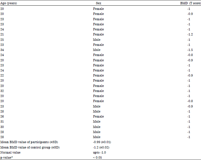 | |
| *p-value <0.05 indicates the results are significant | |
DISCUSSION
Lifestyle modification plays a crucial role in maintaining the bone health (Nahin and Straus, 2001). This type of osteoporosis prevention is very important since, improvements in lifestyle could be applied broadly and may represent a non pharmaceutical approach to improve BMD. Among other factors, hormonal replacement therapy is popular in the prevention of bone loss in postmenopausal women (Anonymous, 1993; Christiansen, 1993). Among the pharmacological treatments, selective estrogen receptor modulators and bisphosphonate have been shown to be effective either in increasing BMD and reducing fracture rates (Goltzman, 2002). But treatment with estrogen has well-known side effects such as breast soreness and nausea and in the long-term, may have increased risks of breast and uterine cancer development and also venous thrombosis (Lindsay et al., 1976). However, the side effects of these medications including gastrointestinal tolerance problems in bisphosphonate and the potential malignancies in hormonal therapy may preclude their long-term use (Reid, 2002). Growing evidence of the benefits of physical exercise for bone health provide an alternative option for prevention and treatment of osteoporosis. This is also effective and useful as it further supports a balanced bone metabolism with additional supplements to avoid other health hazards as a great blessing.
In the present cross-sectional study, there was a significant relationship between more physical activity and higher BMD. In the present study, dance movements was proved to be an activity that has potential to increase the health status of any age group by eliciting positive changes in certain aspects of physical fitness and healthy wellbeing. The result revealed that within a single age group the number of members in control group with osteopenia and osteoporosis was significantly high (Table 1). This T-score is a comparison of BMD of a person to that of a healthy one of the same sex and ethnicity. The lower T-score indicate osteopenia when the value range is less than -1.0 and greater than -2.5 and osteoporosis as -2.5 or lower. The frequency of decreased value of the BMD in control group with increasing age significantly constitutes a high-risk group for osteoporosis and future fractures. This result supports the fact that the density and strength of bone can be increased by exercise (Cole, 2008).
The present investigation suggests that the practice of dance movement in regular basis in proper and controlled way is particularly beneficial for physical wellbeing as was observed by early reports (Chatterjee, 2013c, e; Clippinger, 1997). In case of participants with physical activity, as a complete exercise dance movements play an important role in good health as an early defense system. The immuno-modulation of dancers in the positive direction makes them in proper healthy state and supports the therapeutic use of dance as a complete exercise (Chatterjee, 2011, 2013e). It can prevent from many diseases like cancer, diabetes, cardiac diseases and also has an anti-ageing role (Chatterjee, 2012, 2013c). Osteoporosis may be partially prevented by optimizing peak bone mass in younger years, maintaining bone mass in adult years and minimizing bone loss in later years. Adequate physical activities have been shown to be effective strategies for building, maintaining and slowing loss of bone mass (Cadogan et al., 1997; Suleiman et al., 1997). Therefore, to prevent osteoporosis, women need adequate physical activity level (Heinonen et al., 1996; Snow-Harter et al., 1992). Furthermore, the high level of awareness about the disease and its affordable precautions can reduce osteoporosis in Indian women (Picard et al., 2000; Recker et al., 1996). Since, BMD test is an expensive measurement, a limited number of subjects could be included in the study and the limited number of subjects might be the reason for some inconsistencies between the results of this study and some previous studies.
In general, females are more prone to osteoporosis than males because the involvement of females in physical activity is less than that of males. Further, this involvement of females in direct physical activity decreases from adolescence onwards (Daley and Buchanan, 1999). This observation puts a question mark on the consciousness of general people towards the health science and benefit of exercise when the health of them is in stake. In contrast, the therapeutic use of dance movements in one way may change this spectrum and also help to have a risk free proper healthy state of life in a positive direction. The bone strength can be increased in a positive mode by regular practice of dance movements and other physical activities. Therapeutic approaches are actually aimed at increasing good health status, thus prolonged practice of dance movements, like other sports benefit, in regular and controlled manner may cause betterment in normal life.
CONCLUSION
Physical exercises are actually aimed at increasing healthy response, thus prolonged and controlled exercise may cause betterment in the normal health. This investigation proposes that dance movements as a complete physical exercise plays an important and potential role in developing healthy bones. This may lead to stop the great suffering from diseases like osteopenia or osteoporosis. The participants may reduce their probability of future suffering from such diseases by an altered lifestyle with regular practice of physical exercise. Thus, this study is a new approach to contribution in this direction where dance movement has been admitted to increase bone strength as physical activity.
ACKNOWLEDGMENT
The author is grateful to Prof. Amita Dutt, Director, Performing Arts Therapy Centre, Rabindra Bharati University for her kind advice during the study. The author is thankful to Dr. Sumanta Thakur, Orthopedic Surgeon, for his kind help and suggestion in collection and analysis of data during the present investigation.
REFERENCES
- Aghaei Meybodi, H.R., R. Heshmat, Z. Maasoumi, A. Soltani and A. Hossein-Nezhad et al., 2008. Iranian osteoporosis research network: Background, mission and its role in osteoporosis management. Iran. J. Public Health, 37: 1-6.
Direct Link - Anonymous, 1993. Consensus development conference: Diagnosis, prophylaxis and treatment of osteoporosis. Am. J. Med., 94: 646-650.
PubMedDirect Link - Bailey, D.A., H.A. Mckay, R.L. Mirwald, P.R. Crocker and R.A. Faulkner, 1999. A six-year longitudinal study of the relationship of physical activity to bone mineral accrual in growing children: The university of Saskatchewan bone mineral accrual study. J. Bone Mineral Res., 14: 1672-1679.
CrossRefPubMed - Blair, S.N., Y. Cheng and J.S. Holder, 2001. Is physical activity or physical fitness more important in defining health benefits? Med. Sci. Sports Exerc., 33: S379-S399.
PubMedDirect Link - Cadogan, J., R. Eastell, N. Jones and M.E. Barker, 1997. Milk intake and bone mineral acquisition in adolescent girls: Randomised, controlled intervention trial. Br. Med. J., 315: 1255-1260.
Direct Link - Cashman, K.D., 2002. Calcium intake, calcium bioavailability and bone health. Br. J. Nutr., 87: S169-S177.
CrossRefDirect Link - Cashman, K.D., 2005. Homocysteine and osteoporotic fracture risk: A potential role for B vitamins. Nutr. Rev., 63: 29-36.
CrossRef - Center, J.R., T.V. Nguyen, D. Schneider, P.N. Sambrook and J.A. Eisman, 1999. Mortality after all major types of osteoporotic fracture in men andwomen: An observational study. Lancet, 353: 878-882.
CrossRefDirect Link - Chatterjee, A., 2013. The therapeutic value of Indian classical, folk and innovative dance forms. Rupkatha J. Interdiscipl. Stud. Humanities, 5: 75-83.
Direct Link - Chatterjee, A., 2013. An analytical discussion on the folk and tribal dance forms of Bengal in relation to their effect on health. Indian J. Arts, 1: 29-32.
Direct Link - Chatterjee, A., 2013. Prevention of oxidative stress injury among females by movement therapy in India. J. Med. Sci., 13: 843-846.
CrossRefDirect Link - Chatterjee, A., 2013. Improved health status through prolonged practice of dance as a therapy-a case study. Int. J. Basic Applied Med. Sci., 3: 180-183.
Direct Link - Chrischilles, E., T. Shireman and R. Wallace, 1994. Costs and health effects of osteoporotic fractures. Bone, 15: 377-386.
CrossRefDirect Link - Christiansen, C., 1993. Prevention and treatment of osteoporosis with hormone replacement therapy. Int. J. Fertil. Menopausal. Stud., 38: 45-54.
PubMed - Cole, R.E., 2008. Improving clinical decisions for women at risk of osteoporosis: Dual-femur bone mineral density testing. J. Am. Osteopathic Assoc., 108: 289-295.
Direct Link - Cumming, R.G., M.C. Nevitt and S.R. Cummings, 1997. Epidemiology of hip fractures. Epidemiol. Rev., 19: 244-257.
Direct Link - Cummings, S.R., M.C. Nevitt, W.S. Browner, K. Stone and K.M. Fox et al., 1995. Risk factors for hip fracture in white women. N. Engl. J. Med., 332: 767-774.
CrossRefDirect Link - Daley, A.J. and J. Buchanan, 1999. Aerobic dance and physical self-perceptions in female adolescents: Some implications for physical education. Res. Q. Exercise Sport, 70: 196-200.
CrossRef - Dambacher, M.A., S. Schmitt, E. Schacht, M. Ito and M. Neff et al., 2004. Bone structures In vitro and In vivo in animals and in men-A view into the future. J. Mineralstoffwechsel, 11: 13-19.
Direct Link - Goltzman, D., 2002. Discoveries, drugs and skeletal disorders. Nat. Rev. Drug Discovery, 1: 784-796.
PubMedDirect Link - Heinonen, A., P. Kannus, H. Sievanen, P. Oja and M. Pasanen et al., 1996. Randomised controlled trial of effect of high-impact exercise on selected risk factors for osteoporotic fractures. Lancet, 348: 1343-1347.
CrossRefDirect Link - Johnell, O., 1996. Advances in osteoporosis: Better identification of risk factors can reduce morbidity and mortality. J. Internal Med., 239: 299-304.
PubMed - Keramat, A., B. Patwardhan, B. Larijani, A. Chopra and A. Mithal et al., 2008. The assessment of osteoporosis risk factors in Iranian women compared with Indian women. BMC Musculoskeletal Disorders, Vol. 9.
CrossRefDirect Link - Khan, K., H.A. McKay, H. Haapasalo, K.L. Bennell, M.R. Forwood, P. Kannus and J.D. Wark, 2000. Does childhood and adolescence provide a unique opportunity for exercise to strengthen the skeleton? J. Sci. Med. Sport, 3: 150-164.
PubMed - Kolahi, S., N. Farrin, A.R. Ostadrahim and S.A. Mahboob, 2011. The role of proper food habit and physical activity level in preventing osteoporosis in postmenopausal Iranian women. Int. J. Osteoporosis Metab. Disorders, 4: 37-46.
CrossRefDirect Link - Leung, P.C., E.C.H. Ko, S.W.S. Siu, E.S.Y. Pang, E.L.Y. Wong and K.F. Cheng, 2012. Developing an effective health supplement for the prevention of osteoporosis. Int. J. Osteoporosis Metab. Disorders, 5: 1-12.
CrossRef - Lindsay, R., J.M. Aitken, L.B. Anderson, D.M. Hart, E.B. MacDonald and A.C. Clarke, 1976. Long-term prevention of postmenopausal osteoporosis by oestrogen: Evidence for an increased bone mass after delayed onset of oestrogen treatment. Lancet, 307: 1038-1040.
CrossRefDirect Link - Looker, A.C., L.G. Borrud, B. Dawson-Hughes, J.A. Shepherd and N.C. Wright, 2012. Osteoporosis or low bone mass at the femur neck or lumbar spine in older adults: United States, 2005-2008. NCHS Data Brief No. 93, April 2012, U.S. Department of Health and Human Services, Centers for Disease Control and Prevention, National Center for Health Statistics, USA., pp: 1-8.
- MacDonald, H.M., S.A. New, W.D. Fraser, M.K. Campbell and D.M. Reid, 2005. Low dietary potassium intakes and high dietary estimates of net endogenous acid production are associated with low bone mineral density in premenopausal women and increased markers of bone resorption in postmenopausal women. Am. J. Clin. Nutr., 81: 923-933.
Direct Link - Marshall, S.J., J.A. Sarkin, J.F. Sallis and T.L. McKenzie, 1998. Tracking of health-related fitness components in youth ages 9 to 12. Med. Sci. Sports Exercise, 30: 910-916.
PubMedDirect Link - Nahin, R.L. and S.E. Straus, 2001. Research into complementary and alternative medicine. Problems and potential. Br. Med. J., 322: 161-164.
CrossRefDirect Link - Nordin, B.E.C., 2009. The definition and diagnosis of osteoporosis. Salud Publica Mexico, 51: S132-S133.
CrossRefDirect Link - Picard, D., A. Imbach, M. Couturier, R. Lepage and L.G. Ste-Marie, 2000. Longitudinal study of bone density and its determinants in women in peri-or early menopause. Calcified Tissue Int., 67: 356-360.
CrossRefDirect Link - Reid, I.R., 2002. Pharmacotherapy of osteoporosis in postmenopausal women: Focus on safety. Expert Opin. Drug Saf., 1: 93-107.
CrossRefDirect Link - Riggs, B.L. and L.J. Melton III, 1995. The worldwide problem of osteoporosis: Insights afforded by epidemiology. Bone, 17: S505-S511.
CrossRefPubMedDirect Link - Ross, P.D., 1996. Osteoporosis. Frequency, consequences and risk factors. Arch. Internal Med., 156: 1399-1411.
Direct Link - Snow-Harter, C., M.L. Bouxsein, B.T. Lewis, D.R. Carter and R. Marcus, 1992. Effects of resistance and endurance exercise on bone mineral status of young women: A randomized exercise intervention trial. J. Bone Mineral Res., 7: 761-769.
CrossRefDirect Link - Suleiman, S., M. Nelson, M. Buxton-Thomas and C. Moniz, 1997. Effect of calcium intake and physical activity level on bone mass and turnover in healthy, white, postmenopausal women. Am. J. Clin. Nutr., 66: 937-943.
Direct Link - Tajik, E., F. Ebrahimi, B. Rasouli, E. Tajik, F. Ebrahimi and B. Rasouli, 2013. Bone mineral density contributors, body mass index and calcium intake in postmenopausal women. J. Med. Sci., 13: 684-691.
CrossRefDirect Link - Yoshioka, Y., Y. Mizukami, Y. Kosuge and K. Masuko, 2012. Potential impact of nutritional knowledge on dietary intake and bone mineral density among Japanese women. Int. J. Osteoporosis Metab. Disord., 5: 25-31.
CrossRefDirect Link