
Klever Poaquiza-Caiza
Agroindustrial Engineering Career, Faculty of Agricultural Sciences, State University of Bol�var, Guaranda 020150, Ecuador
Esthefania Escobar-Pungaña
Agroindustrial Engineering Career, Faculty of Agricultural Sciences, State University of Bol�var, Guaranda 020150, Ecuador
Juan Gaibor Chávez
Agroindustrial Engineering Career, Faculty of Agricultural Sciences, State University of Bol�var, Guaranda 020150, Ecuador
Danilo Montero
Agroindustrial Engineering Career, Faculty of Agricultural Sciences, State University of Bol�var, Guaranda 020150, Ecuador
Favian Bayas-Morejón
Agroindustrial Engineering Career, Faculty of Agricultural Sciences, State University of Bol�var, Guaranda 020150, Ecuador
LiveDNA: 593.32480
ORCID: 0000-0003-2920-7155
Asian Journal of Plant Sciences
Year: 2022 | Volume: 21 | Issue: 4 | Page No.: 700-706


ABSTRACT
Background and Objective: Malanga is a tuber introduced in Ecuador, rich in thiamin, riboflavin, flavonoids, protein, fibre and vitamins, ideal for a balanced diet, which today contributes enormously to the socio-economic development of some areas of the country. In this sense, the objective of this work was to know the antioxidant and antimicrobial properties of two varieties of malanga (White and purple) grown in Ecuador. Materials and Methods: For which, the extract of the two malanga varieties was obtained using two extraction methods (soxhlet and supercritical fluids "SCF"). The extracts obtained were analyzed for their antioxidant activity by the DPPH method and their antimicrobial activity by disc-plate diffusion, against strains of Escherichia coli, Salmonella, Arcobacter and Staphylococcus aureus. Gas chromatography analysis coupled to a Mass Spectrometer (to know the bioactive compounds that act) was performed. Results: After these analyses, the SCF extracts showed higher antioxidant activity. With respect to antimicrobial activity, no values greater than 8 mm in halo size were found in any of the extracts, however, it was in the S. aureus strain that there was the greater activity of SCF extracts against the pathogen. After chromatography, the most representative compounds were phthalic acid, bis (2-ethylhexyl), linoleic acid, palmitic acid, linolein, 2-mono, furfural, pyranone, all of these together act as antioxidants, anti-inflammatory and antibacterial. Conclusion: Finally, it was concluded that malanga extracts by SCF could act as inhibitors only against Gram-positive bacteria.
PDF Abstract XML References Citation
Copyright: © 2022. This is an open access article distributed under the terms of the creative commons attribution License, which permits unrestricted use, distribution and reproduction in any medium, provided the original author and source are credited.
How to cite this article
Klever Poaquiza-Caiza, Esthefania Escobar-Pungaña, Juan Gaibor Chávez, Danilo Montero and Favian Bayas-Morejón, 2022. Antioxidant and Antimicrobial Properties Determination of two Varieties of Malanga: White Malanga (Xanthosoma sagittifolium) and Purple Malanga (Xanthosoma violaceum) Cultivated in Ecuador. Asian Journal of Plant Sciences, 21: 700-706.
DOI: 10.3923/ajps.2022.700.706
URL: https://scialert.net/abstract/?doi=ajps.2022.700.706
DOI: 10.3923/ajps.2022.700.706
URL: https://scialert.net/abstract/?doi=ajps.2022.700.706
INTRODUCTION
The tubers are generally underground, accumulating starch in the roots as their main reserve substance, in which they provide nutrients, minerals and are also rich in vitamin E, ideal for a balanced diet, acting as antioxidants that protect us against free radicals, tubers are part of the diet, especially in rural communities, contributing to their socioeconomic development1. Malanga is a little-known tuber that was introduced in Ecuador in 1995, as it is a tropical species, it adapted perfectly to the Amazon region, with the province of Sucumbíos having the largest plantation and being exported to other countries, especially to United States2.
According to Milián-Jiménez3, malanga is an annual herbaceous plant that behaves perennially if not harvested, this species belongs to the monocotyledons within the family of edible herbaceous Araceae and it is produced in the tropical areas of the world. Currently, in Ecuador, two varieties of malanga are produced or also known as taros (malanga) which, due to their physiological, adaptable and nutritious characteristics, the first is white malanga (Xanthosoma sagittifolium), from the Antilles and the second is lilac or purple malanga (Xanthosoma violaceum) of Asian origin4.
The malanga tuber has a high content of thiamine, riboflavin and flavonoids, constituting the daily intake of phenolic antioxidants, it also has proteins, fibre, vitamins A, C, Calcium and a high percentage of Phosphorus, the phytosterols present in the malanga inhibit the absorption of bad cholesterol (LDL) in the body5. Malanga contains high concentrations of polyphenols, flavonoids, condensed tannins and phytic acid that may provide antioxidant activity6.
In addition, the tubers have the inhibitory capacity, elimination of fungi, microorganisms, pathogenic bacteria that cause foodborne illnesses, because it has an antibiotic effect against bacteria, such as Escherichia coli, Staphylococcus, Salmonella, among other bacteria, as well as the fungus Candida albicans and other microorganisms that cause foodborne disease7,8. In the case of the Arcobacter genus, these are not part of the intestinal flora and humans can become infected through the consumption of food of animal origin or contaminated water9. In such a way that one of the guidelines recommended by the WHO has been to improve initiatives to promote research for the development of new drugs and the improvement of existing ones, especially the generation of natural drugs, many of these are based on vegetable extracts and oils obtained using techniques such as Soxhlet, distillation and supercritical fluids10,11.
Considering everything described, the object of the study was to determine the antioxidant and antimicrobial properties of two varieties of malanga (Xanthosoma sagittifolium L. Schott), white malanga (Xanthosoma sagittifolium), purple malanga (Xanthosoma violaceum).
MATERIALS AND METHODS
Research location: The present investigation was carried out in the Research Laboratory of the State University of Bolivarduring the months of January to December, 2021.
Collection of raw material: The white malanga and the purple malanga were collected in the community of San Luis de Ininkis, of the Ethnocultural Agricultural Association (ANENT) belonging to the parish Sevilla Don Bosco, canton Macas, province of Morona Santiago.
Study factors: The present investigation considered two factors of study: Factor A, varieties of tubers and factor B, Extraction Methods (Extraction by Soxhlet and Supercritical Fluids) in Table 1.
Description of obtaining the flour malanga reception, heavy, selection (The malanga corms of uniform size and without bruises were selected), Washing (to separate the remains of earth and roots), Bare, Sliced (2 mm thick), Deep freeze (-80°C), Lyophilization (initial drying lasting 10 hrs at a pressure of 0.030 mbar and a temperature of -57°C and the final drying lasted three days at the same conditions, finally grinding.
Obtaining extracts extraction by the Soxhlet method: To obtain the extracts of the white malanga and the purple malanga, Soxhlet equipment was used, which consists of a heating blanket, a 1000 mL balloon, an extractor body, a cooling body with its inlet and water outlet connected with silicone hoses and universal support that is holding all the equipment.
Extraction by the method of supercritical fluids (SCF): Obtaining the extracts of the two varieties of the tuber under study was carried out in a Helix SFE system, which consists of a CO2 solvent tank, Linde SA with 99.95% purity, a CO2 pump module/control module and a module that contains the vessel, finally, the pump that helps to inject the solvent.
| Table 1: | Study factors | |
| Factors | Code | Levels |
| Varieties of tubers | A | a1: White malanga (Xanthosoma sagittifolium) |
| a2: Purple malanga (Xanthosoma violaceum) | ||
| Extraction methods | B | b1: Extraction by Soxhlet |
| b2: Extraction by supercritical fluid (SCF) |
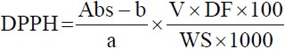
Determination of antioxidant capacity: The antioxidant activity was analyzed by the free radical method 2.2-diphenyl-1-picrylhydrazyl (DPPH), this radical is susceptible to react with antioxidant compounds through a process characterized by the session of a hydrogen atom provided by the antioxidant agent. To determine the antioxidant capacity, a calibration curve was first made with a solution of the reference antioxidant Trolox in a concentration range of 0-800 μmol L–1. The 1450 μL of DPPH plus 50 μL of the sample of the extract diluted in DMCO were taken and carefully homogenized, leaving it to stand for 30 min in the dark, then the samples were read at a wavelength of 517 nm in the UV spectrophotometer (NanoDrop). The results are expressed in μmoL trolox g–1 dry sample, the calculation was made using the following expression recommended by Singh et al.12:
Absreal = BlM-AbsM |
Correction of the actual absorbance of the sample for DPPH
 |
Where:
| Abs | = | Absorbance |
| a and b | = | Slope of the Trolox calibration curve and the cut-off point |
| V | = | Total volume |
| WS | = | Weight of the sample in gram |
| DF | = | Dilution factor |
Determination of antimicrobial activity: For the antimicrobial activity, the diluted extracts in DMCO of the flours of the tubers understudy extracted by the Supercritical Fluids method were used and it was evaluated with a gram-positive battery (Staphylococcus aureus) and three gram-negative bacteria (Arcobacter sp., Escherichia coli sp. and Salmonella sp.), strains previously characterized and conserved in the Bank of Microorganisms of the Laboratory of Molecular Biology of the State University of Bolívar.
Bacterial resuscitation: To resuscitate the Arcobacter sp., strain, three isolates were selected and the blood agar medium was introduced, the Escherichia coli sp., strain in the nutrient agar medium, the Salmonella sp., strain in the XLD agar medium (Xylose, Lysine, Deoxycholate) and the staphylococcus aureus strain were revived in trypticase soy broth. Finally, they were incubated at 37°C for 24 hrs under aerobic conditions, except for Arcobacter sp., which, being a microaerophilic bacterium, requires controlled conditions (10% CO2, 5% O2 and 85% N2) and was incubated for 48 hrs.
Preparation of the inoculum and antimicrobial activity by disk-plate diffusion method: For this analysis, bacterial cultures in the growth phase were used to prepare a bacterial suspension of each strain until a 0.5 McFarland scale was achieved. Subsequently, using a sterile swab, seeding was carried out on Müeller Hinton (MH) Agar plates (Pronadisa, 1058.00, USA), it was kept at rest for 15 min and then the blank discs were immersed in the extracts of malanga obtained. Finally, with the help of a sterile cloth, the discs were placed on the surface of the MH agar.
For supercritical fluid extracts, blank discs were immersed in diluted extracts of 5, 10 and 20 mg of extract diluted with 400 μL of Dimethyl Sulfoxide. In parallel, Levofloxacin disks were tested as a control. Finally, the plates were incubated under controlled conditions at 37°C for 24 hrs. After incubation, the diameters of the inhibition zones of the discs were measured. The results obtained were interpreted according to the criteria of the Clinical Laboratory and Standards Institute.
Determination of the chemical composition by Gas Chromatography The detection of compounds was carried out by Gas Chromatography Coupled to a Mass Spectrometer (GC-MS) in Agilent Technologies equipment (7890A GC system and 5975C inert XL MSD with triple-axis detector). An HP-5MS capillary column (30 m×250 μm, 0.25 μm) with phenylmethyl polysiloxane (0.25 μm film thickness) as stationary phase and helium as carrier gas (0.8 mL min–1) was used. The 1 μL of the derivatized sample (previously filtered) was injected in Split mode using the 1:20 ratio. The injection chamber temperature was 250°C. The oven temperature was maintained from 60-80°C with a 5°C min–1 ramp, then increased to 92°C with a 3°C min–1 ramp for 5 min, then increased to 165°C at a rate of 4°C min–1 and finally at 290°C at a rate of 2°C min–1 for 2 min, 70°C for 2 min and increased to 300°C at 5°C min–1 with a waiting time of 6 min. The compounds were identified by comparison with the mass spectra of the NIST 2011 library. The mass range used was between 40-550 Daltons.
Statistical analysis: The completely random design (DCA) was applied in a factorial arrangement A×B (2×2) with three repetitions. Also, the test of least significant difference (LSD), to determine if the corresponding treatments are significantly different.
RESULTS AND DISCUSSION
Volume and mass of extracts obtained: Soxhlet and supercritical fluid (FSC) methods were analyzed from malanga samples, whereby Soxhlet a larger volume was obtained in purple malanga with 10.7 mL and only 3.3 mL in white malanga in Table 2. This difference is due to its state of maturation and the numerous volatile compounds. Díaz et al.13, in their research, obtained 1.1 mL of hexanic extract from the species Xanthosoma maximiliani Schott. While Guadalupe and Magali14, in his study of Andean tubers obtained 55 mL of ethanolic extract in melloco and 21 mL of ethanolic extract in mashua. These comparisons were made because no bibliographical references to malanga extracts were found. On the other hand, concerning the extracts obtained utilizing FCS, greater mass was given by purple malanga with 1.24 g and white with 1.08 g, as can be seen in Table 2, this difference can be attributed to the starch, the variety of the tuber, to the state of maturation of the same. Regarding the extraction of the extracts by SCF of the malanga, no bibliographical references were found, therefore, the current study was the first to carry out extractions of said tubers by the mentioned method.
Analysis of antioxidant activity, radical capture (DPPH): This method is used to determine the antioxidant capacity of foods and synthetic compounds, the DPPH radical, this free radical is capable of reacting with antioxidant compounds through a process characterized by the transfer of a hydrogen atom provided by the antioxidant agent.
After the analysis of antioxidant activity, a greater DPPH radical scavenging effect was evidenced by the SCF method with values of 207.8 μmoL of trolox g–1 sample in white malanga and 167.6 in purple malanga, being much higher than those obtained by the Soxhlet method in Table 3.
Moncayo et al.15, analyzed the antioxidant capacity of ethanolic extract from leaves of 18 species of plants native to western Ecuador, in which Xanthosoma sagittifolium presents a percentage of 44.99 I% of antioxidant activity by the DPPH method. On the other hand, Kim et al.16, carried out a study on the antioxidant capacity with the DPPH method of extract of Colocasia esculenta and identified a value of 4.52 μmoL of trolox g–1 sample, a value that was considerably lower than that obtained in this work, where the higher the value the higher the antioxidant capacity. The results obtained in this research differ significantly for each variety of tuber, this can be attributed to the extraction method, the analysis performed on starch, the type of tuber and the climatic conditions.
| Table 2: | Volume and mass of extracts obtained |
| Type of sample | Volume extracted from the rota-evaporator (mL) |
| Soxhlet extraction of the two tuber varieties | |
| White malanga | 3.3 |
| Purple malanga | 10.7 |
| Sample | Extract quantity (g) |
| Supercritical fluid extraction | |
| White malanga | 1.0760 |
| Purple malanga | 1.2372 |
| Table 3: | Results of the antioxidant activity content of the DPPH method | |
| Tuber variety | ||
| White malanga | Purple malanga | |
| Extraction methods | Sample (μmoL of trolox g–1) | |
| Soxhlet | 4.61 | 11.32 |
| SCF | 207.87 | 167.64 |
As no previous work on the antioxidant activity of malanga was found, it was contrasted with work carried out on tubers of the same order and family.
Analysis of antimicrobial activity
Antimicrobial activity against Escherichia coli : The results obtained from the antimicrobial activity of the extracts obtained were expressed by measuring the diameter of the inhibition halo in mm against E. coli strains. Where in Table 4 inhibition halos of malanga extracts by supercritical fluids can be seen ranging from 2-4.3 mm in halo size, these values are much lower than those recommended by Ponce et al.17, since this author considers that there is an antimicrobial effect of extracts and vegetable oils, this must be greater than or equal to 8 mm, however it is important to consider that the extract of the purple malanga at a dilution of 1:2 presented a greater inhibitory effect against the Cer3C1 strain with a value of 4.3 mm, concerning the control antibiotic (levofloxacin), the inhibition halo being considerably greater compared to the extracts. On the other hand, Sánchez-Bautista et al.18, in the interpretation of sensitivity, considered the percentage of levofloxacin antibiotic sensitivity of 67.8% in E. coli bacteria. Pérez-Delgado et al.19, in its antibiogram manual, discloses the inhibitory halos of E. coli against levofloxacin from 27-33 mm in diameter. It is important to emphasize that this is the first work on the antimicrobial activity of malanga extracts.
Antimicrobial activity against Salmonella sp.: The following table details the antimicrobial action against the extracts obtained by SCF, at different concentrations. After this analysis, Table 5 shows a poor antimicrobial effect of the extracts obtained against Salmonella strains, with halo size values of 0-1 mm.
| Table 4: | Inhibition halos of extracts of the two malanga varieties at different dilutions against Escherichia coli strains | ||||
| Extraction by SCF | |||||
| White malanga | Purple malanga | ||||
| Dilutions (mg mL–1) | Control antibiotic | ||||
| Escherichia coli strains | 1:2 | 1:4 | 1:2 | 1:4 | Levofloxacin |
| Cer3C1 | 3.3 | 4 | 4.3 | 4 | 28 |
| Res2C1 | 2.7 | 3.3 | 3.7 | 3 | 28 |
| Cer2C1 | 2 | 3.3 | 2.3 | 3 | 28 |
| Table 5: | Inhibition halos of extracts of the two malanga varieties at different dilutions against Salmonella strains | ||||
Extraction by SCF | |||||
White malanga | Purple malanga | ||||
Dilutions (mg mL–1) | Control antibiotic | ||||
| Salmonella strains | 1:2 | 1:4 | 1:2 | 1:4 | Levofloxacin |
| S7 | 0.7 | 0.7 | 0.7 | 0.7 | 11.3 |
| 5M3 | 0 | 0 | 0 | 0 | 11.3 |
| S8 | 2 | 1 | 1 | 1 | 28 |
| Table 6: | Halos of inhibition of extracts of the two varieties of malanga at different dilutions against Arcobacter strains | ||||
Extraction by SCF | |||||
White malanga | Purple malanga | ||||
Dilutions (mg mL–1) | Control antibiotic | ||||
| Arcobacter strains | 1:2 | 1:4 | 1:2 | 1:4 | Levofloxacin |
| Q1BC1 | 3.3 | 2.7 | 1.3 | 1.3 | 30 |
| Q3NC2 | 1.3 | 2.7 | 1 | 1 | 16 |
| Q1NC1 | 1.7 | 1.7 | 2.7 | 1.3 | 13 |
However, the Minimum Inhibitory Concentration (MIC), according to the Clinical Laboratory Standard Institute (CLSI) mentioned by Humphries et al.20 considers that the Enterobacteriaceae family was resistant to the antibiotic levofloxacin when the inhibition halo is <16 mm in diameter. Similarly, Sfeir21 considers levofloxacin resistant if the inhibition halo is <13 mm in diameter against the Enterobacteriaceae family, in such a way that it can be defined that the S7 and 5M3 strains were resistant to the drug.
Antimicrobial activity against Arcobacter sp.: In Table 6, the results of the inhibition halos were presented and low antimicrobial activity against Arcobacter can be observed, where halo size values of 1-3.3 mm are evidenced, being the 1:2 dilution the one that showed the greatest effect against the Q1BC1 strain, however, these values are very low compared to what is recommended by Ponce et al.17. On the other hand, works such as the one developed by Goyes22, in his research on the susceptibility of the different Arcobacter strains, reveal the resistance of the bacteria to levofloxacin with a halo <14 mm in diameter, Humphries et al.20, in the table of cut-off points for the interpretation of MICs and zone diameters, it mentions that the genus Campylobacter is susceptible with a halo >26 mm to the group of fluoroquinolone antibiotics in which levofloxacin is immersed. The interpretation of the results was carried out using the recommendations of the European Committee for Susceptibility Testing according to EUCAST and the CLSI20. In this sense, the Q1NC1 strain showed resistance to the reference antibiotic.
Antimicrobial activity against Staphyloccocus aureus: The results obtained from the antimicrobial action of the extracts of the two varieties of the tuber against the gram+S. aureus bacteria were expressed by measuring the diameter of the inhibition halo. In this analysis, the 1:2 dilution of the purple malanga extract presented the best effect with a halo size of 7 mm, followed by the white malanga extract in the 1:2 dilution with a 6.7 mm halo in Table 7, these results were quite close to the 8 mm recommended by Ponce et al.17.
| Table 7: | Halos of inhibition of extracts of the two varieties of malanga at different dilutions against Staphyloccocus aureus | ||||
Extraction by SCF | |||||
White malanga | Purple malanga | ||||
Dilutions (mg mL–1) | Control antibiotic | ||||
| Staphylococcus aureus strain | 1:2 | 1:4 | 1:2 | 1:4 | Levofloxacin |
| Staphylococcus aureus | 6.7 | 5.3 | 7 | 6 | 26 |
The extract of the white malanga in the different dilutions presents values greater than 5 mm in halo size. EUCAST for S. aureus reports that the halo <22 mm in diameter is resistant to the antibiotic levofloxacin and is susceptible when the inhibition halo is ≥50 mm, while CLSI, reported that inhibitory halos of S. aureus are 25-30 mm in diameter compared to levofloxacin. Concerning the antimicrobial activity of malanga, no reports were found in the literature, so it is the first investigation of the antimicrobial action of these tubers against the S. aureus bacteria. In short, the extracts of the purple and white malanga have low antimicrobial action compared to the control antibiotic used.
Identification of compounds present by gas chromatography (GC-MSD) in purple malanga extract obtained by SCF: After the chromatographic analysis of the obtained extracts, it is important to highlight that some beneficial compounds for health were identified in this work, among these we have: it is, Phthalic acids, bis (2-Ethylhexyl) ester with an area of 36.96% being an acid Phthalic, continuously with the Linoleic acid 13.31% being an essential fatty acid type compound, the Palmitic acid with an area 9.75% forming part of the essential fatty acid, the Linolein, 2-mono with an area 6.65% forming part of the fatty acids and finally Furfural with an area of 0.375 forming an aldehyde-type compound.
The Pyranone compound, which was identified in the extract of the white malanga, was also identified by Zakaria et al.23, in the ethanolic extract of the fresh leaves of Pandanus amaryllifolius, a tropical plant from Southeast Asia, the aforementioned compound has a high capacity to act as an antioxidant in volatile substances and has been much discussed in recent years. The Linoleic acid compound was identified by Jiménez et al.24, in the mashua Tropaeolum tuberosum, whose function is to be anti-inflammatory, in addition, it helps prevent coronary diseases, inhibiting angiogenesis, developing a cytotoxic activity in tumour cells. The Furfural compound was also identified by van der Maas et al.25, this allows to inhibit microbial growth.
CONCLUSION
After this investigation, conclude that the extracts of purple malanga obtained by supercritical fluids can be used as a natural food additive, especially for its antioxidant and antibacterial effect, especially in the Gram-positive group, demonstrated especially by the presence of Furfural, Linoleic acid and Pyranone.
SIGNIFICANCE STATEMENT
This study determines the parallel antioxidant and antimicrobial effect of white and purple malanga extracts, they can be a natural alternative to treat bacterial infections, as well as their broad antioxidant benefit, in such a way that it will be possible in the future to obtain biopharmaceuticals.
REFERENCES
- Lotero, F.M.M., J.E.S. Gil, S.V. Londoño, J.A.S. Jiménez and L.S.T. Valenzuela, 2018. Antimicrobial and antioxidant capacity of andean tubers (Tropaeolum tuberosum and Ullucus tuberosus). Rev. U.D.C.A Actualidad Divulgación Científica, 21: 449-456.
CrossRefDirect Link - Milián-Jiménez, M.D., 2018. Genetic resources of cocoyam of Xanthosoma schott genus in Cuba [In Spanish]. Cultivos Tropicales, 39: 112-126.
Direct Link - Velasquez, D., L.F.M. Andrade and K. Ricardo, 2021. Obtaining Biopolymer Evaluating the Starch of two Varieties of Malanga, white (Colocasia esculenta (L.) Schoot) and Purple (Xanthosoma sagittifolium (L.) Schoot and Endl), two Thickeners and Water-Starch Ratio for the Production of Biodegradable Packaging. PhD Thesis, University of the Armed Forces ESPE, ESPESD, Agricultural Engineering Career.
- Murillo, C.R.F., M.E.P. Pazmiño and A.V. Lopez, 2021. Malanga (Colocasia esculenta) and its effect on cholesterolemia. Proposal for hypocholesterolemic cookies Reciamuc, 5: 327-335.
CrossRefDirect Link - Eleazu, C.O., 2016. Characterization of the natural products in cocoyam (Colocasia esculenta) using GC-MS. Pharm. Biol., 54: 2880-2885.
CrossRefDirect Link - Ehuwa, O., A.K. Jaiswal and S. Jaiswal, 2021. Salmonella, food safety and food handling practices. Foods, Vol. 10.
CrossRefDirect Link - Bayas-Morejón, F., R. Ramón, M. García-Pazmiño and G. Mite-Cárdenas, 2020. Antibacterial and antioxidant effect of natural extracts from Baccharis latifolia (Chilca). Caspian J. Environ. Sci., 18: 489-493.
CrossRefDirect Link - Gonzalez, A. and M.A. Ferrus, 2011. Study of Arcobacter spp. contamination in fresh lettuces detected by different cultural and molecular methods. Int. J. Food Microbiol., 145: 311-314.
CrossRefDirect Link - Pérez, D.Q., 2017. Antimicrobial resistance: Evolution and current perspectives in the context of the "one health" approach. Rev. Cubana Med. Trop., Vol. 69.
Direct Link - Bayas-Chacha, F., M. Bermeo-Sanchez, B. Herrera-Chavez and F. Bayas-Morejon, 2022. Antimicrobial and antioxidant properties of Tropaeolum tuberosum extracts from Ecuador. Asian J. Plant Sci., 21: 321-327.
CrossRefDirect Link - Singh, M., S. Patra, R.K. Singh, 2021. Common Techniques and Methods for Screening of Natural Products for Developing of Anticancer Drugs. In: Evolutionary Diversity as a Source for Anticancer Molecules. Srivastava, A.K., V.K. Kannaujiya and D. Singh (Eds.). Elsevier Inc., Amsterdam, pp: 323-353.
CrossRefDirect Link - Díaz, I.E.C., E.G. Gonçalves, M.O.M. Marques and M. Yoshida, 2011. The incidence of fatty acids in species of Araceae. Rev. Soc. Quím. Perú. 4: 275-284.
Direct Link - Moncayo, S., X. Cornejo, J. Castillo and V. Valdez, 2021. Preliminary phytochemical screening for antioxidant activity and content of phenols and flavonoids of 18 species of plants native to western Ecuador. Trends Phytochem. Res., 5: 92-104.
Direct Link - Kim, Y.S., D. Adeyemi, P. Korovulavula, D.W. Jang and M.K. Park, 2019. Effect of steaming on the functional compounds and antioxidant activity of fijian taro (Colocasia esculenta L. Schott) corms. Korean J. Food Preserv., 26: 449-454.
CrossRefDirect Link - Ponce, A.G., S.I. Roura, C.E. del Valle and M.R. Moreira, 2008. Antimicrobial and antioxidant activities of edible coatings enriched with natural plant extracts: In vitro and in vivo studies. Postharvest Biol. Tech., 49: 294-300.
CrossRefDirect Link - Sánchez-Bautista, A., J. Coy, P. García-Shimizu, J.C. Rodríguez, 2018. From CLSI to EUCAST guidelines in the interpretation of antimicrobial susceptibility: What is the effect in our setting? Enfermedades Infecciosas Microbiología Clín., 36: 229-232.
CrossRefDirect Link - Pérez-Delgado, O., R.L. Alvarado-Pineda, A.E. Yacarini-Martínez, 2021. In vitro antibacterial activity of crude ethanolic extract of Origanum vulgare leaves, against Staphylococcus aureus ATCC 29213, Pseudomonas aeruginosa ATCC 27853 and Escherichia coli ATCC 25922 [In Spanish]. J. Selva Andina Res. Soc., 12: 21-29.
Direct Link - Humphries, R.M., J.A. Hindler, K. Shaffer and S.A. Campeau, 2019. Evaluation of ciprofloxacin and levofloxacin disk diffusion and Etest using the 2019 Enterobacteriaceae CLSI breakpoints. J. Clin. Microbiol.
CrossRefDirect Link - Sfeir, M.M., 2021. Adoption of the updated CLSI fluoroquinolone breakpoints for Gram-negative bacteria in microbiology laboratories. Clin. Microbiol. Infec., 27: 308-310.
CrossRefDirect Link - Zakaria, M.M., U.H. Zaidan, S. Shamsi and S.S.A. Gani, 2020. Chemical composition of essential oils from leaf extract of Pandan, Pandanus amaryllifolius Roxb. Malaysian J. Anal. Sci., 24: 87-96.
Direct Link - Heinert, M.E.J., M.M. Martínez, I.A.C. Guaranda, Y.I.G. Gaitén and J.A. Payrol, 2021. Morphological, chemical characterization and anti-inflammatory activity of Tropaeolum tuberosum (Ruiz & Pav.) Kuntze (Tropaeolaceae) pink variety. Rev. Cubana Farmacia, Vol. 54.
Direct Link - van der Maas, V., J.L.S.P. Driessen and S.I. Mussatto, 2021. Effects of inhibitory compounds present in lignocellulosic biomass hydrolysates on the growth of Bacillus subtilis. Energies, 14: 8419.
CrossRefDirect Link