
A. Jafari
Department of Biology, Faculty of Science, Islamic Azad University, Mashhad Branch, Mashhad, Iran
Y. Nasseh
Research Center for Plant Sciences, Ferdowsi University of Mashhad, Mashhad, Iran
Asian Journal of Plant Sciences
Year: 2009 | Volume: 8 | Issue: 1 | Page No.: 86-88


ABSTRACT
In present research anatomical studies carried out 11 species which growing in Khorassan Province (North-East of Iran). These species were divided to two groups, annual and perennial species. For comparative anatomy studying, cross sections from stem and leaf were prepared using microtome and differential staining. The characters of secondary xylem and axial parenchyma, arrangement of vessel and the arrangement of mesophyll were studied. The results showed the variation of internal structure didn`t have correspondence to taxonomy position.
PDF Abstract XML References Citation
How to cite this article
A. Jafari and Y. Nasseh, 2009. An Internal Structure Investigation on Euphorbia L. Species in North-East of Iran. Asian Journal of Plant Sciences, 8: 86-88.
DOI: 10.3923/ajps.2009.86.88
URL: https://scialert.net/abstract/?doi=ajps.2009.86.88
DOI: 10.3923/ajps.2009.86.88
URL: https://scialert.net/abstract/?doi=ajps.2009.86.88
INTRODUCTION
Euphorbia L. belongs to Euphorbiaceae have annual and perennial species with cyathium inflorcense and laticifer. As the recent report, this genus have 67 species in Iran. The extract of this plant have anti-cancer, activation of tumors, inhibition of HLV-1 multiplication, eye burns, anti bacterial, viral and fungal effect (Hussein et al., 1999; Frustenberger and Hecker, 1985; Frohn et al., 1993; Natarujan et al., 2005; Nawito et al., 1999; Giordani et al., 2001; Glattaar-Saalmuller and Fallier-Becker, 2001).
The earlier anatomy studying on Euphorbiaceae had been reported about formation of laticifer (Fahn, 1990). So, comparative internal structure study on Euphorbia carried out for first time. In present study, stem and leaf anatomy of 11 species in longitudinal and cross sections were investigated. Also wood analysis carried out.
MATERIALS AND METHODS
As for the anatomic study, the examined species were collected from the localities in North-east of Iran during May-June 2002-2003 (Table 1). For preparing of longitudinal and cross section of stem and leaf, base of stem and basal leaves were selected from 6- 7 specimens. The living materials were fixed in FAA then they were dehydrated with ethanol and later, some slices were prepared with microtome. 12-microns thickness was stained with Safranin and Fast-green (Johnson, 1940; Metcalf and Chalk, 1983; Chamberlain, 1990). For the stem maceration, this organ was placed in Jeffery solution for 4 h to soften. The plants were divided to annual and perennial. Annual species were following: E. granulata, E. densa, E. szovitzii, E. helioscopia, E. cheirolepis and perennial species were following: E. microsciadia, E. marshalliana, E. buhsei, E. bungei and E. aucheri.
| Table 1: | The localities of studied Euphorbia species |
 | |
ESULTS AND DISCUSSION
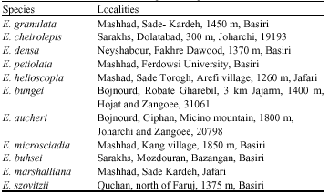
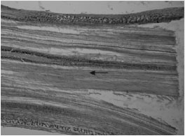
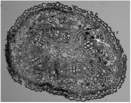
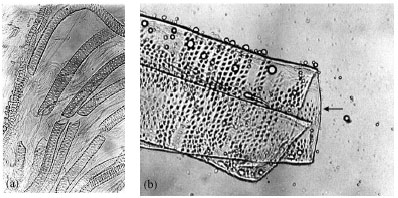
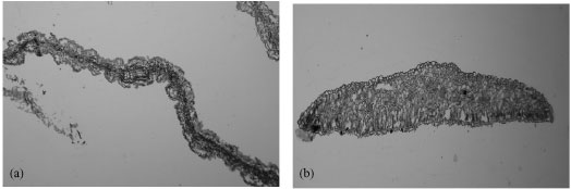
Anatomic results in stem of annual species showed, the laticifer existed around the cortex (e.g., E. cheirolepis and E. petiolata). The laticifer observed above the phloem in E. helioscopia stem (Fig. 1, 2). Decreasing of cortex and pith with dense laticifer and cluster-radial chain pore vessel were observed in E. petiolata (Fig. 3a). The internal structure of stem showed variation in perennial species. The dense gelatinous fiber, tension wood existed in them. Diffuse porous was observed in E. marshaliana (Fig. 3b). All of them had apotracheal boundary axial parenchyma, non- articulated laticifer in phloem. The arrangement of vessel were radial chain pore with solitary in E. microsciadia (Fig. 3c) , radial chain pore in E. marshalliana and E. buhsei and cluster in E. aucheri (Fig. 3d). The vascular rays was uni-seriate or multi- seriate. The vessels had spiral, reticulate and pitted thickening. The vessel was short and wide with simple perforation plate (Fig. 4a, b).
 | |
| Fig. 1: | Longitudinal section of E. cheirolepis stem. The arrow showing laticifer in cortex (x416) |
 | |
| Fig. 2: | Cross section of E. petiolata stem. The arrow showing laticifer above phloem (x416) |
The results of leaf anatomy showed, isolateral mesophyll in E. petiolata, E. marshalliana and dorsi- ventral in E. helioscopia (Fig. 5a, b).
The anatomy results showed variation in internal structure but we can not any correspondence between them. The presence of gelatinous fiber due xerophytic plant. Ring porous and diffuse porous were observed in mesophytic and xerophtic species. Some of them adapted to arid weather. Diffuse porous and cluster vessel are more advanced (Metcalf and Chalk, 1983; Fahn, 1990). On the basis of vessel arrangement, E. microsciadia is primitive by having diffuse porous and solitary vessel. E. buhsei and E. marshalliana more advanced by having diffuse porous and radial chain pore. Finally, E. aucheri is the most advanced by having ring porous and cluster vessel. Also, isolateral and dorsi- ventral mesophyll were in mesophytic and xerophytic species. It sounds the variation in anatomy characters of studied species is related to ecologic factors.
 | |
| Fig. 3: | The arrangement of vessel in: (a) E. petiolata, (b) E. marshalliana, (c) E. microsciadia and (d) E. buhsei (x416) |
 | |
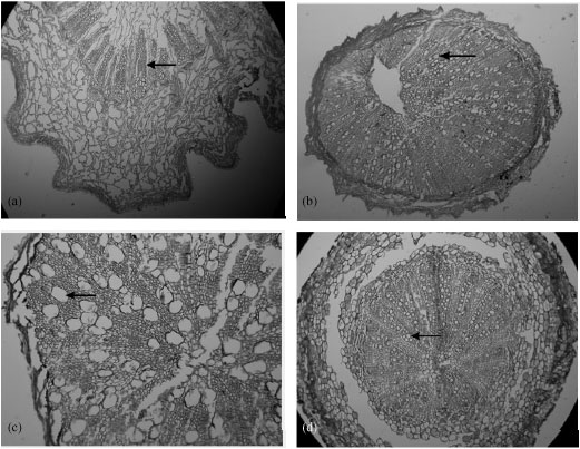
| Fig. 4: | (a) Reticulate thickening vessel in E. aucheri and (b) Pitted vessel in E. granulata. The arrow showing simple perforation plate |
 | |
| Fig. 5: | (a) Isolateral mesophyll in E. petiolata and (b) Dorsi- ventral mesophyll in E. helioscopia (x416) |
REFERENCES
- Frustenberger, G. and E. Hecker, 1985. On the active principles of the spruge family (Euphorbiaceae) XI (1). The skin irritant and tumor promoting ditrepene esters of Euphorbia tricali L. orginatig from South Africa. Z. Naturifrosch., 40: 631-646.
PubMedDirect Link - Frohn, A., C. Frohn, K.P. Steuh and H.J. Thiel, 1993. Eye burns caused by wolfs milk. Ophtalmologe, 90: 58-61.
Direct Link - Giordani, R., J. Trebaux, M. Masi and P. Regli, 2001. Enhanced antifungl activity og ketoconazole by Euphorbia characias latex against Candida albicans. J. Ethnopharmacol., 78: 1-5.
CrossRefDirect Link - Glattaar-Saalmuller, B. and P. Fallier-Becker, 2001. Antiviral action of Euphorbia compositum and its components. Frosch Komplementamed Klass Naturheild, 8: 207-212.
Direct Link - Hussein, G., H. Miyashiro, N. Nakamura, T. Kawahat and T. Otake et al., 1999. Inhibitory effects of Sudanese plant extraction on HLV-1 replication. Phytother Res., 139: 31-36.
Direct Link - Natarujan, D., S.J. Britto, K. Srinivasan, N. Nagamurugen, C. Mohanasudari and G. Perumal, 2005. Anti-bacterial activity of Euphorbia fusiformis: A rare medicinal herb. J. Ethnopharmacol., 102: 123-126.
CrossRefDirect Link