
F.A. Al-Sobayil
College of Agriculture and Veterinary Medicine, Qassim University, Buraydah 51452, P.O. Pox 6622, Al-Qassim, Saudi Arabia
Asian Journal of Animal and Veterinary Advances
Year: 2008 | Volume: 3 | Issue: 5 | Page No.: 298-302


ABSTRACT
The various locations and techniques for performing liver biopsy in dromedary camels (Camelus dromedarius) were described in the present study. Five cadavers of adult healthy camels preserved with 10% formaline solution were used to determine the topographical anatomy of the liver on the right side. Liver biopsies were collected from 10 adult healthy camels. Each camel was sedated with 2% solution of xylazine HCL (Rompun® 0.1 mg kg-1, IV) and locally anesthetized with lidocaine HCL at the sites of collecting liver biopsy. Liver biopsies were collected from five locations from each camel: 7-11th intercostals spaces. The exact site of each location was described. At each site, ten attempts of liver biopsies were performed and the success attempts were recorded. The result showed that the liver of the dromedary lies under cover the ribs mainly on the right side between the cranial aspect of the 6th rib and the caudal end of the 12th rib. The technique of liver biopsy was significantly easier from the 8 and 9th intercostal spaces compared to the other locations.
PDF Abstract XML References Citation
How to cite this article
F.A. Al-Sobayil, 2008. The Different Locations for Collecting Liver Biopsies in Dromedary Camels (Camelus dromedarius). Asian Journal of Animal and Veterinary Advances, 3: 298-302.
DOI: 10.3923/ajava.2008.298.302
URL: https://scialert.net/abstract/?doi=ajava.2008.298.302
DOI: 10.3923/ajava.2008.298.302
URL: https://scialert.net/abstract/?doi=ajava.2008.298.302
INTRODUCTION
The liver is the largest gland in the body that has a complex organ in terms of structure, function and pathology. It is located in the most cranial part of the abdomen, immediately behind the diaphragm. Although the liver extends across the median plane, the bulk lies to the right in almost all species. In most species, the liver is grossly divided into lobes by a series of fissures that extend inward from the ventral margin. The size of the liver varies depending on its metabolic functions and generally the average values ranges from 1 to 4% of body weight (Dyce et al., 1987). Anatomy of the liver has been reported in domestic animals including dromedary camel (Dyce et al., 1987; Smuts and Bezuidenhout, 1987).
The liver is an important organ that performs many essential functions for life. These functions include metabolism (amino acids, carbohydrates, lipids), synthesis (albumin, cholesterol, plasma protein, clotting factors), digestion and absorption of nutrients (related to bile formation), secretion of bilirubin (bile) and elimination (detoxification of toxins, catabolism of certain drugs) (Pratt, 1992; Tennant, 1997). In fact, the liver is considered the primary organ for drug metabolism and drug residuals that might have an effect on the safety of the consumption of the animal carcass by humans. In clinical practice, liver disease is not uncommon. There are several methods that have been used to diagnose hepatic disorders. In general, these methods involve the evaluation of clinical history, physical examination, biochemical tests, hepatic imaging and histopathologic examination of hepatic biopsies. Laboratory tests imaging examinations are used for confirming the history and clinical signs suggestive of hepatic disease. Laboratory tests are used to assess the severity and to monitor the progress of hepatic disease during the stage of treatment. Hepatic sonography is a common noninvasive imaging techniques used in determining certain morphologic changes in the liver.
The diagnosis of hepatic disease through the histopathological evaluation of a liver tissue is also a very useful method. It can be performed after taking a liver biopsy from the patient. Liver histopathology can often define the liver disease as infectious, toxic, or obstructive/congestive. It is usually performed to diagnose hepatic tumors or to evaluate the extent of damage that has occurred to the liver because of chronic disease. The procedure of collecting liver biopsy have been reported in domestic animals including horses (Orsini and Kreuder, 2002), cattle (Rebhun, 1995), sheep (Navarre and Pugh, 2002), goats (Smith and Sherman, 1994) and pet animals (Fossum, 1997). Only one report has described the technique of liver biopsy in dromedary camels using only one location (Bucci et al., 1982).
The present study was carried out to describe the technique for performing liver biopsy in dromedary camels (Camelus dromedarius) using various locations.
MATERIALS AND METHODS
Camels and Procedure
The topographical anatomy of the liver was determined using five cadavers of adult healthy camels preserved with 10% formaline solution as per Grossman`s (1959) technique. Liver biopsy was collected from 10 adult (6-9 years-old) healthy camels. Each camel was sedated with 2% solution of xylazine HCL (Rompun® 0.1 mg kg-1, IV). While the animal was in a setting position, the hair over the right side was shaved and the locations of liver biopsy were aseptically prepared. Each site of liver biopsy was locally infiltrated with local anesthetic, lidocaine HCL (0.5-1 mL). A scalpel puncture of the skin was made over the determined position of collecting liver biopsy. A Tru-Cut biopsy needle (Tru-Cut Biopsy Needle, Baxter Healthcare Corp., Valencia, CA, USA) was introduced and advanced slightly cranial and ventral to the selected site. In each animal, liver biopsies were collected from five locations. The first location was in the 7th intercostal space (between the 7 and 8th costal bones) about 15-20 cm proximal to the costal arch. The second location was in the 8th intercostal space (between the 8 and 9th costal bones) about 5-10 cm proximal to the costal arch. The third location was in the 9th intercostal space (between the 9 and 10th costal bones) about 5-10 cm proximal to the costal arch. The fourth location was in the 10th intercostal space (between the 10 and 11th costal bones) approximately 30-35 cm proximal to the costal arch. The fifth location was in the 11th intercostal space (between the 11 and 12th costal bones) approximately 35-40 cm proximal to the costal arch. At each site, ten attempts of liver biopsies were performed and the success attempts were recorded. After the liver biopsies were taken, the skin puncture at each site was sutured with only single interrupted suture using No. 1 silk and the suture was removed 14 days post suturing.
Statistical Analysis
Analysis of Variance (ANOVA) method was used to determine the difference between sites. Tukey`s Honset Significant Difference was used to determine the multiple comparisons of means. The significance level was set at p<0.05 and SAS software (Statistical Analysis System) was used to perform all statistical calculations.
RESULTS AND DISCUSSION
Liver of the dromedary lies under cover the ribs mainly on the right side between the cranial aspect of the 6th rib and the caudal end of the 12th rib. At the 6th rib, the liver extends 18-30 cm dorsal to the sternal pad. At the 8 and 9th intercaostal spaces, the liver extends 2-8 cm distally to the costal arch (Fig. 1). At the 10 and 11th intercaostal spaces, the liver was located on the most proximal third of the intercostal spaces (Fig. 1).
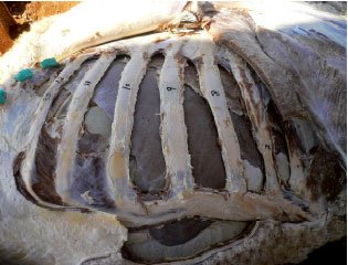 | |
| Fig. 1: | A photograph showing the location of the liver on the right side of the dromedary camels. The numbers 7, 8, 9, 10, 11, 12 are 7th to 12th ribs |
| Table 1: | The location of liver biopsy in the intercostals spaces and the number of successes and fails of liver biopsy in each site |
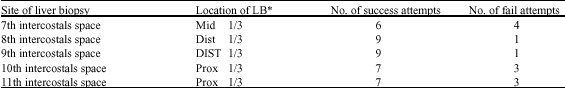 | |
LB: Liver biopsy | |
Table 1 shows the site of liver biopsy in each intercostal space and the number of successes and fails of liver biopsy in each site. Both the 8th and 9th intercostals sites were significantly easier to obtain liver biopsy compared to the other locations. The facility of performing liver biopsy from the 8th intercostal space was not significantly different from that in the 9th intercostal space (p<0.05). Obtaining liver biopsy in both the 10 and 11th intercostal sites were significantly easier than that in the 7th intercostal site (p<0.05). The most difficult sit of obtaining liver biopsy was in the 7th intercostal space.
The present study showed that the major part of the liver is located on the right side of the body and it extends from the cranial aspect of the 6th rib to the caudal end of the 12th rib. Smuts and Bezuidenhout (1987) have reported similar result except that they have reported that the right liver extends from the 5th to the 12th ribs.
Results obtained showed that performing liver biopsy in sitting position was relatively easy in dromedary camels. Liver biopsy of dromedary camels can be obtained from different locations with no adverse effects from the procedures. The degree of difficulty of performing liver biopsy was not the same for the different locations. The technique of liver biopsy was relatively easy from the 8 and 9th intercostal spaces. This was expected because approximately the distal two-third of the 8th intercostal space and the distal three quarters of the 9th intercostal space are completely covered with liver tissues. In addition, it was relatively easy to identify the last four ribs in dromedary camels. Bucci et al. (1982) have used the 9th right intercostal space to successfully collect liver biopsy from dromedary camels. In the present study, the successful attempts of obtaining liver biopsies at these locations were 9 out of 10 attempts. In Bucci et al. (1982) study, 99 successful rates were obtained out of 120 attempts of liver biopsies.
The most difficult location of obtaining liver biopsy was in the 7th intercostal space. This finding was not surprising because of the small area of liver tissue located in the 7th intercostal space and also because of the difficulty of determining the 7th intercostal space especially in fatty camels. The liver in the 7th intercostal space covers almost the middle third of the space. However, the right lung might partially superimposes with the liver which makes it relatively difficult to perform liver biopsy in that intercostal space. The site of liver biopsy was in the distal area of the middle third of the intercostal space. The successful attempts of obtaining liver biopsies at this location were 6 out of 10 attempts. The successful percentage of collection of liver biopsies from the 10 and 11th intercostal spaces was 70% (7 successes out of 10 attempts). It was very easy to identify the last three ribs in camels. The liver in the 10 and 11th intercostal spaces covers almost the proximal one third of the space (Fig. 1).
The location and technique of performing liver biopsy have been described in domestic animals. In horses, liver biopsy can be performed blindly from the right 14th intercostal space in a line drawn from the point of the shoulder to the tuber coxae (Orsini and Kreuder, 2002). Liver biopsy in adult cattle is performed from the right 11th intercostal space at a level of mid-paralumbar fossa (Rebhun, 1995). In goat, the preferred site for performing liver biopsy is in the right 9th intercostal space (Smith and Sherman, 1994). The site of entry is dorsal to a line drawn from the point of the elbow to the craniodorsal angle of the paralumbar fossa. The entry at the 8th intercostal space may cause a puncture of the caudal lung lobe, while entering the 10th intercostal space may put the biopsy instrument behind the caudal edge of the liver (Smith and Sherman, 1994). Other study has reported that liver biopsy in sheep and goats can be performed in the 9th or the 10th intercostal spaces slightly above an imaginary line from the tuber coxae to the point of the elbow (Navarre and Pugh, 2002). In pet animals, liver biopsy is usually performed under the guidance of sonography. Liver biopsy can be obtained after making a small incision in the skin on the left side between the costal arch and xiphoid process and then the biopsy needle is inserted through the skin incision in a craniodorsal direction with slight angling toward the left of midline (Fossum, 1997). The present study has showed that the best two sites of collecting liver biopsy in dromedary camels were in the 8 and 9th intercostal spaces. This is similar to what has been reported in sheep and goats (Smith and Sherman, 1994; Navarre and Pugh, 2002).
In conclusion, liver biopsy can be performed in sedated sitting dromedary camels in different locations. The 8 and 9th right intercostal spaces were the most easier locations that can be used for collecting liver biopsy in dromedary camels, whereas the 7th intercostal space was relatively the most difficult location for doing that.
ACKNOWLEDGMENTS
The author wish to acknowledge the contribution of Professors M.A. Al-Halag and S.M. Hagrus (Department of Veterinary Medicine) for their assistance in preparing the cadavers of camels.