
B. I. Onyeanusi
Faculty of Veterinary Medicine, Ahmadu Bello University, Zaria, Nigeria
A. A. Adeniyi
Faculty of Veterinary Medicine, Ahmadu Bello University, Zaria, Nigeria
J. O. Ayo
Faculty of Veterinary Medicine, Ahmadu Bello University, Zaria, Nigeria
C. S. Ibe
Faculty of Veterinary Medicine, Ahmadu Bello University, Zaria, Nigeria
C. G. Onyeanusi
Faculty of Pharmaceutical Sciences, Ahmadu Bello University, Zaria, Nigeria
Pakistan Journal of Nutrition
Year: 2009 | Volume: 8 | Issue: 7 | Page No.: 1043-1047


ABSTRACT
A comparative study was carried out on the urinary system of the African Giant Rat (AGR) and the Wistar Rat (WR) using standard laboratory procedures. The mean liveweight of the AGR and WR were 863.590±33.740 and 140.625±6.078 g, respectively. The mean kidney weight of the male and female AGR and WR were 2.119±0.062, 2.053±0.009, 0.633±0.091 and 0.572±0.132 g, respectively. It was observed that the mean kidney weight in the male was higher than that of the female in both AGR and WR but the difference was not significant (p>0.05). The mean weight of the right kidney was heavier than that of the left kidney in both the AGR and WR. The mean weight of the right kidney of the AGR was 2.21±0.051 g while, the left was 2.00±0.055 g. The mean weight of the right kidney in the WR was 0.633±0.012 g while, the left was 0.596±0.022 g. No significant difference (p>0.05) was obtained in the thickness of the bladder and its length in both AGR and WR but there was a high significant difference (p<0.001) between the ureter length of the AGR and the WR. The length of the right ureter was longer than the left and the female had a slightly longer ureter than the male in both rats. The relative thickness of the medulla, which is an indicator of the length of the loop of Henle, was 4.297 in the AGR while, that of the WR was 5.6. The higher relative thickness of the medulla (5.6) signifies that the kidney of the WR has an anatomical adaptation for the concentration of urine and thus, better conservation of water in the arid zone while, the lower relative thickness of the medulla (4.2) of the AGR suggests lack of appropriate anatomical adaptation in the kidney for conservation of water. The study has also provided a baseline morphometric data on the urinary system of both the AGR and WR in the Northern Guinea Savannah zone of Nigeria.
PDF Abstract XML References Citation
How to cite this article
B. I. Onyeanusi, A. A. Adeniyi, J. O. Ayo, C. S. Ibe and C. G. Onyeanusi, 2009. A Comparative Study on the Urinary System of the African Giant Rat (Cricetomys Gambianus Waterhouse) and the Wistar Rat. Pakistan Journal of Nutrition, 8: 1043-1047.
DOI: 10.3923/pjn.2009.1043.1047
URL: https://scialert.net/abstract/?doi=pjn.2009.1043.1047
DOI: 10.3923/pjn.2009.1043.1047
URL: https://scialert.net/abstract/?doi=pjn.2009.1043.1047
INTRODUCTION
The African Giant Rat (AGR) and the Wistar Rat (WR) are two wild rodents that belong to the order Rodentia and are quiet common in Nigeria. They are well distributed but are more dominant in the Savannah areas of the country. The AGR and WR have good potentials as laboratory animals but the AGR is better sought after as meat delicacy by the local people.
Both species of rat are very similar in their anatomical dispositions but the adult rats can readily be differentiated by the difference in their sizes. An adult AGR is known to have attained an enormous weight of about 1,400 g (Ajayi, 1975) with a total length of 83.0 cm from the tip of the nose to the tip of the tail (Happlod, 1987). An adult WR was known to have weighed up to 340 g (Rytand, 1938).
Both rats conserve water and thus, survive very well in the arid zone. Their urinary system may be playing an important role in this water conservation process by its ability to concentrate urine (Moffat, 1975). The kidney is that part of the urinary system that is directly responsible for the concentration of the urine and the loop of Henle has been identified as forming an important part of the concentrating mechanisms of the kidney.
There is a good correlation between the length of the loops and therefore the thickness of the medulla and the ability to concentrate urine in animals (Moffat, 1975).
Comparative studies have been made on the haemogram of the Wistar rat and the African giant rat (Durotoye and Oke, 1990) as well as on the testicular and epididymal proteins of the AGR and WR (Adeyemo and Oke, 1990) in the Southern part of Nigeria. It is our aim to improve further on the knowledge of the biology of these rats by comparing notes on the urinary system to see its anatomical adaptation at contributing to water conservation.
MATERIALS AND METHODS
A total of 46 African Giant Rats (AGR) and 40 Wistar Rats (WR) were used for this study. The AGR were caught alive from Bomo village, while the WR were caught from Chikaji village-all in Zaria Local Government Area of Kaduna State, Nigeria. Sex differences were taken into consideration during the study. All rats were kept in the Department of Veterinary Anatomy Laboratory and fed with commercially formulated feed. Water was given ad libitum while, the study lasted.
The rats were weighed alive using a weighing balance (P1210, Mettler Instruments AG, Switzerland) with a sensitivity of 0.1 g before they were sacrificed. A mid-ventral abdominal incision was made on each sacrificed animal, the peritoneum reflected and the intestine displaced to gain access to the urinary system. All organs were then exteriorized. The length, weight, thickness and width of the organs were measured using a ruler, weighing balance and a vernier calliper, respectively.
The kidney volume was determined by standard water displacement method (Scherle, 1970). Samples were taken from the kidney, ureter and bladder and fixed in buffered neutral formaldehyde. These tissues were later processed for histological examinations. Sections were cut at 5.0 μ and stained with Haematoxylin and Eosin (H and E). A calibrated micrometer eyepiece was used to measure the depth of the medulla and cortex. The data obtained were subjected to statistical analysis using Student’s t-test and correlation analysis. Values of p<0.05 were considered significant.
RESULTS
Macroscopic observations: The kidneys of the African Giant Rat (AGR) and Wistar Rat (WR) were reddish-brown with the AGR having a darker red colour in vivo. The kidneys of both rats were bean-shaped and smooth and covered with a thin fibro-muscular capsule but those of the AGR were bigger in size. Each kidney had dorsal and ventral surfaces, medial and lateral borders, an upper and a lower pole. The hilus and sides were surrounded by adipose tissue.
The lateral borders were convex while, the medial borders were concave and indented at the hilus. The major renal vessels entered and left the hilus while, the ureter also originated from the hilus. The kidneys lay alongside the vertebral column in the abdominal cavity and at their upper poles was the suprarenal gland. The right kidney was situated more cranially than the left. The right kidney was related to the liver while, the left was related to the stomach, pancreas, descending colon, spleen and small intestine.
The ureter entered dorso-laterally into the urinary bladder. The urinary bladder was pear-shaped when empty but appeared more spherical when full in both rats. Urinary bladder in the AGR was bigger in size than in the WR and a cross-section of the kidney in both rats revealed the cortex and the medulla.
Morphometric observations: The mean liveweight of the AGR and WR were 863.590±33.740 and 140.625±6.078 g, respectively (Table 1). It was observed that the mean weight of the right kidney was heavier than that of the left in both rats. The mean weight of the right kidney in the AGR was 2.21±0.05g while, the left was 2.00±0.055g. The mean weight of the right kidney in the WR was 0.633±0.012g while, the left was 0.596±0.022g.
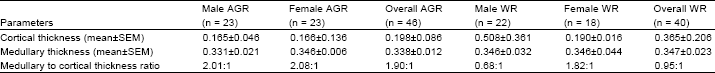
The ratio of the medullary thickness to the cortical thickness in the AGR was 1.90:1 while, the value in the WR was 0.95:1. The ratios of the liveweight to kidney and bladder weights in the AGR and WR were 414.2:1, 1292.7:1, 232.4:1 and 1326.7:1, respectively (Table 1). The ratio of the kidney weight to bladder weight in the AGR and WR were 3.72:1 and 5.07:1, respectively. The ratios of the liveweight to the kidney and bladder weights in the male and female AGR and WR were 405.3:1, 1427.6:1, 423.3:1, 1220.6:1, 231:1, 1037.2:1, 233.7:1 and 1453.1:1, respectively (Tables 2 and 3).
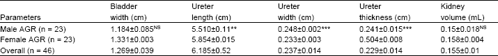
The ratios of the kidney weight to bladder weight in the male and female AGR and WR were 3.52:1, 2.88:1, 4.52:1 and 6.22:1, respectively (Tables 2 and 3). The mean kidney weights of the male and female AGR and WR were 2.119±0.062g, 2.053±0.009g, 0.633±0.091g and 0.572±0.132g, respectively (Tables 2 and 3). The mean kidney weight in the male was higher than that of the female in both AGR and WR but the difference was not significant.
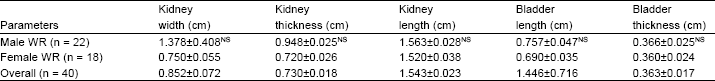
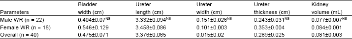
The mean kidney length and width of the AGR were 2.482±0.278cm and 1.443±0.01cm, respectively, while, those of the WR were 1.543±0.023cm and 0.852±0.072cm, respectively (Table 4). No significant difference (P>0.05) was obtained in the thickness of the bladder and its length in both AGR and WR (Table 4). There was a high significant difference (P<0.001) between the ureter length of the AGR and WR (Table 4).
| Table 1: | Liveweight, kidney and bladder weights and their ratios in African giant and Wistar rats (mean±SEM) |
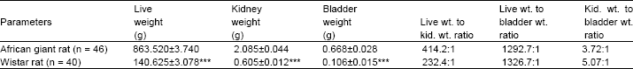 | |
| *** = Highly significant difference (p<0.001) | |
| Table 2: | Liveweight, kidney and bladder weights and their ratios in African giant rat (mean±SEM, n = 46) |
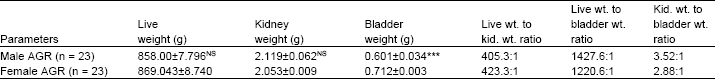 | |
| NS = Non-Significant difference (p>0.05), *** = Highly significant difference (p<0.001) | |
| Table 3: | Liveweight, kidney and bladder weights and their ratios in Wistar rat (Mean±SEM, n = 40) |
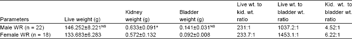 | |
| NS = Non-Significant difference (p>0.05), * = Significant difference (p<0.5) | |
| Table 4: | Comparative morphometric values of the urinary organs in African giant and Wistar rats (mean±SEM) |
 | |
| Table 4: | Continue |
 | |
| NS = Non-Significant difference (p>0.05), * = Significant difference (p<0.05), *** = Highly significant difference (p<0.001) | |
| Table 5: | Morphometric parameters of the urinary organs in African giant rat (Mean±SEM, n = 46) |
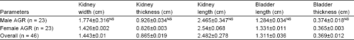 | |
| Table 5: | Continue |
 | |
| NS = Non-Significant difference (p>0.05), ** = Very significant difference (p<0.01), *** = Highly significant difference (p<0.001) | |
| Table 6: | Morphometric parameters of the urinary organs in Wistar rat (mean±SEM, n = 40) |
 | |
| Table 6: | Continue |
 | |
| NS = Non-Significant difference (p>0.05) | |
There was no significant sex difference (P>0.05) in the morphometric parameters in the WR (Table 5).
However, a significant sex difference (P<0.001) was obtained in the ureter width and thickness in the AGR (Table 6). The length of the right ureter was longer than that of the left and the female had a slightly longer ureter than the male in both rats. The ratios of medullary thickness to cortical thickness for the male and female AGR and WR were 2.01:1, 2.08:1, 0.68:1 and 1.82:1, respectively (Table 7). The relative thickness of the medulla, which is an indicator of the length of the loop of Henle, was 4.297 in the AGR while that of the WR was 5.546 (Table 8). For all rats under study, high significant correlations were obtained between the liveweight and kidney weight, kidney length, kidney width, bladder width and ureter width. No significant relationship was obtained between liveweight and bladder weight in the Wistar rat.
| Table 7: | Renal medullary and cortical thickness and their ratios in African giant and Wistar rats (Mean±SEM) |
 | |
| Table 8: | Renal medullary thickness of African giant and Wistar rats |
 | |
| NS = Non-Significant difference (p>0.05), ** = Very significant difference (p<0.01) | |
DISCUSSION
The results obtained in this study showed that most features of the kidneys of the AGR and WR were similar in many aspects and agreed with those parameters reported by Webster et al. (1947) and Hebel and Stromberg (1976) on the WR. One obvious difference between both rats, however, was in their sizes. The kidneys of the AGR were bigger than those of the WR and this was commensurate with the sizes and weights of the adult animals studied. The mean liveweight of the AGR in this study (863.520±33.740g) was within the range (800-1400g) reported by Ajayi (1975), while that of the WR (140.625±6.078g) was lower than the values (180-250 g) of Rytand (1938) and that (185.5 g) of Abdalla and Tawfik (1969). There was a slight variation between the values of the liveweight of the male AGR (858.00±27.796 g) and that of the female (869.04±8.74 g) though it was not significant.
These findings disagreed with those of Dunns (1967) and Kozma et al. (1974) who reported that the male rats had heavier liveweight than the female. This disagreement could have arisen due to some factors like age differences. In the Wistar rat, however, the liveweight of the male (146.252±8.225 g) and the female (133.683±6.283 g) were in agreement with those of Dunns (1967) and Kozma et al. (1974).
The mean kidney weights of the AGR and WR were significantly different, with values of 2.085±0.044 and 0.605±0.012 g, respectively. There is a dearth of information on the renal system of the AGR but values obtained in this study on kidney weights of the WR were similar to the value reported by Rytand (1938). The right (2.21±0.051 g) was heavier than that of the left (2.00±0.055 g). This finding agreed with those obtained in the camel (Tayeb, 1971), the rat and mouse (Dunns, 1967) and the rabbit (Kozma et al., 1974). It also agreed with that of Webster et al. (1947) who showed that the weight of the kidney of the adult male WR was heavier than that of the female of the same liveweight.
In this study also, the right kidney was slightly heavier than the left and also the kidney of the male was generally larger than that of the female. The kidney weights represented 0.25 and 0.24% of the liveweight for the male and female AGR, while in the WR the value of 0.43% was for both the male and female. The value of 0.43% recorded for the WR was lower than the values of 0.71 and 0.76% recorded by Dunns (1967) and Hebel and Stromberg (1976), respectively, for the WR. Again, these differences in values might have arisen due to variations in age, breed and some environmental factors, including the diets. In both the AGR and WR, there was a significant difference (p<0.001) in the kidney length, width and thickness (Table 3).
The results on the WR from this study were similar to those of Hebel and Stromberg (1976) who obtained a length of 2.0±0.5 cm, a width of 1.0-1.5 cm and a thickness of 1.0 cm for the kidney in WR.
The length of the left ureter was shorter than that of the right for both the male and female AGR and WR. These are also in agreement with those of Hebel and Stromberg (1976) who, demonstrated a shorter length for the left ureter in the WR. Similar finding has been reported in the dog also (Miller et al., 1965). There was no difference in the bladder shape and thickness, though the bladder width in the AGR (1.269±0.039 cm) was bigger than that of WR (0.412±0.013 cm) and this difference was highly significant.
The shape and thickness of the bladder in the WR were similar to those obtained by Hebel and Stromberg (1976). They reported values of 0.5cm for the bladder width and 1.0cm for the bladder length in the WR while, the present study indicated 0.412±0.013 cm for the width and 1.446±0.036 cm for the length. The bladder length of WR showed a slightly greater value (1.446±0.036 cm) than that of the AGR (1.311±0.036 cm).
The relative thickness of the AGR medulla was 4.3. This value was similar to 4.2 for the dog and 4.8 for the cat (Schmidt-Nielsen and O’Dell, 1961). The relative thickness of the WR medulla was 5.6 and similar to 5.9 and 6.0 recorded for WR by Ruckebuch et al. (1991) and Schmidt-Nielsen and O’Dell (1961), respectively. The Jeroboa and Desert rats had relative thickness values of 9.0 and 11.0, respectively (Schmidt-Nielsen and O’Dell, 1961).
The relative thickness of the medulla is an index of both the length of the loop of Henle and the vasa recta, which act as counter-current multiplier system. The relative thickness of medulla varies directly with the ability to produce hypertonic urine. The present study has shown that the relative thickness of the WR is higher than that of AGR and may indicate that the WR is capable of producing more hypertonic urine than the AGR. The AGR, on the other hand, has a relative thickness of 4.3 that is less than that of the cat (4.8) and the cat is not known to belong to animals that produce hypertonic urine due to any anatomical specialty in their urinary system.
Conclusion: It can be concluded that the AGR apparently lacks anatomical adaptation in its urinary system for water economy. However, studies by Tisher (1971) and Tisher et al. (1972) have shown that the rhesus monkey produces concentrated urine in the absence of a well-developed inner medulla and loop of Henle. But the rhesus monkey has been known also to have a protein binding mechanism involving calbidin that is responsible for production of this concentrated urine (Moutairou et al., 1996). It might be appropriate here to suggest that such binding mechanism is responsible for the ability of AGR to live with restricted drinking water and not necessarily due to any anatomical adaptation.
REFERENCES
- Scherle, W., 1970. A simple method for volumetry of organs in quantitative stereology. Mikroscopic, 26: 57-60.
PubMedDirect Link - Schmidt-Nielsen, B. and R.O. Dell, 1961. Structure and concentrating mechanism in the mammalian kidney. Am. J. Physiol., 200: 1119-1124.
Direct Link - Tisher, C.C., 1971. Relationship between renal structure and concentration ability in the rhesus monkey. Am. J. Physiol., 220: 1100-1106.
Direct Link - Tisher, C.C., R.W. Schrier and J.S. McNeil, 1972. Nature of urine concentrating mechanism in the Macaque monkey. Am. J. Physiol., 223: 1128-1137.
Direct Link - Webster, S.H., E.T. Liljegreen and D.J. Zimicen, 1947. Body weight ratios for liver, kidney and spleen of laboratory animals: 1 Albino rat. Am. J. Anatomy, 81: 477-513.
PubMedDirect Link