
Zhihao Zhang
Department of Pharmacy, The First Affiliated Hospital, Sun Yat-sen University, Guangzhou, 510080, China
Binghua Wei
Department of Pharmacy, The First Affiliated Hospital, Sun Yat-sen University, Guangzhou, 510080, China
Yanzhe Xia
Department of Pharmacy, The First Affiliated Hospital, Sun Yat-sen University, Guangzhou, 510080, China
Chenshu Xu
Department of Pharmacy, The First Affiliated Hospital, Sun Yat-sen University, Guangzhou, 510080, China
Xiao Chen
Department of Pharmacy, The First Affiliated Hospital, Sun Yat-sen University, Guangzhou, 510080, China
International Journal of Pharmacology
Year: 2015 | Volume: 11 | Issue: 7 | Page No.: 681-688


ABSTRACT
Danmo Capsule (DMC) is a patent Chinese botanic drug, widely used in combinational therapy with prednisone in China to treat renal diseases. The combined therapy regimen can reduce the dosage of glucocorticoids and relieve symptoms of glucocorticoid related adverse drug effects. Previous study from our laboratory showed that co-administration of the DMC significantly increased the plasma Cmax and AUC of glucocorticoid in rats. However, the effect of DMC on prednisone/prednisolone tissue distribution remains unclear. Therefore, the purpose of the current research was to study the tissue distribution of prednisone/prednisolone in rats after DMC was co-administered. Rats were treated with control vehicle or DMC (0.432 g kg–1) for 14 days before prednisone acetate (42 mg kg–1) was orally given. Samples from the heart, liver, spleen, lung, kidney, brain, fat, muscle, testes, small intestine and thymus were collected at different time points. The concentrations of prednisone and prednisolone were determined by high-performance liquid chromatography. Compared with prednisone alone group, the prednisone concentrations in rat lung, kidney, heart, muscle, small intestine and fat were increased in DMC treated groups, while those in liver, spleen, brain and thymus were decreased. However, the prednisolone concentrations in liver, lung, small intestine and thymus were increased, while those in kidney, fat, muscle, testes and heart were decreased following DMC pre-treatment. These results indicated that the alteration of prednisone/prednisolone levels in important tissues, when DMC was co-administered, may contribute to the effect of DMC in reducing adverse drug reactions of glucocorticoid and lowering its dosage administered.
PDF Abstract XML References Citation
Received: May 31, 2015;
Accepted: July 28, 2015;
Published: August 25, 2015
How to cite this article
Zhihao Zhang, Binghua Wei, Yanzhe Xia, Chenshu Xu and Xiao Chen, 2015. Tissue Distribution of Prednisone/Prednisolone is Affected by TCM Danmo Capsule in Rats. International Journal of Pharmacology, 11: 681-688.
DOI: 10.3923/ijp.2015.681.688
URL: https://scialert.net/abstract/?doi=ijp.2015.681.688
DOI: 10.3923/ijp.2015.681.688
URL: https://scialert.net/abstract/?doi=ijp.2015.681.688
INTRODUCTION
Prednisone, a synthetic corticosteroid, is commonly prescribed as an immunosuppressive agent in the treatment of immunological diseases, such as systemic lupus erythematosus, rheumatoid arthritis, polymyositis and nephrotic syndrome. Long term therapy with prednisone has been shown to result in various adverse effects, such as myopathy, osteoporosis, hyperglycaemia, electrolyte abnormalities, hypertension and truncal obesity.
As an ester pro-drug, prednisone can be rapidly and extensively converted by 11β-hydroxysteroid dehydrogenase to prednisolone, its active 11β-hydroxylmetabolite (Conti et al., 1994; Garg and Jusko, 1994; Jusko and Rose, 1980). Pharmacokinetic profiles of prednisone and prednisolone are complex, which involve hepatic hydroxylation by drug metabolizing enzyme CYP3A4, efflux by drug transport protein P-glycoprotein (P-gp), inter-conversion between prednisone and prednisolone and renal excretion of unmodified drugs (Chan et al., 2009; Frey and Frey, 1990; Karssen et al., 2002; Pichard et al., 1992; Yates et al., 2003). Therefore, it presents an increasing possibility of the potential drug-drug or herb-drug interaction between prednisone/prednisolone and other co-administered drugs, which share the same routes of drug metabolism and transport.
In China, Traditional Chinese Medicine is employed with western medicine to treat lots of chronic diseases, such as non-acute bronchial asthma (Zhao et al., 2012), myelosuppression induced by chemotherapy or radiotherapy (Jia et al., 2015), functional dyspepsia (Yang et al., 2013, 2000), as well as chronic kidney disease (Wang et al., 2012) and immunoglobulin A nephropathy (Wei et al., 2013). Danmo capsule (DMC) is a patent Chinese botanic drug, prepared with the extract of Eclipta prostrata and Radix salviae miltiorrhizae. It is widely used in combinational therapy with prednisone in China to treat renal diseases, such as glomerulonephritis, nephrotic syndrome and lupus nephritis. The combined therapy regimen can reduce the dosage of glucocorticoids and relieve symptoms of glucocorticoid related adverse drug effects (Jinping et al., 2003; Lo et al., 1992; Wang et al., 2010; Wu and Yeung, 2010; Zhou et al., 2012).
Previous study from our laboratory showed that the plasma Cmax and AUC of glucocorticoid were significantly increased, when DMC was co-administered in rats (Ren et al., 2013). However, the effect of DMC on prednisone/prednisolone tissue distribution has not yet been clarified. Therefore, the purpose of the current study was to study the tissue distribution of prednisone/prednisolone in rats after DMC was co-administered.
MATERIALS AND METHODS
Experimental animals: Male Sprague-Dawley rats weighing between 220 and 310 g were supplied by the Laboratory of Animal Service Center of Guangdong Province (Guangzhou, China). The animals were kept in a 22-24°C room with a light/dark cycle of 12:12 h and 55-60% relative humidity. They had free access to standard rodent food and water. All studies were conducted in accordance with the Regulations of Experimental Animal Administration issued by the State Committee of Science and Technology of People’s Republic of China.
Chemical and reagents: Prednisone and prednisolone with a purity of 98% and dexamethasone (internal standard, IS) with a purity of 95% as determined by HPLC with ultraviolet (UV) detection was provided by Sigma Aldrich Corporation (St. Louis, MO, USA). Prednisone acetate tablets (5 mg per tablet) were produced by Huanan Pharmaceutical Inc. (Guangdong, China) and Danmo Capsule (0.4 g per capsule) were produced by the First Affiliated Hospital of Sun Yat-Sen University (Guangdong, China). Methanol and acetonitrile of HPLC grade were purchased from TEDIA Company Inc., Beijing, China. All other reagents were of analytical grade or HPLC grade, when appropriate. Ultra-pure water was obtained from a Milli Q-plus system (Billerica, MA, USA).
Preparation of standard and quality control samples: The stock standard solutions of prednisone, prednisolone and dexamethasone were prepared by dissolving the accurately weighed individual compounds in methanol-water (55:45, v/v) to give a final concentration of 200 μg mL–1. The solutions were then serially diluted with methanol-water (55:45, v/v) to obtain working solutions at concentrations over 10-5000 ng mL–1 for prednisone and prednisolone and a working solution of dexamethasone at 500 ng mL–1. The stock solutions of the analyte or IS were stored at -80°C and the working solutions were stored at 4°C and were brought to room temperature before use. The analytical standard and Quality Control (QC) samples were prepared by spiking blank rat tissue homogenates with standard working solutions during validation and each experimental run for tissue distribution study. Calibration samples were made at concentrations of 10, 20, 50, 100, 200, 500, 1000, 2000 and 5000 ng mL–1 for prednisone and prednisolone. Quality control samples were at concentrations of 15, 250 and 4500 ng mL–1.
Sample preparation: The 20 μL of dexamethasone was added to 100 μL of rat tissue homogenate in a 1.5 mL test tube and vortexed. Sodium hydroxide solution (0.1 mol L–1, 20 μL) was added and vortexed for 1 min. The 600 μL Ethyl acetate was then added and vortex-mixed for another 1 min. After centrifugation at 10800 rpm for 10 min, the supernatant was transferred to a clean 1.5 mL centrifuge tube and evaporated to dryness. The residues were dissolved in 20 μL of acetonitrile and vortexed for 1 min. After centrifugation at 10800 rpm for 5 min, an aliquot (10 μL) of the reconstitute was injected onto the HPLC for analysis.
Equipment and chromatographic conditions: A reverse phase HPLC system consisting of water pump 600 and auto-sampler 717 coupled to a 2489 UV/V is detector (Milford, MA, USA) was used for solvent and sample delivery (Gai et al., 2005). Chromatographic separation was achieved by using a NucleodurTM 100-5C 18ec column (5 μm particle size, i.d. 4.6Χ250 mm) at 40°C. The mobile phase consisted of methanol: 0.2% phosphoric acid in deionized water (55:45, v/v, A) and acetonitrile (B). The low rate was set at 1.0 mL min–1, with 100-100% (v/v) A at 0.0-15.0 min, 100-20% A at 15.0-18.0 min, 20-20% A at 18.0-23.0 min, 20-100% A at 23.0-28.0 min and 100-100% A at 28.0-35.0 min. The detection wavelength was set at 240 nm. The total running time was 35 min for each sample.
Method validation
Specificity: The selectivity of the method was demonstrated by comparing chromatograms of blank rat tissue homogenate (without IS), rat tissue homogenate spiked with the analytes and IS and tissue homogenate from drug treated rats. All blank tissue homogenates were prepared and analyzed to ensure the absence of interfering peaks.
Calibration curves: Calibration standard was prepared as described above in triplicate and analyzed on three consecutive days. The linearity of the method was assessed by analyzing prednisone and prednisolone samples over 10-5000 ng mL–1 concentration ranges in rat tissues. The peak area ratio of analyte to IS against the concentration of analyte was plotted using 1/X2 as the weighting factor. The slope, intercept and the correlation coefficient (r2) for each standard curve from each analytical run were determined automatically by Massllynx version 4.0 software program.
Precision and recovery: Intra-and inter-batch precision and recoveries of analyte were evaluated at low, medium and high level of QC samples. The precision was defined as, the Relative Standard Deviation (RSD%), while recoveries were calculated by comparing the observed peak area ratios in samples to those non-processed standard solutions at the same concentrations.
Tissue distribution study: Rats were randomly divided into two groups. In group 1, rats orally received DMC by gavage at a dose of 0.432 g kg–1 day–1. In group 2, 0.1% kg–1 day–1 of sodium carboxymethyl cellulose was given to rats orally. All rats were dosed for 14 days and sacrificed at 0.5, 0.75, or 4 h after oral administration of 42 mg kg–1 prednisone acetate. Major organs and tissues including the heart, liver, spleen, lung, kidney, brain, fat, muscle, testes, small intestine and thymus were removed. The tissues were quickly excised, rinsed well with ice-cold saline, blotted dry and weighed. The samples were homogenized in ice-cold saline to prepare 0.5 g mL–1 homogenates. Blank tissue homogenates were prepared in a similar manner using rats without prior exposure to prednisone or DMC. The tissue homogenates were stored at -80°C until assay.
Statistical analysis: Data are presented as the Mean±SD. Student’s t-test was performed for statistical comparison of the results. The criterion of significance was set at p<0.05 and tests were performed using SPSS version 12.0 software (SPSS Inc, Chicago, IL, USA).
RESULTS
Method validation: Representative chromatograms of the analytes in rat liver and kidney are shown in Fig. 1. The retention times for prednisone, prednisolone and IS were 8.7, 11.3 and 16.2 min, respectively. No significant interference from endogenous substances or metabolites was observed at the expected retention times of the analytes and IS, indicating the reliability of the method.
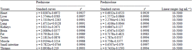
The calibration curves showed good linearity over the concentration range of 10-5000 ng mL–1 in rat tissue homogenates with a correlation coefficient (r2) larger than 0.99. Representative regression equations of the standard curve, correlation coefficient values and linear ranges for prednisone and prednisolone in rat tissues are listed in Table 1.
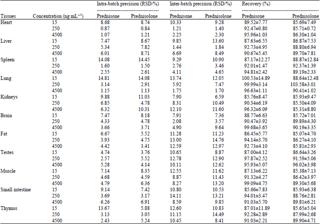
The intra-and inter-batch precision and recovery data for prednisone and prednisolone in representative rat tissues are summarized in Table 2. Intra-batch precision of prednisone and prednisolone ranged between 0.84 and 14.81% and the inter-batch precision of prednisone and prednisolone was between 1.21 and 13.74%. The recoveries of prednisone and prednisolone from rat tissue homogenates ranged over 85.66-100.53 and 85.31-103.58%, respectively and were similar at all concentrations without concentration dependence. These results indicate that the precision values and extraction efficiency for the analytes are acceptable.
Tissue distribution study: The tissue distribution study was conducted in rats receiving oral administration of 42 mg kg–1 prednisone acetate with or without 0.432 g kg–1 day–1 DMC pre-treatment. The rats were then sacrificed at 0.5, 0.75 and 4 h post-dose of prednisone acetate at 14 days according to our previous research (Ren et al., 2013). The results of tissue concentration of prednisone and prednisolone were presented in Fig. 2 and 3.
| Table 1: | Standard curves, correlation coefficients and linear ranges of prednisone and prednisolone in tissue samples (concentration of IS = 500 ng mL–1) |
 | |
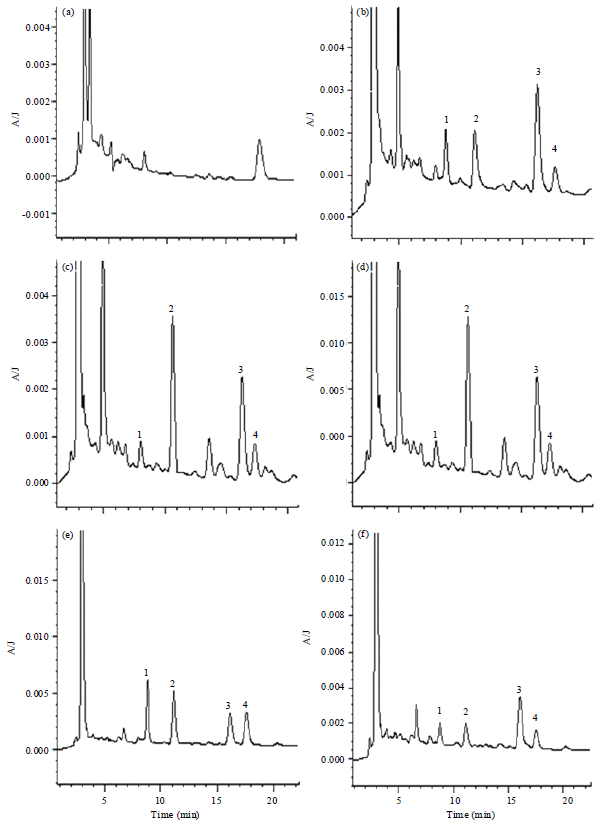 | |
| Fig. 1(a-f): | Chromatograms of the analytes in rat liver and kidney, (a, d) Representative chromatograms of blank tissues, (b, e) Blank tissues spiked with 1: Prednisone, 2: Prednisolone, 3: Dexamethasone (I.S.) and 4: Corticosterone and (c, f) Tissue samples from a rat after receiving oral dose of DMC and prednisone acetate |
Prednisone and prednisolone were widely distributed and detected at 0.5 h post-dose (the earliest time point of sampling), reaching peak levels in most tissues within 0.75 h after oral administration.
Prednisone was detectable in various tissues except for the testes 0.5 h after prednisone acetate administration in prednisone treated alone group and lungs, 4 h after prednisone acetate administration in prednisone+DMC treated group.
| Table 2: | Intra- and inter-batch precision and recovery for determination of prednisone and prednisolone in rat tissue samples (n = 5) |
 | |
The small intestine, thymus, brain, fat and muscle had higher prednisone concentration among all the detected tissues. Compared with prednisone treated alone group, the prednisone level was increased by DMC pre-treatment in lung (0.5 and 4 h), kidney (0.5 h), heart (0.75 h), muscle (0.75 and 4 h), small intestine (0.75 h), fat (4 h). On the other hand, pre-treatment of DMC decreased the prednisone level in liver (0.75 and 4 h), spleen (0.75 h), thymus (0.75 h) (Fig. 2).
Prednisolone is the active 11β-hydroxylmetabolite of prednisone converted by 11β-hydroxysteroid dehydrogenase (11β-HSD ). Prednisolone was presented in most of the tissues, but undetectable in kidney and brain 4 h post-dose of prednisone acetate in both group and lung 4 h post-dose of prednisone acetate in prednisone treated alone group. High levels of prednisolone were observed in small intestine, thymus, liver and lung. The DMC pre-treatment also changed the tissue distribution of prednisolone in various tissues. Compared with prednisone treated alone group, the prednisolone level was increased by DMC pre-treatment in liver (0.5 h), lung (0.5, 0.75 and 4 h), thymus (0.5 h), in small intestine (0.75 and 4 h). On the other hand, pre-treatment of DMC decreased the prednisolone level in kidney (0.75 h), fat (0.75 h), muscle (0.75 h) (Fig. 3).
DISCUSSION
The DMC is used in combinational therapy with prednisone to treat renal diseases, which may reduce the dosage of glucocorticoids and relieve symptoms of glucocorticoid related adverse drug effects and also significantly increased the plasma Cmax and AUC of glucocorticoid in rats (Ren et al., 2013). The aim was to study the tissue distribution of prednisone/prednisolone in rats after DMC was co-administered.
The DMC is the extract of 50% Radix Salviae miltiorrhizae and 50% Eclipta prostrate (weight/weight), which is manufactured by a pharmaceutical company. The quality control test of the batch of capsules is to determine the salvianolic acid B by HPLC. The amount of salvianolic acid B is not less than 0.9 mg for each capsule, which keep the consistent quality of different batch of the capsule formulation. In China DMC is usually used in combinational therapy with prednisone to treat glomerulonephritis, nephrotic syndrome and lupus nephritis. When DMC is combined of prednisone to treat 68 patients with adult primary nephritic syndrome, patients had significantly more improvement than with prednisone alone (85.3 vs., 56.1%), lower adverse drug reactions in combined treatment patients were also observed (14.8 vs., 48.4%) (Ye et al., 1993).
 | |
| Fig. 2(a-c): | Tissue distributions of prednisone at different times. Concentration of prednisone in the tissues of rats at (a) 0.5 h, (b) 0.75 h and (c) 4 h after receiving a single oral dose of prednisone acetate (42 mg kg–1) with or without pre-treatment of DMC (0.432 g kg–1) for 14 days (Mean±plusmn;SD, n = 3) |
Higher effective percentage of DMC with prednisone was also obtained in 148 patients with lupus nephritis than in 142 patients with western medicine treatment (85.1 vs., 73.9%), different adverse drug reactions were also observed in the two group patients (14.8 vs., 48.4%) (Luo et al., 1998).
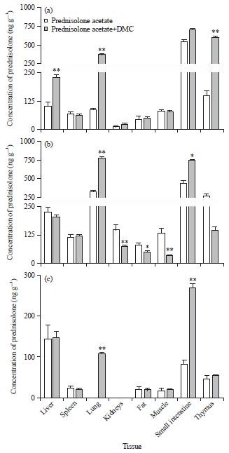 | |
| Fig. 3(a-c): | Tissue distributions of prednisolone at different times. Concentration of prednisolone in the tissues of rats at (a) 0.5 h, (b) 0.75 h and (c) 4 h after receiving a single oral dose of prednisone acetate (42 mg kg–1) with or without pre-treatment of DMC (0.432 g kg–1) for 14 days (Mean±SD, n = 3) |
The metabolism and transport of prednisone/prednisolone mainly involve CYP 3A4 enzyme, P-gp transporter and 11β-hydroxysteroid dehydrogenase (11β-HSD). 11β-HSD, which consists of two distinct enzymes, 11β-HSD I and 11β-HSD II, catalyzes the inter-conversion between prednisone and prednisolone (Kimura et al., 2010; Liu et al., 2006; Quintieri et al., 2008). The 11β-HSD I is expressed in target organs, such as liver, kidney, testicle, ovary and fat tissue, where prednisone is activated and extensively converted to prednisolone (Brozic et al., 2009; Odermatt and Nashev, 2010; Schnackenberg, 2008; Staab and Maser, 2010). The 11β-HSD II, however, is a powerful inactivator of glucocorticoid and presents in kidney, pancreas and rectum (Ferrari, 2010; Saiah, 2008). Previous studies indicate that the level of active glucocorticoids increased with increasing activity of 11β-HSD I or decreasing activity of 11β-HSD II in kidney, leading to adverse drug reaction, such as hypertension (Staab and Maser, 2010). Moreover, increased activity of 11β-HSD I in adipose tissues resulted in higher levels of active glucocorticoids, accelerated the breakdown of fat and caused fat redistribution and central obesity. Therefore, the local concentration of 11β-HSD in different tissues may contribute to the glucocorticoid associated adverse drug reactions. In the current study, the prednisolone concentration was decreased in kidney, testes, muscle and fat tissues in DMC pre-treated rats, indicating the effect of DMC in regulating tissue distribution of prednisolone, which may attribute to the reduced adverse drug reactions of glucocorticoid by DMC. Recently reported data from our laboratory reveal that the enzyme activity, gene and protein expression of rat kidney 11β-HSD II are reduced by oral administration of Eclipta prostrata, one of the major constituents of DMC (Xu et al., 2014). The effect of DMC in reducing adverse drug reactions of glucocorticoid may thus be regulated through 11β-HSD enzymes.
CONCLUSION
In summary, the prednisone/prednisolone levels in important rat tissues are affected by oral administration of DMC alters, which may attribute to the effect of DMC on reducing adverse drug reactions of glucocorticoid and lowering its dosage administered in patients undergoing combinational therapy.
ACKNOWLEDGMENTS
We thank the financial support from Guangdong Provincial Department of Science and Technology (No. 2011A080300004. and No. 2013A022100024) and Traditional Chinese Medicine Bureau of Guangdong Province (No. 20131145).
REFERENCES
- Brozic, P., P. Kocbek, M. Sova, J. Kristl and S. Martens et al., 2009. Flavonoids and cinnamic acid derivatives as inhibitors of 17β-hydroxysteroid dehydrogenase type 1. Mol. Cell. Endocrinol., 301: 229-234.
CrossRefDirect Link - Chan, K.F., Y. Zhao, T.W.S. Chow, C.S.W. Yan and D.L. Ma et al., 2009. Flavonoid dimers as bivalent modulators for p-glycoprotein-based multidrug resistance: Structure-activity relationships. Chem. Med. Chem., 4: 594-614.
CrossRefDirect Link - Conti, M., F.J. Frey, G. Escher, C. Marone and B.M. Frey, 1994. Renal handling of prednisolone/prednisone: effect of steroid dose and llβ-hydroxysteroid dehydrogenase. Nephrol. Dial. Transplant, 9: 1622-1628.
PubMedDirect Link - Ferrari, P., 2010. The role of 11β-hydroxysteroid dehydrogenase type 2 in human hypertension. Biochim. Biophys. Acta (BBA)-Mol. Basis Dis., 1802: 1178-1187.
CrossRefDirect Link - Frey, B.M. and F.J. Frey, 1990. Clinical pharmacokinetics of prednisone and prednisolone. Clin. Pharmacokinetics, 19: 126-146.
CrossRefPubMedDirect Link - Gai, M.N., E. Pinilla, C. Paulos, J. Chavez, V. Puelles and A. Arancibia, 2005. Determination of prednisolone and prednisone in plasma, whole blood, urine and bound-to-plasma proteins by high-performance liquid chromatography. J. Chromatogr. Sci., 43: 201-206.
CrossRefDirect Link - Garg, V. and W.J. Jusko, 1994. Bioavailability and reversible metabolism of prednisone and prednisolone in man. Biopharm. Drug Dispos., 15: 163-172.
PubMedDirect Link - Jia, Y., H. Du, M. Yao, X. Cui, Q. Shi, Y. Wang and Y. Yang, 2015. Chinese Herbal medicine for myelosuppression induced by chemotherapy or radiotherapy: A systematic review of randomized controlled trials. Evidence-Based Compl. Altern. Med. 10.1155/2015/690976.
Direct Link - Jinping, Q., H. Peiling, L. Yawei and Z. Abliz, 2003. Effects of the aqueous extract from Salvia miltiorrhiza Bge on the pharmacokinetics of diazepam and on liver microsomal cytochrome P450 enzyme activity in rats. J. Pharm. Pharmacol., 55: 1163-1167.
CrossRefDirect Link - Jusko, W.J. and J.Q. Rose, 1980. Monitoring prednisone and prednisolone. Ther. Drug Monit., 2: 169-176.
PubMedDirect Link - Karssen, A.M., O.C. Meijer, I.C. van der Sandt, A.G. De Boer, E.C. De Lange and E.R. De Kloet, 2002. The role of the efflux transporter P-glycoprotein in brain penetration of prednisolone. J. Endocrinol., 175: 251-260.
CrossRefDirect Link - Kimura, Y., H. Ito, R. Ohnishi and T. Hatano, 2010. Inhibitory effects of polyphenols on human cytochrome P450 3A4 and 2C9 activity. Food Chem. Toxicol., 48: 429-435.
CrossRefDirect Link - Liu, D.Y., M. Yang, H.J. Zhu, Y.F. Zheng and X.Q. Zhu, 2006. [Human pregnane x receptor-mediated transcriptional regulation of cytochrome P450 3A4 by some phytochemicals]. Zhejiang Da Xue Xue Bao Yi Xue Ban, 35: 8-13.
PubMedDirect Link - Lo, A.C.T., K. Chan, J.H.K. Yeung and K.S. Woo, 1992. The effects of Danshen (Salvia miltiorrhiza) on pharmacokinetics and pharmacodynamics of warfarin in rats. Eur. J. Drug Metab. Pharm., 17: 257-262.
CrossRefDirect Link - Odermatt, A. and L.G. Nashev, 2010. The glucocorticoid-activating enzyme 11β-hydroxysteroid dehydrogenase type 1 has broad substrate specificity: Physiological and toxicological considerations. J. Steroid Biochem. Molecular Biol., 119: 1-13.
CrossRefDirect Link - Pichard, L., I. Fabre, M. Daujat, J. Domergue, H. Joyeux and P. Maurel, 1992. Effect of corticosteroids on the expression of cytochromes P450 and on cyclosporin A oxidase activity in primary cultures of human hepatocytes. Mol. Pharmacol., 41: 1047-1055.
Direct Link - Quintieri, L., P. Palatini, A. Nassi, P. Ruzza and M. Floreani, 2008. Flavonoids diosmetin and luteolin inhibit midazolam metabolism by human liver microsomes and recombinant CYP 3A4 and CYP3A5 enzymes. Biochem. Pharmacol., 75: 1426-1437.
CrossRefDirect Link - Ren, B., B. Wei, R. Li, L. Huang, X. Hong, X. Fu and X. Chen, 2013. Study on the pharmacokinetics drug-drug interaction of danmo capsules with prednisone in rats. Int. J. Pharmacol., 9: 164-169.
CrossRefDirect Link - Saiah, E., 2008. The role of 11β-hydroxysteroid dehydrogenase in metabolic disease and therapeutic potential of 11β-HSD1 inhibitors. Curr. Med. Chem., 15: 642-649.
CrossRefDirect Link - Schnackenberg, C.G., 2008. 11β-hydroxysteroid dehydrogenase type 1 inhibitors for metabolic syndrome. Curr. Opin. Invest. Drugs, 9: 295-300.
PubMedDirect Link - Staab, C.A. and E. Maser, 2010. 11β-Hydroxysteroid dehydrogenase type 1 is an important regulator at the interface of obesity and inflammation. J. Steroid Biochem. Molecular Biol., 119: 56-72.
CrossRefDirect Link - Wang, X., W.Y.W. Lee, X. Zhou, P.M.Y. Or and J.H.K. Yeung, 2010. A pharmacodynamic-pharmacokinetic (PD-PK) study on the effects of Danshen (Salvia miltiorrhiza) on midazolam, a model CYP3A probe substrate, in the rat. Phytomedicine, 17: 876-883.
CrossRefDirect Link - Wang, Y.J., L.Q. He, W. Sun, Y. Lu and X.Q. Wang et al., 2012. Optimized project of traditional Chinese medicine in treating chronic kidney disease stage 3: A multicenter double-blinded randomized controlled trial. J. Ethnopharmacol., 139: 757-764.
CrossRefDirect Link - Wei, M., P. Xiong and L. Zhang, 2013. Effect of Chinese herbs on immunoglobulin A nephropathy: A randomized controlled trial. J. Tradit. Chin. Med., 33: 65-69.
CrossRefDirect Link - Wu, W.W.P. and J.H.K. Yeung, 2010. Inhibition of warfarin hydroxylation by major tanshinones of Danshen (Salvia miltiorrhiza) in the rat In vitro and In vivo. Phytomedicine, 17: 219-226.
CrossRefDirect Link - Xu, C., B. Wei, X. Fu, M. Luo and S. Liu et al., 2014. Effect of Eclipta prostrate on 11β-Hydroxysteroid dehydrogenase in rat liver and kidney. Evidence-Based Complementary Altern. Med.
CrossRef - Yang, N., X. Jiang, X. Qiu, Z. Hu, L. Wang and M. Song, 2013. Modified Chaihu Shugan powder for functional dyspepsia: meta-analysis for randomized controlled trial. Evidence-Based Compl. Altern. Med.
CrossRefDirect Link - Yang, X., R.G. Ye, G.X. Liu, L.B. Wei, G.M. Huang, Y.J. Li, Z.H. Zheng and G.Q. Zhang, 2000. The clinic and experimental researches on treatment of nephritic diseases with the integrating traditional chinese medicine and western medicine for 38 years. Acad. J. Sums, 21: 401-408.
Direct Link - Yates, C.R., C. Chang, J.D. Kearbey, K. Yasuda and E.G. Schuetz et al., 2003. Structural determinants of P-glycoprotein-mediated transport of glucocorticoids. Pharm. Res., 20: 1794-1803.
PubMedDirect Link - Zhao, Y.H., Z.L. Liu, L.H. Li, S.H. Jiang and C.H. Shi, 2012. Systematic review of randomized controlled trials of traditional Chinese medicine treatment of non-acute bronchial asthma complicated by gastroesophageal reflux. J. Tradit. Chin. Med., 32: 12-18.
CrossRefDirect Link - Zhou, X., K. Chan and J.H.K. Yeung, 2012. Herb-drug interactions with Danshen (Salvia miltiorrhiza): A review on the role of cytochrome P450 enzymes. Drug Metabol. Drug Interact., 27: 9-18.
CrossRefDirect Link