
M. Badrul Alam
Department of Pharmacy, Atish Dipankar University of Science and Technology, Dhaka, Bangladesh
M. Saifur Rahman
Department of Pharmacy, Atish Dipankar University of Science and Technology, Dhaka, Bangladesh
M. Hasan
Department of Pharmacy, Atish Dipankar University of Science and Technology, Dhaka, Bangladesh
M. Maruf Khan
Department of Pharmacy, Atish Dipankar University of Science and Technology, Dhaka, Bangladesh
Kamrun Nahar
Department of Pharmacy, Atish Dipankar University of Science and Technology, Dhaka, Bangladesh
Sheema Sultana
Department of Pharmacy, Atish Dipankar University of Science and Technology, Dhaka, Bangladesh
International Journal of Pharmacology
Year: 2012 | Volume: 8 | Issue: 4 | Page No.: 243-251


ABSTRACT
Dillenia indica Linn. (Dilleniaceae) is widely used as food and reputed in the folk medicine of Bangladesh and India in particular, to relieve pain associated with gastrointestinal disturbance. Methanolic extract of the bark of Dillenia indica Linn. (MDI) was subjected to evaluate for its analgesic and antioxidant properties. The analgesic activity of MDI (200 and 400 mg kg-1 b.wt. p.o.) was determined for its central and peripheral pharmacological actions using hotplate as well as tail immersion method and acetic acid-induced writhing test with formalin induced pain in mice, respectively. To evaluate antioxidant potential of MDI, total antioxidant activity, scavenging of 1,1-diphenyl-2-picrylhydrazyl (DPPH) radical as well as total Reactive Oxygen Species (ROS) and assessment of reducing power were used. The extract, at the dose of 200 and 400 mg kg-1, produced a significant (p<0.05) increase in pain threshold in hotplate and tail immersion methods whereas significantly (p<0.05) reduced the writhing caused by acetic acid and the number of licks induced by formalin in a dose dependent manner. In DPPH and total ROS scavenging method, MDI showed good antioxidant potentiality with the IC50 value of 12.32±0.16 and 34.72±0.48 μg mL-1, respectively with a good reducing power. The findings of the study suggested that MDI has strong analgesic and antioxidant effects, conforming the traditional use of this plant for pain alleviation to its antioxidant potentiality.
PDF Abstract XML References Citation
Received: November 05, 2011;
Accepted: February 17, 2012;
Published: March 27, 2012
How to cite this article
M. Badrul Alam, M. Saifur Rahman, M. Hasan, M. Maruf Khan, Kamrun Nahar and Sheema Sultana, 2012. Antinociceptive and Antioxidant Activities of the Dillenia indica Bark. International Journal of Pharmacology, 8: 243-251.
DOI: 10.3923/ijp.2012.243.251
URL: https://scialert.net/abstract/?doi=ijp.2012.243.251
DOI: 10.3923/ijp.2012.243.251
URL: https://scialert.net/abstract/?doi=ijp.2012.243.251
INTRODUCTION
Pain transmission is a mechanism that involves a very complex interaction of peripheral and central structures from the skin surface to the central cerebral cortex (Susanna, 1999). According to the International Association for the Study of Pain, pain has been defined as an unpleasant sensory and emotional experience associated with actual or potential tissue damage or described in terms of such damage (Merskey and Bogduk, 1994). There are several types of pain, namely ‘nociceptive’, ‘neurogenic’, ‘neuropathic’ and ‘psychogenic’ which are associated with a stimulation of nociceptors, damage to neuronal tissue, dysfunction of a nerve or psychological factors, respectively (Millan, 1999). The direct and indirect action of chemical mediators, such as arachidonic acid metabolites (prostaglandins and leukotrienes), peptides, serotonin, acetylcholine, cytokines, nitric oxide, among others which can be produced or released following tissue injury or by exogenous irritants (formalin, acetic acid), are responsible for the multiplicity of events that occur during pain transmission, in both the peripheral and central nervous systems (Belfrage et al., 1995; Dray, 1997; Sawynok, 1998; Besson, 1999). Moreover, various free radicals as well as reactive oxygen species (ROS) are also responsible for the induction of short-term algesia (Chung, 2004) and triggers some second messengers, are involved in sensitization of dorsal horn neurons that plays a fundamentally important role in neuropathic pain (Ali and Salter, 2001; Zhang et al., 2003). Oxidative damage caused by the free radicals or ROS is considered to play a causative role in aging and several stress related diseases including heart disease, inflammation, diabetes, cognitive dysfunction, cancer and Alzheimer’s disease (Gulcin et al., 2002). Undoubtedly, in vivo suppression of these reactive species is important for the human body to eliminate the toxicity induced by these reactive species. For several years, many researchers have been investigated powerful and nontoxic antioxidants from natural sources, especially edible or medicinal plants to prevent the above reactive species related disorders in human as well as replace the synthetic compounds which are in use may have carcinogenic activity and harmful to the lungs and liver (Rechner et al., 2002).
Plant extracts have been used for centuries, as popular remedies against several health disorders. The study of plants that have been traditionally used as pain killers should still be seen as a fruitful and logical research strategy in the search for new analgesic drugs and pain mechanisms (Calixto et al., 2000). Despite the severe adverse effects of morphine, steroidal or non steroidal anti-inflammatory agents are used for the treatment of pain clinically; naturally occurring agents with reduced side effects are required to substitute this chemical therapeutics. The genus Dillenia has sixty species, of which Dillenia indica Linn., belongs to the family Dilleniaceae, is the most common edible species. Originated from Indonesia, this evergreen tropical tree is now found from Bangladesh, India and Nepal to China. The common names include Chulta (Bengali, Hindi), Bhavya (Sanskrit) and Elephant apple (English). The leaf, bark and fruit of this plant are used as traditional medicine. The juice of D. indica leaves, bark and fruits are mixed and given orally (5-15 mL, two to five times daily) in the treatment of cancer and diarrhea (Sharma et al., 2001). The fruit juice of this plant has anti-leukemic effect (Kumar et al., 2009), cardiotonic effect, used as cooling beverage in fever and also employed in cough mixture. The leaves and bark are used as a laxative and astringent. Bruised bark is applied as a cataplasm for patients with arthritis (Shome et al., 1980). The solvent extracts of fruits and leaves of D. indica are reported to have antioxidant activity (Abdille et al., 2005). CNS depressant activities (Bhakuni et al., 1969) and anti-inflammatory activity (Yeshwante et al., 2009) in mice were found from the alcoholic extract of the leaves of D. indica. Literature reviews indicated that no studies combining the analgesic and antioxidant of the bark of Dillenia indica have so far been undertaken. Taking this in view and as a part of our ongoing research (Alam et al., 2011; Rahman et al., 2011) on Bangladeshi medicinal plants, the present study aimed to evaluate the analgesic activity of the bark extracts of Dillenia indica (MDI) along with their in vitro antioxidant activity.
MATERIALS AND METHODS
Plant materials: The bark of the plant of Dillenia indica Linn. was collected from the botanical garden of Pharmacy department, Jahangirnagar University, Bangladesh during January 2009. The plant material was taxonomically identified by the National Herbarium of Bangladesh whose voucher specimen No. JU/32234 is maintained in our laboratory for future reference.
Chemicals: Ammonium molybdate, Folin-Chiocaltu phenol reagent, were purchased from E. Merck (Germany). 1,1-diphenyl-2-picryl-hydrazyl (DPPH), ascorbic acid, quercetin and potassium ferric cyanide and 2',7'-dichlorfluorescein-diacetate (DCFH-DA) were purchased from Sigma Chemical Company (St. Louis, MO, USA). Nalbuphine, Diclofenac-Na was collected from Square Pharmaceuticals Ltd., Bangladesh. All other chemicals and reagents were of analytical grade.
Preparation of plant extract: The plant material was shade-dried with occasional shifting and then powdered with a mechanical grinder, passing through sieve No. 40 and stored in a tight container. The dried powder material (1.5 kg) was refluxed with MeOH for 3 h. The total filtrate was concentrated to dryness, in vacuo at 40°C to render the MeOH extract (490 g).
In vivo analgesic activity
Animal: Albino mice (25-30 g) of both sexes were used for assessing biological activity. The animals were maintained under standard laboratory conditions and had free access to food and water ad libitum. The animals were allowed to acclimatize to the environment for 7 days prior to experimental session. The animals were divided into different groups, each consisting of five animals which were fasted overnight prior to the experiments. Experiments on animals were performed in accordance with guidelines of the Institutional Animal Ethics Committee, Atish Dipankar University of Science and Technology, Dhaka, Bangladesh. Animal treatment and maintenance for acute toxicity and analgesic effects were conducted in accordance with the Principle of Laboratory Animal Care (NIH publication No. 85-23, revised 1985) and the Animal Care and Use Guidelines of Atish Dipankar University of Science and Technology, Dhaka, Bangladesh.
Acute toxicity study: Acute oral toxicity assay was performed in healthy nulliparous and non pregnant adult female albino Swiss mice (25-30 g) divided into different groups. The test was performed using increasing oral dose of the MDI in water (50, 100, 200, 500, 1000 mg kg-1 b.wt.), in 20 mL kg-1 volume to different test groups. Normal group received water. The mice were allowed to feed ad libitum, kept under regular observation for 48 h, for any mortality or behavioral changes (Sanmugapriya and Venkataraman, 2006).
Hot plate method: The animals were divided into four groups with five mice in each group. Group I animals received vehicle (1% Tween 80 in water, 10 mL kg-1 b.wt.), animals of Group II received nalbuphine at 10 mg kg-1 b.wt. while animals of Group III and Group IV were treated with 200 and 400 mg kg-1 b.wt. (p.o.) of the MDI. The animals were placed on Eddy’s hot plate kept at a temperature of (55±0.5)°C. A cut off period of 15 sec, was observed to avoid damage to the paw (Franzotti et al., 2000). Reaction time was recorded when animals licked their fore or hind paws or jumped prior to 0, 30, 60 and 90 min after oral administration of the samples (Eddy and Leimback, 1953; Malairajan et al., 2006; Toma, 2003).
Tail immersion test: The procedure is based on the observation that morphine like drugs selectively prolongs the reaction time of the typical tail withdrawal reflex in mice (Toma, 2003). The animals were treated as discussed above. From 1-2 cm of the tail of mice was immersed in warm water kept constant at 55°C. The reaction time was the time taken by the mice to deflect their tails. The first reading was discarded and the reaction time was recorded as a mean of the next three readings. A latency period of 20 sec was defined as complete analgesia and the measurement was then stopped to avoid injury to mice. The latent period of the tail-flick response was determined before and 0, 30, 60 and 90 min after the administration of drugs.
Acetic acid-induced writhing test: The analgesic activity of the samples was also studied using acetic acid-induced writhing model in mice. Test samples and vehicle were administered orally 30 min before intraperitoneal administration of 0.7% v/v acetic acid but Diclofenac-Na was administered intraperitoneally 15 min before injection of acetic acid. After an interval of 5 min, the mice were observed for specific contraction of body referred to as ‘writhing’ for the next 10 min (Ahmed et al., 2004).
Formalin test: The antinociceptive activity of the drugs was determined using the formalin test described by Dubuisson and Dennis (1977). Control group received 5% formalin. Twenty microliter of 5% formalin was injected into the dorsal surface of the right hind paw 60 min after administration of MDI (200 and 400 mg kg-1, p.o.) and 30 min after administration of diclofenac Na (10 mg kg-1, i.p.). The mice were observed for 30 min after the injection of formalin and the amount of time spent licking the injected hind paw was recorded. The first 5 min post formalin injection is referred to as the early phase and the period between 15 and 30 min as the late phase. The total time spent licking or biting the injured paw (pain behavior) was measured with a stop watch.
In vitro antioxidant activity
Determination of total antioxidant capacity: The antioxidant activity of the MDI was evaluated by the phosphomolybdenum method according to the procedure of Prieto et al. (1999).
Free radical scavenging activity measured by 1,1-diphenyl-2-picryl-hydrazyl (DPPH): The free radical scavenging activity of MDI, based on the scavenging activity of the stable 1,1-diphenyl-2-picrylhydrazyl (DPPH) free radical, was determined by the method described by Braca et al. (2001).
Measurement of the inhibition of the total ROS generation: Mice kidney homogenates, prepared from the kidneys of freshly killed male Swiss albino mice, weighing 30-39 g, were mixed with or without a suspension of extracts and then incubated with 12.5 μM DCFH-DA, at 37°C for 30 min. Phosphate buffer (50 mM, pH 7.4) was used. DCFH-DA is a stable compound which easily diffuses into cells and is hydrolyzed by intracellular esterase to yield a reduced non-fluorescent compound, DCFH which is trapped within the cells. The ROS produced by cells oxidize the DCFH to the highly fluorescent 2',7'-dichlorodihydrofluorescein (DCF). The fluorescence intensity of the oxidized DCF was monitored on a microplate fluorescence spectrophotometer (Bio-Tek Instruments Inc., Winooski, VT), with excitation and emission wavelengths of 460 and 530 nm, respectively (Label and Bondy, 1990). IC50 value was calculated from the equation of line obtained by plotting a graph of concentration versus % inhibition.
Reducing power activity: The reducing power of MDI was determined according to the method previously described (Oyaizu, 1986). Extracts at different concentrations in 1 mL of 10% DMSO were mixed with 2.5 mL of phosphate buffer (0.2 M, pH 6.6) and 2.5 mL potassium ferricyanide [K3Fe (CN)6] (1%) and then the mixture was incubated at 50°C for 30 min. Afterwards, 2.5 mL of trichloroacetic acid (10%) was added to the mixture which was then centrifuged at 3000 rpm for 10 min. Finally, 2.5 mL of upper layer solution was mixed with 2.5 mL distilled water and 0.5 mL FeCl3 (0.1%) and the absorbance was measured at 700 nm. Increased absorbance of the reaction mixture indicated increased reducing power.
Statistical analysis: All values were expressed as the Mean±SEM of three replicate experiments. The analysis was performed by using SPSS statistical package for WINDOWS (version 16.0; SPSS Inc, Chicago). Results related to the reducing power activities were statistically analyzed by applying the Student t-test and p<0.001 were considered to be statistically significant. All in vivo data are subjected to ANOVA followed by Dunnett’s test and p<0.05 were considered to be statistically significant.
RESULTS
In vivo analgesic activity
Hot plate method: Both doses of the extract produced a dose dependent increase in latency time when compared with the vehicle (Fig. 1). The result was found to be statistically significant (p<0.05-0.001).
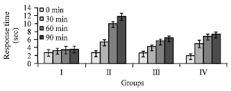 | |
| Fig. 1: | Effects of the MDI on latency to hotplate test. Values are Mean±SEM (n = 5); *p<0.05, Dunnett test as compared to control. Group I: Vehicle (1% Tween 80 in water), Group II: Nalbuphine 10 mg kg-1 b.wt., Group III and Group IV : 200 and 400 mg kg-1 b.wt. (p.o.) of the crude extract of D. indica |
| Table 1: | Effects of the MDI on acetic acid-induced writhing in mice |
 | |
| Values are Mean±SEM (n = 5), *p<0.05, Dunnett test as compared to vehicle control. Group I: Vehicle (1% Tween 80 in water), Group II: Diclofenac Na 10 mg kg-1 b.wt., Group III and Group IV: 200 and 400 mg kg-1 b.wt. (p.o.) of the MDI | |
| Table 2: | Effect of MDI in hindpaw licking in the formalin test in mice |
 | |
| Values are Mean±SEM (n = 5), * p<0.05, Dunnett test as compared to vehicle control. Group I: Vehicle (1% Tween 80 in water), Group II: Diclofenac Na 10 mg kg-1 b.wt., Group III and Group IV: 200 and 400 mg kg-1 b.wt. (p.o.) of the MDI | |
Tail immersion test: The tail withdrawal reflex time following administration of the MDI was found to increase with increasing dose of the sample. The result was statistically significant (p<0.05-0.001) and was comparable to the reference drug nalbuphine (Fig. 2).
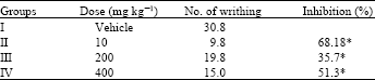
Acetic acid-induced writhing test: Table 1 shows the effects of the extract of on acetic acid-induced writhing in mice. The oral administration of both doses of MDI significantly (p<0.001) inhibited writhing response induced by acetic acid in a dose dependent manner.
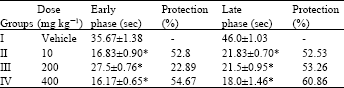
Formalin test: MDI (200 and 400 mg kg-1, p.o.) significantly (p<0.001) suppressed the licking activity in either phase of the formalin-induced pain in mice in a dose dependant manner (Table 2).
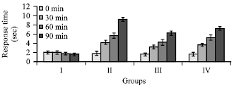 | |
| Fig. 2: | Effects of the MDI on tail withdrawal reflex of mice induced by tail immersion method. Values are Mean±SEM (n = 5); *p<0.05, Dunnett test as compared to control. Group I: Vehicle (1% Tween 80 in water), Group II: Nalbuphine 10 mg kg-1 b.wt., Group III and Group IV: 200 and 400 mg kg-1 b.wt. (p.o.) of the crude extract of D. indica |
| Table 3: | Antioxidant activities of the MDI on total antioxidant capacity, DPPH and total ROS assay methods |
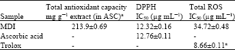 | |
| Ascorbic acid equivalents (ASC) (mg g-1 of each extract) for the total antioxidant capacity, Values are expressed as Means±SEM of triplicate experiments | |
MDI, at the dose of 400 mg kg-1 b.wt., showed the more licking activity against both phases of formalin-induced pain than that of the standard drug diclofenac Na.
In vitro antioxidant activity
Total antioxidant capacity: Total antioxidant capacity of D. indica is expressed as the number of equivalents of ascorbic acid. Total antioxidant capacity of MDI was found to be 213.9±0.69 mg g-1 m equivalent of ascorbic acid (Table 3).
DPPH radical scavenging activity: The percentage (%) scavenging of DPPH radical was found to be concentration dependent with the IC50 value of 12.32±0.16 μg mL-1, while IC50 value of standard ascorbic acid was found to be 12.76±0.11 μg mL-1 (Table 3).
Inhibition of total ROS generation: The percentage inhibition of ROS generation was illustrated in Table 3 and it is observed that scavenging of ROS by the extract is also concentration dependent with the IC50 value of 34.72±0.48 μg mL-1, while IC50 value of standard trolox was found to be 8.66±0.11 μg mL-1.
Reducing power ability: For the measurement of the reductive ability, we investigated the Fe3+ to Fe2+ transformation in the presence of MDI and compared with standards (galic acid, quercetin and ascorbic acid) (Fig. 3).
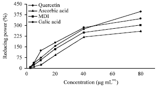 | |
| Fig. 3: | Reducing power of MDI, quercetin, ascorbic acid and galic acid by spectrophotometric detection of Fe3+ to Fe2+ transformation |
Like the antioxidant activity, the reducing power of MDI was found to be concentration dependent and statistically significant (p<0.001).
DISCUSSION
The hotplate and tail immersion methods are commonly used for assessing central antinociceptive response. Both methods are further distinguished by their tendency to respond to the pain stimuli conducting through neuronal pathways as tail immersion mediates a spinal reflex to nociceptive stimuli, while the hot plate involves higher brain functions and is a supraspinally organized response (Chapman et al., 1985). Narcotic analgesics inhibit both peripheral and central mechanism of pain, while non steroidal anti-inflammatory drugs inhibit only peripheral pain (Elisabetsky et al., 1995; Pal et al., 1999). As noted, nalbuphine, the reference narcotic analgesic drug (5 mg kg-1, p.o.) exhibited significant and paramount analgesic effects in both the hot plate (supra spinal) as well as the tail immersion (spinal) test; whereas, MDI (200 and 400 mg kg-1, p.o.) also produced a statistically significant but lesser in degree antinociceptive response to that of nalbuphine in both test suggesting that the plant extract may act as a narcotic analgesic. However, the mechanism(s) behind the central analgesic response of MDI in both tested methods is not completely understood and may need further investigation.
On the otherhand, acetic acid induced writhing response is a sensitive procedure to evaluate peripherally acting analgesics and represents pain sensation by triggering localized inflammatory response. Such pain stimulus leads to the release of free arachidonic acid from the tissue phospholipid (Ahmed et al., 2006). The response is thought to be mediated by peritoneal mast cells (Ribeiro et al., 2000), acid sensing ion channels (Voilley, 2004) and the prostaglandin pathways (Hossain et al., 2006). The organic acid has also been postulated to act indirectly by inducing the release of endogenous mediators which stimulates the nociceptive neurons that are sensitive to NSAIDs and narcotics (Adzu et al., 2003). It is well known that non-steroidal anti-inflammatory and analgesic drugs mitigate the inflammatory pain by inhibiting the formation of pain mediators at the peripheral target sites where prostaglandins and bradykinin are proposed to play a significant role in the pain process (Hirose et al., 1984). In addition, it was suggested that non narcotic analgesics produce their action by interfering with the local reaction to peritoneal irritation thereby reducing the intensity of afferent nervous stimulation in the acetic acid induced writhing test, a model of visceral pain (Vogel and Vogel, 1997). Therefore, it is likely that MDI might have exerted its peripheral antinociceptive action by interfering with the local reaction caused by the irritant or by inhibiting the synthesis, release and/or antagonizing the action of pain mediators at the target sites and this response in agreement with the previous studies with other parts of D. indica, leaves (Bose et al., 2010). The above findings clearly demonstrated that both central and peripheral mechanisms are involved in the antinociceptive action of MDI. The analgesic activity of MDI could also be linked to the mechanism of action either on central nervous system or peripheral nervous system. Interestingly, compounds like flavonoids (Kim et al., 2004a) and steroids, triterpenes in part, have been shown to possess anti-inflammatory, analgesic activity and the claim made by Pritam et al. (2011). Based on the classes of compounds detected in MDI extract, several mechanisms of action could be used to explain the observed activities of MDI extract.
The formalin model normally postulates the site and the mechanism of action of the analgesic (Chau, 1989). This biphasic model is represented by neurogenic (0-5 min) and inflammatory pain (15-30 min), respectively (Hunskaar and Hole, 1987). Drugs that act primarily on the central nervous system such as narcotics inhibit both as steroids and NSAIDs suppress mainly the late phase (Adzu et al., 2003). The suppression of neurogenic and inflammatory pains by the extract might imply that it contains active analgesic principle that may be acting both centrally and peripherally. This is an indication that the extract can be used to manage acute as well as chronic pain. The mechanism by which formalin triggers C-fibers activation remained unknown for a relatively long time. Recently, however, McNamara et al. (2007) demonstrated that formalin activates primary afferent neurons through a specific and direct on TRPA1, a member of the transient receptor potential family of cation channels, expressed by a subset of C-fiber nociceptors and this effect is accompanied by increased influx of Ca2+ ions. TRPA1 cation channels at primary sensory terminals were also reported to mediate noxious mechanical stimuli (Kerstein et al., 2009). These experiments suggest that Ca2+ mobilization through TRPA1 cation channels is concomitant with noxious chemicals and mechanical stimuli as they produce their analgesic action. It is likely that the inhibitory effect of MDI to pain response is due to inhibit the increase of the intracellular Ca2+ through TRPA1, presumably evoked by formalin. So, the bark extract of D. indica may contain substances that affect the metabolism of Ca2+. Literature survey revealed that tannins, triterpenoids and flavonoid are the major phytoconstituents of D. indica (Abdille et al., 2005; Parvin et al., 2009; Muhit et al., 2010). Flovonoids, for example, have been found to suppress the intracellular Ca2+ ion elevation in a dose dependent manner, as well as the release of proinflammatory mediators such as TNFα (Kempuraj et al., 2005).
To determine the efficacy of natural antioxidants either as pure compounds or as plant extract, a great number of in vitro methods have been developed in which antioxidant compounds act by several mechanisms. Determination of specific antioxidant species might be less useful than the knowledge of the total antioxidant capacity of a sample. The knowledge of total antioxidant activity can be useful in the analysis of changes in plasma antioxidant activity related to oxidative stress or the understanding of structure–activity relationships of pure antioxidant species. Because of its simplicity and the cheap reagents it uses, the phosphomolybdenum method is an alternative to the methods already available for the evaluation of total antioxidant capacity. The phosphomolybdenum method was based on the reduction of Mo(VI) to Mo(V) by the compounds having antioxidant property and is successfully used to quantify vitamin E in seeds (Prieto et al., 1999). DPPH• is a stable free radical that accepts an electron or hydrogen radical to become a stable diamagnetic molecule (Nakayama, 1994) and is usually used as a substrate to evaluate the antioxidant activity of a compound (Chang et al., 2002). Based on the data obtained from this study, DPPH radical scavenging activity of MDI (IC50 12.32±0.16 μg mL-1) was similar to the standard ascorbic acid (IC50 12.76±0.11 μg mL-1). These findings agree with previous reports on scavenging of free radicals with other parts of D. indica, leaves and fruits. (Abdille et al., 2005; Parvin et al., 2009). Moreover, it was revealed that MDI did show the proton donating ability and could serve as free radical inhibitor or scavenger. A direct correlation between antioxidant capacity and reducing power of certain plant extracts has been reported (Tanaka et al., 1988). The reducing properties are generally associated with the presence of reductones which have been shown to exert antioxidant action by breaking the free radical chain by donating a hydrogen atom (Duh et al., 1999). Because a substance may act as an antioxidant due to its ability to reduce ROS by donating hydrogen atom (Jayaprakasha et al., 2001; Khanam et al., 2004), the ferric reducing property of plant extracts (Fig. 3) implies that they are capable of donating hydrogen atom in a dose dependent manner. Polyphenolic compounds, like flavonoids, tannins and phenolic acids, commonly found in plants have been reported to have multiple biological effects, including antioxidant activity (Kahkonen et al., 1999). Phenolic compounds are understood to induce the cellular antioxidant system; increase approximately 50% cellular glutathione concentration. Flavonoids are important in the modulation of γ-glutamylcysteine synthase in both cellular antioxidant defenses and detoxification of xenobiotics (Muchuweti et al., 2007). A flavonoid dillenetin have been isolated from the MDI (Muhit et al., 2010) and the total phenol and flavonoid content was found to be the highest in methanolic extract (Abdille et al., 2005) and this may be the cause for the highest antioxidant activity in different model.
Flavonoids may increase the amount of endogenous serotonin or may interact with 5-HT2A and 5-HT3 receptors which may be involved in the mechanism of central analgesic activity (Annegowda et al., 2010). Previous researchers reported the presence of several therapeutically valued flavonoids from the D. indica (Arbianti et al., 2007; Bose et al., 2010). Moreover, MDI showed significant analgesic activity in the entire experimental model which may be due to its high flavonoid content as well as free radical scavenging activity, as these free radicals are involved during pain stimulation and antioxidants showed reduction in such pain (Kim et al., 2004b). It is well established that sensitization dorsal horn cells in the spinal cord (central sensitization) plays a fundamentally important role in neuropathic pain. Excessive ROS affects central sensitization and triggers second messengers system that involved in sensitization of dorsal horn neurons and also activates spinal glial cells which in turn play an important role in chronic pain (Raghavendra et al., 2003). In addition, recent studies suggest that the inflammatory tissue damage is due to the liberation of reactive oxygen species form phagocytes invading the inflammation sites (Parke and Sapota, 1996). There are also reports on the role of flavonoid, a powerful antioxidant (Vinson et al., 1995; Brown and Rice-Evans, 1998), in analgesic activity primarily by targeting prostaglandins (Ramesh et al., 1998; Rajnarayana et al., 2001). Moreover, flavonoids have the ability to inhibits eicosanoid biosynthesis. Eicosanoids, such as prostaglandins are involved in various immunological responses and are the end products of the cyclooxygenase and lipoxygenase pathways (Jothimanivannan et al., 2010). Tannins are also found to have a contribution in antinociceptive activity (Ramprasath et al., 2006). Again the plant extract demonstrated good antioxidant action in the tested models. So it can be assumed that cyclooxygenase (COX) inhibitory activity along with antioxidant activity may reduce the production of free arachidonic acid from phospholipid or may inhibit the enzyme system responsible for the synthesis of prostaglandins and ultimately relieve pain-sensation.
CONCLUSION
Based on the results of the present study, we conclude that the plant extract possesses strong analgesic and antioxidant potential. However, further studies are necessary to examine underlying mechanisms of analgesic and antioxidant effects and to isolate the active compound(s) responsible for these pharmacological activities.
REFERENCES
- Abdille, M.H., R.P. Singh, G.K. Jayaprakasha and B.S. Jena, 2005. Antioxidant activity of the extracts from Dillenia indica fruits. Food Chem., 90: 891-896.
CrossRefDirect Link - Ahmed, F., M.S.T. Selim, A.K. Das and M.S.K. Choudhuri, 2004. Anti-inflammatory and antinociceptive activities of Lippia nodiflora Linn. Pharmazie, 59: 329-330.
Direct Link - Ali, D.W. and M.W. Salter, 2001. NMDA receptor regulation by Src kinase signalling in excitatory synaptic transmission and plasticity. Curr. Opin. Neurobiol., 11: 336-342.
Direct Link - Annegowda, H.V., M.N. Mordi, S. Ramanathan and S.M. Mansor, 2010. Analgesic and antioxidant properties of ethanolic extract of Terminalia catappa L. Leaves. Int. J. Pharmacol., 6: 910-915.
CrossRefDirect Link - Arbianti, R., T.S. Utami, A. Kurmana and A. Sinaga, 2007. Comparison of antioxidant activity and total phenolic content of Dillenia indica leaves extracts obtained using various techniques. Proceedings of the 14th Regional Symposium on Chemical Engineering, December 4-5, 2007, Yogyakarta, Indonesia.
- Belfrage, M., A. Sollevi, M. Segerdahl, K.F. Sjolund and P. Hansson, 1995. Systemic adenosine infusion alleviates spontaneous and stimulus evoked pain in patients with peripheral neuropathic pain. Anesth. Analg., 81: 713-717.
PubMed - Bhakuni, D.S., M.L. Dhar, M.M. Dhar, B.N. Dhawan and B.N. Mehrotra, 1969. Screening of Indian plants for biological activity: Part II. Indian J. Exp. Biol., 7: 250-262.
Direct Link - Bose, U., K. Gunasekaran, V. Bala and A.A. Rahman, 2010. Evaluation of phytochemical and pharmacological properties of Dillenia indica Linn. leaves. J. Pharmacol. Toxicol., 5: 222-228.
CrossRef - Braca, A., N. De Tommasi, L. Di Bari, C. Pizza, M. Politi and I. Morelli, 2001. Antioxidant principles from Bauhinia tarapotensis. J. Nat. Prod., 64: 892-895.
PubMed - Brown, J.E. and C.A. Rice-Evans, 1998. Luteolin rich artichoke extract protects low density lipoprotein from oxidation in vitro. Free Radic. Res., 29: 247-255.
CrossRef - Calixto, J.B., D.A. Cabrini, J. Ferreira and M.M. Campos, 2000. Kinins in pain and inflammation. Pain, 87: 1-5.
PubMedDirect Link - Chang, L.W., W.J. Yen, S.C. Huang and P.D. Duh, 2002. Antioxidant activity of sesame coat. Food Chem., 78: 347-354.
CrossRefDirect Link - Chapman, C.R., K.I. Casey, R. Dubner, K.M. Foley, R.H. Gracely and A.E. Reading, 1985. Pain measurement: An overview. Pain, 22: 1-31.
PubMed - Chung, J.M., 2004. The role of reactive oxygen species (ROS) in persistent pain. Mol. Interv., 4: 248-250.
CrossRefPubMedDirect Link - Duh, P.D., Y.Y. Tu and G.C. Yen, 1999. Antioxidant activity of water extract of Harng Jyur (Chrysanthemum morifolium Ramat). LWT-Food Sci. Technol., 32: 269-277.
CrossRefDirect Link - Eddy, N.B. and D. Leimbach, 1953. Synthetic analgesics. II. Dithienylbutenyl- and dithienylbutylamines. J. Pharmacol. Exp. Ther., 107: 385-393.
PubMedDirect Link - Elisabetsky, E., T.A. Amador, R.R. Albuquerque, D.S. Nunes and A.D.C.T. Carvalho, 1995. Analgesic activity of Psychotria colorata (Willd. ex R. and S.) Muell. Arg. alkaloids. J. Ethnopharmacol., 48: 77-83.
CrossRefDirect Link - Franzotti, E.M., C.V.F. Santos, H.M.S.L. Rodrigues, R.H.V. Mourao, M.R. Andrade and A.R. Antoniolli, 2000. Anti-inflammatory, analgesic activity and acute toxicity of Sida cordifolia L. (Malva-branca). J. Ethnopharmacol., 72: 273-277.
CrossRefPubMedDirect Link - Furst, S., 1999. Transmitters involved in antinociception in the spinal cord. Brain Res. Bull., 48: 129-141.
CrossRefDirect Link - Gulcin, I., M.E. Buyukokuroglu, M. Oktay and O.I. Kufrevioglu, 2002. On the in vitro antioxidative properties of melatonin. J. Pineal Res., 33: 167-171.
CrossRefDirect Link - Hossain, M.M., M.S. Ali, A. Saha and M. Alimuzzaman, 2006. Antinociceptive activity of whole plant extracts of Paederia foetida. Dhaka Univ. J. Pharm. Sci., 5: 67-69.
Direct Link - Hunskaar, S. and K. Hole, 1987. The formalin test in mice: Dissociation between inflammatory and non-inflammatory pain. Pain, 30: 103-114.
CrossRefDirect Link - Jayaprakasha, G.K., R.P. Singh and K.K. Sakariah, 2001. Antioxidant activity of grape seed (Vitis vinifera) extracts on peroxidation models in vitro. Food Chem., 73: 285-290.
CrossRefDirect Link - Jothimanivannan, C., R.S. Kumar and N. Subramanian, 2010. Anti-inflammatory and analgesic activities of ethanol extract of aerial parts of Justicia gendarussa Burm. Int. J. Pharmacol., 6: 278-283.
CrossRefDirect Link - Kahkonen, M.P., A.I. Hopia, H.J. Vuorela, J.P. Rauha, K. Pihlaja, T.S. Kujala and M. Heinonen, 1999. Antioxidant activity of plant extracts containing phenolic compounds. J. Agric. Food Chem., 47: 3954-3962.
CrossRefPubMedDirect Link - Kempuraj, D., B. Madhappan, S. Christodoulou, W. Boucher and J. Cao et al., 2005. Flavonols inhibit proinflammatory mediator release, intracellular calcium ion levels and protein kinase C theta phosphorylation in human mast cells. Br. J. Pharmacol., 145: 934-944.
CrossRefDirect Link - Kim, H.P., K.H. Son, H.W. Chang and S.S. Kang, 2004. Anti-inflammatory plant flavonoids and cellular action mechanism. J. Pharmacol. Sci., 96: 229-245.
PubMed - Kumar, D., S. Mallick, J.R. Vedasiromoni and B.C. Pal, 2009. Anti-leukemic activity of Dillenia indica L. fruit extract and quantification of betulinic acid by HPLC. Phytomedicine, (In Press).
CrossRefDirect Link - Label, C.P. and S.C. Bondy, 1990. Sensitive and rapid quantization of oxygen reactive species formation in rat synaptosomes. Neurochem. Int., 17: 435-440.
PubMed - Malairajan, P., G. Gopalakrishnan, S. Narasimhan and K.J.K. Veni, 2006. Analgesic activity of some Indian medicinal plants. J. Ethnopharmacol., 106: 425-428.
CrossRefPubMedDirect Link - McNamara, C.R., J. Mandel-Brehm, D.M. Bautista, J. Siemens and K.L. Deranian et al., 2007. TRPA1 mediates formalin-induced pain. Proc. Natl. Acad. Sci. USA, 104: 13525-13530.
PubMed - Millan, M.J., 1999. The induction of pain: An integrative review. Prog. Neurobiol., 57: 1-164.
CrossRefDirect Link - Muchuweti, M., E. Kativu, C.H. Mupure, C. Chidewe, A.R. Ndhlala and M.A.N. Benhura, 2007. Phenolic composition and antioxidant properties of some spices. Am. J. Food Technol., 2: 414-420.
CrossRefDirect Link - Nakayama, T., 1994. Suppression of hydroperoxide-induced cytotoxicity by polyphenols. Cancer Res., 54: 1991s-1993s.
PubMedDirect Link - Oyaizu, M., 1986. Studies on products of browning reaction: Antioxidative activities of products of browning reaction prepared from glucosamine. Jpn. J. Nutr. Diet., 44: 307-315.
CrossRefDirect Link - Pal, S., T. Sen and A.K.N. Chaudhuri, 1999. Neuropsychopharmacological profile of the methanolic fraction of Bryophyllum pinnatum leaf extract. J. Pharm. Pharmacol., 51: 313-318.
CrossRefPubMedDirect Link - Parke, D.V. and A. Sapota, 1996. Chemical toxicity and reactive Oxygen species. Int. J. Occup. Med. Environ. Health, 9: 331-340.
PubMedDirect Link - Parvin, M.N., M.S. Rahman, M.S. Islam and M.A. Rashid, 2009. Chemical and biological investigations of Dillenia indica Linn. Bangledesh J. Pharmacol., 4: 122-125.
CrossRef - Prieto, P., M. Pineda and M. Aguilar, 1999. Spectrophotometric quantitation of antioxidant capacity through the formation of a phosphomolybdenum complex: Specific application to the determination of vitamin E. Anal. Biochem., 269: 337-341.
CrossRefDirect Link - Pritam, S.J., T. Amol, B.B. Sanjay and J.S. Sanjay, 2011. Analgesic activity of Abelmoschus monihot extracts. Int. J. Pharmacol., 7: 716-720.
CrossRefDirect Link - Raghavendra, V., F. Tanga, M.D. Ruthowshi and J.A. Deleo, 2003. Anti-hyperalgesic and morphine-sparing actions of propentofylline following peripheral nerve injury in rats: Mechanistic implications of spinal glia and proinflammatory cytokines. Pain, 104: 655-664.
PubMed - Rahman, M.H., M.B. Alam, N.S. Chowdhury, M.K. Jha and M. Hasan et al., 2011. Antioxidant, analgesic and toxic potentiality of Stephania japonica (Thunb.) Miers. Leaf. Int. J. Pharmacol., 7: 257-262.
CrossRefDirect Link - Rajnarayana, K., M.S. Reddy, M.R. Chaluvadi and D.R. Krishna, 2001. Biflavonoids classification, pharmacological, biochemical effects and therapeutic potential. Ind. J. Pharmacol., 33: 2-16.
Direct Link - Ramesh, M., Y.N. Rao, A.V.N.A. Rao, M.C. Prabhakar, C.S. Rao, N. Muralidhar and B.M. Reddy, 1998. Antinociceptive and anti-inflammatory activity of a flavonoid isolated from Caralluma attenuata. J. Ethnopharmacol., 62: 63-66.
CrossRefDirect Link - Rechner, A.R., G. Kuhnle, P. Bremner, G.P. Hubbard, K.P. Moore and C.A. Rice-Evans, 2002. The metabolic fate of dietary polyphenols in humans. Free Radic. Biol. Med., 33: 220-235.
CrossRef - Ribeiro, R.A., M.L. Vale, S.M. Thomazzi, A.B.P. Paschoalato, S. Poole, S.H. Ferreira and F.Q. Cunha, 2000. Involvement of resident macrophages and mast cells in the writhing nociceptive response induced by zymosan and acetic acid in mice. Eur. J. Pharmacol., 387: 111-118.
CrossRefPubMedDirect Link - Sanmugapriya, E. and S. Venkataraman, 2006. Toxicological investigations on Strychnos potatorum L. Seeds in experimental animal models. J. Health Sci., 52: 339-343.
Direct Link - Sharma, H.K., L. Chhangte and A.K. Dolui, 2001. Traditional medicinal plants in Mizoram, India. Fitoterapia, 72: 146-161.
CrossRefPubMedDirect Link - Shome, U., K.R. Khanna and P.H. Sharma, 1980. Pharmacognostic studies on Dillenia indica Linn. II. Fruit and seed. Proc. Indian Acad. Sci. (Plant Sci.), 89: 91-104.
Direct Link - Tanaka, M., C.W. Kuie, Y. Nagashima and T. Taguchi, 1988. Applications of antioxidative Maillard reaction products from histidine and glucose to sardine products. Nippon Suisan Gakkaishi, 54: 1409-1414.
Direct Link - Ramprasath, V.R., P. Shanthi and P. Sachdanandam, 2006. Immunomodulatory and anti-inflammatory effects of Semecarpus anacardium Linn. Nut milk extract in experimental inflammatory conditions. Biol. Pharm. Bull., 29: 693-700.
CrossRefDirect Link - Vinson, J.A., Y.A. Dabbagh, M.M. Serry and J. Jang, 1995. Plant flavonoids, especially tea flavonols, are powerful antioxidants using an in vitro oxidation model for heart disease. J. Agric. Food Chem., 43: 2800-2802.
Direct Link - Voilley, N., 2004. Acid-Sensing Ion Channels (ASICs): New targets for the analgesic effects of Non-Steroid Anti-Inflammatory Drugs (NSAIDs). Curr. Drug Targets Inflamm. Allergy, 3: 71-79.
PubMedDirect Link - Yeshwante, S.B., A.R. Juvekar, D.M. Nagmoti, S.S. Wankhede, A.S. Shah, R.B. Pimprikar and D.S. Saindane, 2009. Anti-inflammatory activity of methanolic extracts of Dillenia indica L. leaves. Pharmacology, 1: 63-66.
CrossRef - Zhang, X., J. Wu and W.D. Willis, 2003. The effects of protein phosphatase inhibitions on nociceptive behavioral responses of rats following intradermal injection of capsaicin. Pain, 106: 443-451.
CrossRef - Alam, M.B., M.S. Hossain, N.S. Chowdhury, M. Asadujjaman and R. Zahan et al., 2011. Antioxidant, anti-inflammatory and anti-pyretic activities of Trichosanthes dioica Roxb. fruits. J. Pharmacol. Toxicol., 6: 440-453.
CrossRefDirect Link - Toma, W., J.S. Graciosa, C.A. Hiruma-Lima, F.D.P. Andrade, W. Vilegas and A.R.M.S. Brita, 2003. Evaluation of the analgesic and antiedematogenic activities of Quassia amara bark extract. J. Ethnopharmacol., 85: 19-23.
CrossRefDirect Link