
Rajesh Mandade
Department of Pharmacology, S.N. Institute of Pharmacy, Pusad, Nagpur Road, Pusad, Dist. Yavatmal, Maharashatra, 445204, India
S. A. Sreenivas
Guru Nanak Institute of Pharmacy, Ibrahimpattnam, Hyderabad, India
International Journal of Pharmacology
Year: 2011 | Volume: 7 | Issue: 3 | Page No.: 363-369


ABSTRACT
Medicinal plants play a major role in the management of Diabetes mellitus especially in developing countries. The present study investigated the possible therapeutic effects of Hibiscus rosasinensis (H. rosasinensis) extract on certain biochemical markers in Streptozotocin (STZ)-induced diabetes mellitus in rats. The effects of an aqueous ethanolic extract of H. rosasinensis Aerial part on blood glucose, albumin, albumin/globulin ratio, urea, insulin, C-peptide, uric acid and creatinine and the activities of diagnostic marker enzymes aspartate aminotransferase, alanine aminotransferase, alkaline phosphatase and gamma-glutamyl transpeptidase were examined in the plasma, liver and kidney tissues of control and experimental groups. Oral administration of H. rosasinensis (500 mg kg-1) aqueous extract to diabetic rats for 4 weeks significantly reduced blood glucose, urea, uric acid and creatinine but increased the activities of insulin, C-peptide, albumin, albumin/globulin ratio and restored all marker enzymes to near control levels. The present results shown that H. rosasinensis extract has an antihyperglycaemic effect and consequently may alleviate liver and renal damage associated with STZ-induced diabetes mellitus in rats.
PDF Abstract XML References Citation
Received: November 07, 2010;
Accepted: March 18, 2011;
Published: May 18, 2011
How to cite this article
Rajesh Mandade and S. A. Sreenivas, 2011. Anti-Diabetic Effects of Aqueous Ethanolic Extract of Hibiscus rosasinensis L. on Streptozotocin-Induced Diabetic Rats and the Possible Morphologic Changes in the Liver and Kidney. International Journal of Pharmacology, 7: 363-369.
DOI: 10.3923/ijp.2011.363.369
URL: https://scialert.net/abstract/?doi=ijp.2011.363.369
DOI: 10.3923/ijp.2011.363.369
URL: https://scialert.net/abstract/?doi=ijp.2011.363.369
INTRODUCTION
Diabetes is a disease associated with glucose metabolism resulting from defects in insulin secretion and action (WHO, 1999). It is characterized by hyperglycemia, glucosuria and several microvascular and macrovascular complications (Brownlee, 2001; Virella-Lopes and Virella, 2003). The complications of diabetes are linked to oxidative stress induced by hyperglycemia which overcomes the body’s natural anti-oxidant system (Dandu and Inamdar, 2008; Kikkawa et al., 2003; Udoh et al., 2007). In the later stages of diabetes, lipid metabolism is affected and seen as hyperlipidema and hypercholesterolemia which are risk factors in artherosclerosis (Ross, 1999; Schwartz, 2006; Krishnakumar et al., 1999). There is also possibility of liver damage in diabetes due to increased gluconeogenesis and ketogenesis.
DM is grossly reflected by profound changes in protein metabolism and by a negative nitrogen balance and loss of nitrogen from most organs. Increased urea nitrogen production in DM may be accounted for by enhanced catabolism of both liver and plasma proteins. Management of DM without any side effects is still a challenge to the medical system. There is an increasing demand by patients to use natural products with antidiabetic activity, because insulin and oral hypoglycaemic drugs have undesirable side effects. (Rao and Rao, 2001). Previous studies have demonstrated that flavonoids have remarkable inhibiting effects on protein glycosylation (Asgary et al., 1999, 2002).
Currently, the treatment of diabetes mainly involves a sustained reduction in hyperglycemia by the use of biguanides, thiazolidinediones, sulphonylureas, Diphenylalanine derivatives, meglitinides and α-glucosidase inhibitors in addition to insulin. However, due to unwanted side effects the efficacies of these compounds are debatable and there is a demand for new compounds for the treatment of diabetes (Thirunavukkarasu et al., 2003). Hence, plants have been suggested as a rich, as yet unexplored source of potentially useful anti diabetic drugs. However, only a few have been subjected to detailed scientific investigation due to a lack of mechanism-based available in vitro assays (Saxena and Vikram, 2004). Medicinal plants are widely used in management of diseases all over the world (Rahman et al., 2005; Aliyu et al., 2007) historically; the use of medicinal plants is as old as mankind and medicine. The herb Hibiscus rosa-sinensis Linn (Malvaceae) is a glabrous shrub widely cultivated in the tropics as an ornamental plant and has several forms with varying colours of flowers. In medicine, however the red flowered variety is preferred (Adhirajan et al., 2003) The leaves and flowers are observed to be promoters of hair growth and aid in healing of ulcers (Jadhav et al., 2009). Flowers have been found to be effective in the treatment of arterial hypertension and to have significant antifertility effect (Sethi et al., 1986). Flowers are considered as aphrodisiac, emollient and emmenagogue and the decoction of flowers is used in bronchial catarrh (Pullaiah, 2002) and diarrhoea (Kasture et al., 2000). And also has calcium channel blocking action (Gilani et al., 2005).
The present study was performed to assess the antidiabetic effects of extract of H. rosasinensis on streptozotocin-induced diabetic rats and the possible changes in the liver and kidney.
MATERIALS AND METHODS
Plant material: Aerial part of Hibiscus rosasinensis collected from the botanical garden of S.N. Institute of Pharmacy, Pusad, India. Identification and authentication of the samples was done by using standard botanical monographs. They were further confirmed with the Department of Botany, R.S.T.M University Nagpur.
Preparation of crude extract and fractionation: The plant material was cleaned off adulterants; shade dried and was coarsely grounded. The powdered material (1 kg) was soaked in 80% aqueous-ethanol for 3 days with occasional shaking. It was filtered through a muslin cloth and then through a filter paper. This procedure was repeated thrice and the combined filtrate was evaporated on a rotary evaporator under reduced pressure (-760 mmHg) to a thick, semi-solid mass of dark brown color; i.e., the crude extract (Hr.Cr) with a yielding of approximately 10% (Gilani et al., 2005).
For the purpose of fractionation, 20 g of the crude extract was dissolved in a minimum amount of 80% aqueous-ethanol and loaded on silica gel as inert support in the proportion of 1:20. Dried silica gel was packed in a chromatographic column and successively eluded with solvents of increasing polarity to get petroleum ether, ethyl acetate and aqueous fractions. Individually collected fractions were evaporated on rotary evaporator to give the fractions with yield of 9.2, 6.1 and 66%, respectively.
Phytochemical investigation: The preliminary phytochemical studies (Rathi et al., 2003) were conducted for the above extracts of H. rosasinensis to find out the presence of Sterols, Carbohydrates and glycosides, Tannins, Flavonoid was carried out using standard test (Gupta et al., 2009).
Selection of animals: Wistar albino rats of either sex weighing between 160-180 gm were used. Institutional Animal Ethics Committee approved the experimental protocol; animals were maintained under standard conditions in an animal house approved by Committee for the Purpose of Control and Supervision on Experiments on Animals (CPCSEA). Albino rats were used in this project was obtained from the Animal House of S.N.Institute of Pharmacy, Pusad. The animals were housed in Poly propylene cages and maintained at 24±2°C under 12 h light/dark cycle and were feed ad libitum with standard pellet diet and had free access to water.
Experimental procedure: The animals, irrespective of sex, between 2-3 months of age with body weight ranging between 160 to 180 g were distributed into four groups (with eight animals in each group) as follows: I control group, II diabetic control group and III diabetic group treated with 500 mg kg-1 body weight extract of H. rosasinensis IV diabetic group treated with Insulin 6 units/kg body weight of rats/day. Animals of group II, III and IV were rendered diabetic by a single intraperitoneal (i.p.) injection of 65 mg kg-1 of Streptozotocin (STZ) (Al-Attar and Zari, 2007) freshly prepared in 0.1M of citrate buffer (pH 4.5). Group I was injected with buffer alone. (Sharma et al., 2006). After 72 h, of STZ injection blood was drawn from the tail of conscious rats and the glucose content was estimated with glucometer. Only those rats with blood glucose above 250 mg dL-1 were selected for the study. 15 days after the STZ injection, animals of group III and IV received extract of H. rosasinensis (500 mg kg-1, respectively) and insulin (6 unit kg-1) for 4 weeks.
At the end of the experiment, blood was collected into heparinised tubes and the plasma and serum were separated by centrifugation. The liver and kidney were quickly removed, washed in ice-cold, isotonic saline and blotted individually on ash-free filter paper and the organ weights were measured (Atangwho et al., 2007). The tissues were then homogenised in 0.1 M Tris-HCl buffer, pH 7.4. The homogenate was used for the estimations of proteins, enzymes and other parameters. Blood glucose, urea, uric acid and creatinine were estimated using a commercial diagnostic kit (Ranbaxy Laboratories, New Delhi, India). The albumin and globulin contents were estimated by the method described by Reinhold (1980).
The enzymes, AST, ALT and ALP, were assayed by the method of King and Armstrong (1988) and γ-glutamyl transpeptidase (γ-GT) was assayed by the method of Rosalki and Rau (1972). The protein content in the plasma, liver and kidney were estimated by the method of Lowry et al. (1951). All spectrophotometric measurements were carried out in a UV-visible spectrophotometer.
Hypothesis testing methods included one way Analysis of Variance (ANOVA) followed by least significant differences test. P-values of less than 0.05 were considered to indicate statistical significance. All the results were expressed as Mean±SD for eight animals in each group.
RESULTS
A significant increase in the level of blood glucose, a decrease in plasma insulin and C-peptide were observed in diabetic rats when compared to control rats. Administration of H. rosasinensis (500 mg kg-1) and insulin to diabetic rats significantly decreased the level of blood glucose to near control level. Table 1 demonstrates the levels of protein, plasma albumin and albumin/globulin ratio in control and STZ-diabetic rats. The level of protein in plasma was found to be reduced in diabetic animals (p<0.05) when compared to control animals. The lowered level of protein, after H. rosasinensis treatment, increased to near control. The levels of albumin and albumin/globulin ratio in plasma were decreased in diabetic animals. These lowered levels of plasma albumin and albumin/globulin ratio level were restored significantly in H. rosasinensis-treated diabetic rats.
Urea, uric acid and creatinine levels were significantly elevated in STZ-DM in rats (p<0.05) when compared to control animals. Oral administration of H. rosasinensis extract for 4 weeks significantly lowered urea, uric acid and creatinine levels in STZ-diabetic rats. Table 2 shows the activities of AST, ALT, ALP and γ-GT in plasma, liver and kidney of control and STZ-diabetic rats. The activities of these enzymes were found to be significantly increased (p<0.05) in the plasma and liver of diabetic rats. In the kidney of diabetic animals, the activities of ALP and γ-GT were increased while the activities of AST and ALT were not altered. Oral administration of H. rosasinensis for 30 days resulted in the near normalisation of the activities of AST, ALT, ALP and γ-GT in the plasma, liver and kidney of diabetic rats.
| Table 1: | Different levels ratio in control and STZ diabetic |
 | |
| Values are given as Mean±SD for groups of eight animals each. Values are statistically significant at *p<0.05. Diabetic rats were compared with control rats, H. rosasinensis-treated diabetic rats were compared with diabetic rats, insulin-treated diabetic rats were compared with diabetic rats | |
| Table 2: | The activities of plasma, kidney and liver |
 | |
| Values are given as Mean±SD for groups of eight animals each. Values are statistically significant at *p<0.05. Diabetic rats were compared with control rats, H. rosasinensis-treated diabetic rats were compared with diabetic rats, insulin-treated diabetic rats were compared with diabetic rats. Units of measurement (per L) for AST and ALT: μmol of pyruvate liberated/hr, ALP: μmol of phenol liberated/min, γ-GT: mol of pnitroaniline liberated/min | |
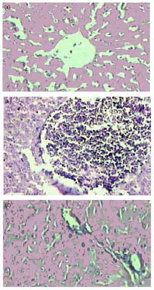 | |
| Fig. 1: | Photomicrograph of liver section of rats, control (a) and STZ (b), H. rosasinensis extract treated (c). The specimens were stained with Hematoxylin and Eosin |
Histological results
Liver: By light microscopy, liver of the STZ treated diabetic rats showed 2-3 foci of interlobular lymphocytes predominant inflammatory cells infiltration per x100 magnifications as compared by necrosis and apoptosis of few hepatocytes. Mild lymphocytic infiltration and congested vessel intlemajority of portal spaces were noted. Histological examination of livers of the diabetic rats treated by H. rosasinensis extract, showed gradual significant reduction in parenchymal and portal inflammation and lymphocytes were replaced by few eosinophils, the hepatic tissue appeared somewhat like the control and the Insulin treated groups (Fig. 1a-c).
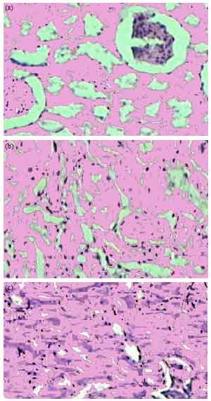 | |
| Fig. 2: | Photomicrograph of Kidney section of rats, control (a) and STZ (b), H. rosasinensis extract treated (c). The specimens were stained with Hematoxylin and Eosin |
Kidney: Histological examination of the STZ-induced diabetic rats’ renal tissue compared to the controls groups revealed mild increase in mesangial cells and matrix of glumeroli. Hyaline thickening of some arteriole wall was noted. By H. rosasinensis extract these pathologic changes improved toward to the Insulin treated groups (Fig. 2a-c).
DISCUSSION
The present investigation indicates the hypoglycaemic and protective effects of H. rosasinensis leaves in the liver and kidney of STZ-diabetic rats. We have observed a significant decrease in blood glucose in H. rosasinensis-treated diabetic rats, when compared with diabetic control rats. The optimum dosage (500 mg kg-1) was standardized and confirmed by a previous study with significant hypoglycaemic activity. The possible mechanism of H. rosasinensis hypoglycaemic action may be through potentiation of pancreatic secretion of insulin from β-cell of islets or due to enhanced transport of blood glucose to the peripheral tissue (Saravanan and Pari, 2008). This was clearly evidenced by the increased level of insulin in diabetic rats treated with H. rosasinensis.
Reduction in plasma total protein and albumin level was observed in diabetic rats and this is consistent with the results obtained by Bakris (1993) and Tuvemo et al. (1997). The decrease in protein and albumin may be due to microproteinuria and albuminuria which are important clinical markers of diabetic nephropathy (Mauer et al., 1981) and/or may be due to increased protein catabolism (Almdal and Vilstrup, 1988). The results of the present study demonstrated that the treatment of diabetic rats with the extract of H. rosasinensis caused a noticeable elevation in the plasma total protein and albumin levels as compared with their normal levels (Safiyeh et al., 2007). Such improvement of serum protein and albumin was previously observed after the oral administration of Balanites aegyptiaca (B. aegyptiaca) to experimentally diabetic rats (Mansour and Newairy, 2000). It has been established that insulin stimulates the incorporation of amino acids into proteins (Almdal and Vilstrup, 1988).
C-peptide and insulin are the products of the enzymatic cleavage of proinsulin and secreted into the circulation in equimolar concentrations. The measurement of both C-peptide and insulin levels have been reported to be a valuable index of insulin secretion rather than insulin alone. In this study, the plasma C-peptide and insulin levels were significantly higher in the H. rosasinensis than in the DM group.
The plasma levels of urea, uric acid and creatinine levels were measured, as DM also causes renal damage due to abnormal glucose regulation, including elevated glucose and glycosylated protein tissue levels, haemodynamic changes within the kidney tissue and increased oxidative stress (Aurell and Bjorck, 1992). The STZ-induced diabetic rats exhibited significantly higher plasma urea, uric acid and creatinine levels compared to the DM group. However, the H. rosasinensis supplement lowered these plasma values to a control range. A significant elevation in serum creatinine and urea levels indicate an impaired renal function of diabetic animals (Shinde and Goyal, 2003). Thus, it would appear that the H. rosasinensis leaves supplement lowered the plasma urea, uric acid and creatinine levels by enhancing the renal function that is generally impaired in diabetic rats. These results are in agreement with other previous studies on the mesocarp extract of B. aegyptiaca (Saeed et al., 1995) and herbal formulation D-400 (Dubey et al., 1994).
The increase in the activities of plasma AST, ALT and ALP indicated that DM may induce hepatic dysfunction. The enzymes directly associated with the conversion of amino acids to keto acids are AST and ALT and are increased in the diabetic condition. Begum and Shanmugasundaram (1978) also reported an increase in the activities of AST and ALT in the liver of diabetic animals. Treatment with H. rosasinensis or insulin normalised these enzyme activities. Similarly, increased activities of AST and ALT in the diabetic liver were also reported by Jorda et al. (1982). The increased protein catabolism accompanying gluconeogenesis and urea formation that are seen in the diabetic state might be responsible for the elevation of these tissue transaminases. The rise in the activity of ALT is due to hepatocellular damage and is usually accompanied by a rise in AST (Rao et al., 1989). This might be the reason for the elevated activities of these enzymes which were brought back to near normal value by H. rosasinensis treatment. This result shows the normalizing effects of H. rosasinensis on hepatocellular damage and suppression of gluconeogenesis. Elevated activity of ALP was observed in Elevated activity of ALP was observed in STZ-diabetic rats. Prince et al. (1997) have also reported increased ALP activity in experimentally diabetic rats. The increased activity of this enzyme in plasma may be a result of diabetes-induced damage to the tissues. H. rosasinensis treatment restored the activity of this enzyme to near normal by reducing its induction in DM. γ-GT catalyses the transfer of the γ-glutamyl group from γ-glutamyl peptides to another peptide or L-amino acids or to water. The assay of γ-GT is a helpful adjunct in detecting hepatic damage. A highly significant elevation in the activity of γ-GT was observed in plasma, liver and kidney of STZ-induced diabetic rats. This is in accord with earlier investigations (McLennan et al., 1991), where in a dramatic increase in γ-GT expression was found in the liver of diabetic rats. Elevated activity of γ-GT in plasma takes place as a result of hepatic induction of the enzyme. In addition, hepatocellular damage or cholestasis may also contribute to the elevation in the activity. Increased activity of γ-GT in STZ-induced diabetic rats was lowered to near normal by H. rosasinensis treatment that indicates the possible prevention of necrosis by H. rosasinensis treatment.
In conclusion, H. Rosasinensis Aerial part extract lowered blood glucose with a simultaneous increase in the plasma insulin and C-peptide levels. In addition, H. Rosasinensis extract could influence protein metabolism and marker enzymes in STZ-induced diabetic rats. Extract also protect liver and Kidney from damage due to diabetes (Zanna et al., 2008).
REFERENCES
- Aliyu, R., A.H. Adebayo, D. Gatsing and I.H. Garba, 2007. The effects of ethanolic leaf extract of Commiphora africana (Burseraceae) on rat liver and kidney functions. J. Pharmacol. Toxicol., 2: 373-379.
CrossRefDirect Link - Almdal, T.P. and H. Vilstrup, 1988. Strict insulin therapy normalises organ nitrogen contents and the capacity of urea nitrogen synthesis in experimental diabetes in rats. Diabetlogia, 31: 114-118.
CrossRefDirect Link - Gilani, A.H., S. Bashir, K.H. Janbaz and A.J. Shah, 2005. Presence of cholinergic and calcium channel blocking activities explains the traditional use of Hibiscus rosasinensis in constipation and diarrhoea. J. Ethnopharmacol., 102: 289-294.
CrossRefPubMedDirect Link - Asgary, S., G.A. Naderi, S.N. Zadegan and R. Vakili, 2002. The inhibitory effects of pure flavonoids on in vitro protein glycosylation. J. Herbal Pharmacother., 2: 47-55.
PubMedDirect Link - Bakris, G.L., 1997. Diabetic nephropathy. What you need to know to preserve kidney function. Postgrad. Med., 93: 89-100.
PubMed - Brownlee, M., 2001. Biochemistry and molecular cell biology of diabetic complications. Nature, 414: 813-820.
CrossRefPubMedDirect Link - Dubey, G.P., S.P. Dixit and S. Alok, 1994. Alloxan-induced diabetes in rabbits and effect of herbal formulation D-400. Indian J. Pharmacol., 26: 225-226.
Direct Link - Jorda, A., M. Gomez, J. Cabo and S. Grisolia, 1982. Effect of streptozotocin diabetes on some urea cycle enzymes. Biochem. Biophys. Res. Commun., 106: 37-43.
CrossRef - Kasture, V.S., C.T. Chopde and V.K. Deshmukh, 2000. Anticonvulsive activity of Albizzia lebbeck, Hibiscus rosa sinesis and Butea monosperma in experimental animals. J. Ethnopharmacol., 71: 65-75.
CrossRefPubMedDirect Link - Krishnakumar, K., K.T. Augustii and P.L. Vijayammal, 1999. Hypoglycaemic and anti-oxidant activity of Salacia oblonga Wall. extract in streptozotocin-induced diabetic rats. Indian J. Physiol. Pharmacol., 43: 510-514.
Direct Link - Lowry, O.H., N.J. Rosebrough, A.L. Farr and R.J. Randall, 1951. Protein measurement with the folin phenol reagent. J. Biol. Chem., 193: 265-275.
CrossRefPubMedDirect Link - Mansour, H.A. and A.A. Newairy, 2000. Amelioration of impaired renal function associated with diabetes by Balanites aegyptiaca fruits in streptozotocin induced diabetic rats. J. Med. Res. Inst., 21: 115-125.
Direct Link - Mauer, S.M., M.W. Steffes and D.M. Brown, 1981. The kidney in diabetes. Am. J. Med., 70: 603-612.
CrossRefPubMedDirect Link - Prince, P.S.M., V.P. Menon and L. Pari, 1997. Effect of Syzigium cumini extracts on hepatic hexokinase and glucose-6-phosphatase in experimental diabetes. Phytother. Res., 2: 529-531.
Direct Link - Saxena, A. and N.K. Vikram, 2004. Role of selected Indian plants in management of type 2 diabetes: A review. J. Altern. Complement Med., 10: 369-378.
CrossRefDirect Link - Shinde, U.A. and R.K. Goyal, 2003. Effect of chromium picolinate on histopathological alterations in STZ and neonatal STZ diabetic rats. J. Cell. Mol. Med., 7: 322-329.
CrossRef - Thirunavukkarasu, V., C.V. Anuradha and P. Viswanathan, 2003. Protective effect of fenugreek (Trigonella foenum graecum) seeds in experimental ethanol toxicity. Phytother. Res., 17: 737-743.
CrossRefPubMedDirect Link - Tuvemo, T., U. Ewald, M. Kobbah and L.A. Proos, 1997. Serum magnesium and protein concentrations during the first five years of insulin-dependent diabetes in children. Acta Paediatrica, 86: 7-10.
CrossRefPubMedDirect Link - Udoh, A.E., I. Ntu, O. Essien and M. Ndon, 2007. Red cell catalase activity in diabetics. Pak. J. Nutr., 6: 511-515.
CrossRefDirect Link - Virella-Lopes, M.F. and G. Virella, 2003. The role of immune and inflammatory processes in the development of macrovascular disease in diabetes. Fron. Biosci., 8: 750-768.
Direct Link - Dandu, A.M. and N.M. Inamdar, 2008. Protective effects of Andrographis paniculata against endothelial dysfunction in diabetic wistar rats. J. Pharmacol. Toxicol., 3: 311-317.
CrossRefDirect Link - Atangwho, J.J., P.E. Ebong, M.U. Eteng, E.U. Eyong and A.U. Obi, 2007. Effect of Vernonia amygdalina del leaf on kidney function of diabetic rats. Int. J. Pharmacol., 3: 143-148.
CrossRefDirect Link - Al-Attar, A.M. and T.A. Zari, 2007. Modulatory effects of ginger and clove oils on physiological responses in streptozotocin-induced diabetic rats. Int. J. Pharmacol., 3: 34-40.
CrossRefDirect Link - Rahman, M.W., M. Mostofa, S.A. Sardar, M.R. Sultana, M.M. Haque and M.E. Choudhury, 2005. Investigation of comparative hypoglycemic effect of neem (Azadirachta indica), karala (Momordica charantea) and nayantara (Cathranthus roseus) with glibenclamide on rat. Int. J. Pharmacol., 1: 257-260.
CrossRefDirect Link - Saravanan, G. and L. Pari, 2008. Hypoglycaemic and antihyperglycaemic effect of Syzygium cumini bark in streptozotocin-induced diabetic rats. J. Pharmacol. Toxicol., 3: 1-10.
CrossRefDirect Link - Safiyeh, S., F. Fathallah, N. Vahid, S.S. Habib and N. Nabat, 2007. Effect of Equisetum arvense L. (Equisetaceae) in microalbuminuria and creatinine excretion in streptozotocin-induced diabetes in male rats. Int. J. Pharmacol., 3: 155-159.
CrossRefDirect Link - Zanna, H., S. Adeniji, B.B. Shehu, S. Modu and G.M. Ishaq, 2008. Effects of aqueous suspension of the root of Hyphaene thebaica (L.) mart on some indicators of liver and kidney function in rats. J. Pharmacol. Toxicol., 3: 330-334.
CrossRefDirect Link - Rathi, A., C.V. Rao, S. Khatoon and S. Mehrotra, 2003. Ethnopharmacological evaluation of Peristrophe bicalyculata Nees for anti-inflammatory and analgesic activity. Nat. Prod. Sci., 9: 195-199.
Direct Link - Rao, G.M., L.O. Morghmom, M.N. Kabur, B.M. Ben Mohamed and K. Ashibani, 1989. Serum Glutamic Oxaloacetic Transaminase (GOT) and Glutamic Pyruvic Transaminase (GPT) levels in diabetes mellitus. Indian J. Med. Sci., 43: 118-121.
PubMed - Rao, B.K. and C.H. Rao, 2001. Hypoglycemic and antihyperglycemic activity of Syzygium alternifolium (Wt.) Walp. seed extracts in normal and diabetic rats. Phytomedicine, 8: 88-93.
CrossRefPubMedDirect Link