
K. Nirmala
Food and Drug Toxicology Research Centre, National Institute of Nutrition (ICMR), PO-Jamai Osmania, Hyderabad-500 007, India
T. Prasanna Krishna
Food and Drug Toxicology Research Centre, National Institute of Nutrition (ICMR), PO-Jamai Osmania, Hyderabad-500 007, India
K. Polasa
Food and Drug Toxicology Research Centre, National Institute of Nutrition (ICMR), PO-Jamai Osmania, Hyderabad-500 007, India
International Journal of Cancer Research
Year: 2007 | Volume: 3 | Issue: 1 | Page No.: 13-24


ABSTRACT
The present study involved the recognition of the effect of aqueous extract of ginger to prevent carcinogen-induced genotoxicity in human peripheral blood lymphocytes ex-in vivo by single cell gel electrophoresis technique in the presence and absence of ginger extract. Blood was obtained from 30 human volunteers of which 10 were apparently normal male non-smokers, 10 male smokers and 10 females. Aqueous ginger extract at three concentrations namely 2.5, 5 and 10 μg were tested for protective effect against B(a)P (300 μM) induced DNA damage. The results indicated that DNA damage expressed as comet ratio was decreased in samples treated with only B(a)P. When ginger extracts were present in incubation mixtures the comet ratios have increased depending on the dose of ginger extract. At the highest concentration of ginger extract namely 10 μg, the comet ratios were similar to their respective controls demonstrating the protective effect of ginger on carcinogen induced DNA damage.
PDF Abstract XML References Citation
How to cite this article
K. Nirmala, T. Prasanna Krishna and K. Polasa, 2007. Protective Effect of Ginger Against Benzo(a)pyrene Induced DNA Damage. International Journal of Cancer Research, 3: 13-24.
DOI: 10.3923/ijcr.2007.13.24
URL: https://scialert.net/abstract/?doi=ijcr.2007.13.24
DOI: 10.3923/ijcr.2007.13.24
URL: https://scialert.net/abstract/?doi=ijcr.2007.13.24
INTRODUCTION
Considerable attention has been focused on identifying naturally occurring chemopreventive substances capable of inhibiting, retarding or reversing the multistage carcinogenesis. Wide ranges of phenolic substances, particularly those present in dietary and medicinal plants, have been reported to possess substantial anticarcinogenic and antimutagenic activities. Cancer chemo-preventive potential of naturally occurring phytochemicals present in cruciferous, alliums and other medicinal plants is of great interest. These substances have great potential to protect against human cancer (Park and Pezzuto, 2002).
The increasing use of molecular genetic techniques, micro arrays, proteomics, development of human biomonitoring methods help in understanding the relative role of different dietary factors and genetic factors in human cancers (Ferguson, 2002). Some of the spices are rich sources of phytochemicals which have chemopreventive properties.
Spices are the dried aromatic parts of plants generally the seeds,berries,roots,pods and sometimes leaves. Given the wide range of species and plant parts from which spices are derived, they can contribute significant variety and complexity to the human diet. In the past uses of spices and herbs were often indistinguishable and they were recognized for their value of phytochemicals in relation to health (Lampe, 2003). For thousands of years spices have been used as flavourants, medicines and dyes. In The Ginger Book, Fulder (1996) lists nine reasons for adding spices to foods and hence it is important that spices can be considered the royal family of food and in this hot and passionate family, ginger sits next to the throne.
Today, ginger is considered as one of the world’s favourite spice. Ginger is one of the 1400 species of the Zingiberaceae family which includes spices such as turmeric and cardamom. Ginger (Zingiber officinale roscoe) is one of the most heavily consumed dietary substances in the world (Surh, 1999). Some pungent constituents present in ginger showed potent antioxidant and anti-inflammatory activity in experimental carcinogenesis (Katiyar et al., 1996; Lee and Surh, 1998; Park et al., 1998).
The prevention of diet related cancer is an important area in light of the frequency and severity of the disease (Surh, 2003). It is expected that antiproliferative and antimutagenic properties of specified food ingredients may play an inhibitory role during the process of carcinogenesis. The anticarcinogenic role of nutrients as well as non-nutrient dietary components needs to be explored more extensively (De and Ganguly, 2003).
In this context, we have studied the effect of ginger to prevent carcinogen-induced genotoxicity in human peripheral blood lymphocytes ex-in vivo by single cell gel electrophoresis technique (SCGE) also called as comet assay. Using this assay system, it is possible not only to detect potential antigenotoxins, but also more closely study the conditions under which protective activities can be preferably achieved. The antioxidative status of all the subjects was assessed by quantification of vitamin C, vitamin A and vitamin E. Since GST Mu is one of the determinants in host susceptibility to some types of cancers, its activity was also measured in all the subjects recruited for the study.
MATERIALS AND METHODS
Ten grams of cleaned fresh ginger was made into paste using two volumes of water and kept for shaking overnight. Later it was filtered and the supernatant was collected and the aqueous extract was lyophilized. This extract was used in the genotoxicity assay by diluting it to 1 mg mL-1 and from this 2.5, 5 and 10 μL was taken for the assay which corresponds to 2.5, 5 and 10 μg of ginger extract, respectively.
Chemicals and reagents
Normal Melting Point Agarose (NMPA), Low Melting Point Agarose (LMPA), proteinase-K, sodium lauroyl sarcosinate, D-glucose, ethidium bromide, benzo(a)pyrene, tris hydroxy methyl amino methane (Sigma Chemical Co.), RPMI 1640 medium, potassium chloride (KCl), dihydrogen potassium phosphate (KH2PO4), sodium chloride (NaCl), disodium hydrogen phosphate (Na2HPO4), sodium hydroxide (NaOH), Ethylene Diamine Tetra Acetic Acid (EDTA), sodium hydrogen carbonate (NaHCO3), 8-hydroxy guanosine, trypan blue, Dimethyl Sulfoxide (DMSO) and absolute alcohol were obtained from Qualigens and Spectrochem Ltd. India. Fully frosted slides and coverslips were obtained from Blue star Ltd., India.
Retinol (Vit.A), Retinyl acetate (internal standard), α- tocopherol (Vit. E), Ascrobic acid (Vit C) and α-bipyridyl were purchased from Sigma Chemical Co.
HPLC grade methanol, water and n-Hexane, Trichloroacetic acid (TCA), orthophosporic acid and ferric chloride were obtained from Qualigens and Spectrochem Ltd., India. Radioactive trans stilbene oxide (3H TSO) was obtained from American Radio Labelled Chemicals, Inc., St. Louis, MO, USA. Trans Stilbene Oxide (TSO), glutathione (GSH), hexanol and histopaque were obtained from Sigma Chemical Co. Napthalene, PPO and Dioxan were purchased from E.Merck Ltd.
Study Design
Blood was obtained from 30 human volunteers of which 10 were apparently normal male non-smokers,10 male smokers and 10 females aged between 35-55 years. Whole blood was used for treatment with benzo(a)pyrene in the absence and presence of various concentrations of ginger extract namely 2.5, 5 and 10 μg, respectively. Besides solvent control, normal controls were also included for each treatment and for each subject. A concentration of 300 μM of B(a)P was observed to produce significant damage and so the test concentration was fixed at 300 μM. Plasma was separated and used for the estimation of vitamin A, vitamin E and vitamin C. Lymphocytes were isolated for the estimation of GST Mu isozyme activity.
Comet Assay
The comet assay technique (Singh et al., 1988) was followed with slight modifications. Frosted slides were prelayered with 0.5% normal agarose. Forty microliter of heparinised blood in RPMI medium was incubated for 20 min with 300 μM of benzo(a)pyrene in the absence and presence of 2.5, 5 and 10 μg of ginger extract and later mixed with 75 μL of 0.5% low melting agarose and spread on the prelayered slides. After the solidification of the second layer a third layer of 100 μL of low melting agarose was added. After solidification of third layer the slides were immersed in lysing solution.
Testing for Cell Viability
Cell suspension from all the treatment groups were mixed with trypan blue (0.4% solution W/V in 0.85% saline). They were allowed to stand for 5-10 min and placed in haemocytometer. The percentage of cells that excluded trypan blue indicated viability.
The percentage viability = No. of unstained cells /total no.of cells countedx100 |
This test was done before proceeding the assay.
Micronutrients
Preparation of Standards
Vitamin A, vitamin E and internal standard were prepared by dissolving a minute quantity of the standard in a small volume of absolute alcohol. Absorbances were read in the spectrophotometer and their concentrations were calculated using the Molar extinction coefficients E1%1 cm (for vitamin A 1780 at 325 nm, internal standard 1550 at 326 nm and vitamin E 75.8 at 290 nm, respectively).
Analysis of Micronutrients in Plasma
Fresh plasma was obtained from the heparinised blood and immediately was frozen in liquid nitrogen and stored at -80°C until analysis. Each sample was analysed in duplicates for vitamin A and vitamin E and vitamin C. Retinol (Vitamin A) and α-tocopherol (Vitamin E) was measured simultaneously using HPLC by the method of Bieri et al. (1979).
Estimation of Vitamin A and Vitamin E
Vitamin A and vitamin E were estimated simultaneously using Shimadzu HPLC-7A at wave lengths 326 and 290nm respectively. Retinyl acetate was used as internal standard. The plasma samples were processed by precipitating the proteins with absolute alcohol and extracted with n-Hexane. After centrifugation, the hexane layer was removed and evaporated and finally reconstituted in mobile phase (95:5 methanol:water) and injected on to HPLC column (Reverse phase μ Bondapak c-18) with a solvent flow rate of 1.5 mL min-1. Peak height ratios of standard/internal standard were calculated with the peak areas. Concentrations of vitamin A and vitamin E were calculated against the standards.
Estimation of Vitamin C
Vitamin C was estimated in plasma according to the method of Zannoni et al. (1974). Fresh plasma (0.1 mL) was deproteinised with cold TCA (40%) in a ratio of 0.1 mL plasma to 0.012 mL of TCA (40%). It was allowed to stand for 10 min in ice followed by centrifugation at 15000 g for 15 min. The acidified supernatant fraction was removed for analysis. A reagent blank was run along with the standard and samples. Ascorbic acid standard concentrations ranging from 0.6 to 3.0 μg was taken and the standard was prepared in 5% TCA. The following reagents were added to both blank and samples along with standards in the following order: 0.01 mL of 85% orthophosphoric acid, 0.08 mL of 1% aqueous α,α-bipyridyl and 0.01 mL of 3% aqueous ferric chloride. The final volume in blank, standards and samples was 0.25 mL. The tubes were mixed thoroughly after each addition and were kept for nearly 15 min. for the colour to develop and were finally read at 525 nm in Perkin Elmer spectrophotometer.
Lymphocyte GST Mu Activity
GST isozyme class Mu from human lymphocytes have been shown to be dominantly inherited and can be determined by activity measurement directed towards the substrate Trans Stilbene Oxide (TSO). Cytochrome P450 enzymes catalyses the oxidation of PAH’s to electrophilic intermediates capable of possibly initiating the carcinogenic process. Alternatively the electrophilic intermediates may undergo conjugation or hydration reactions catalysed by GST and epoxide hydrolase producing hydrophilic compounds that are excreted from the body (Warholm et al., 1983).
Human Lymphocyte Isolation
From peripheral blood drawn by vein puncture, mononuclear leucocyte fraction was isolated by gradient separation using a Ficoll-paque procedure. To 3 mL of histopaque in a test tube, 3 mL of whole blood was added slowly through the sides without disturbing. The tubes were centrifuged at 1500 rpm for 15 min. The lymphocyte layer was drawn into a tube and PBS (pH 7.2) was added and centrifuged to get the lymphocyte sediment.
2x10⁶ lymphocytes (in 200 μL PBS) were sonicated in a Branson sonifier for sometime. 50 μL of lymphocytes were incubated at 37°C with 40 μL of 250 mM of sodium phosphate buffer, pH, 7.2 containing 4mM reduced glutathione and 250 μM tritiated TSO. After incubation of 30 min. the reaction was terminated by extraction with hexanol. 50 μL of aqueous phase was mixed with 10 mL of Brays mixture and counted in a liquid scintillation counter. Protein estimation was done. GST Mu activity was expressed as p.mol.conjugated/ min/mg prot. Levels of enzyme activity whose values were>50 pmoles were classified as GST Mu+ subjects.
Statistical Analysis
The data was analyzed by the analysis of variance (ANOVA). Testing of mean values in different groups was done by Duncan’s multiple range test. SPSS 11.5 Window version was used for the statistical analysis. (Middle Brooks, 1977).
RESULTS
Comet Assay
The length of the comet (L) and the cell diameter (D) was measured in 50 cells for each treatment and for each individual and the group means±SD were calculated (Table 1). The protective effect of ginger against B(a)P induced DNA damage in male smokers is given in Fig. 1. There was a significant difference (p<0.001) between the basal value and B(a)P treatment. In the presence of ginger the extent of DNA damage was decreasing with the increasing concentrations of ginger.
| Table 1: | Micronutrients in smokers, non smokers and females and GST μ status |
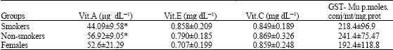 | |
| Values are Mean±SD, No. of subjects: 10 in each group, *Smokers Vs Non-smokers p<0.05 | |
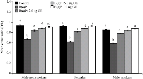 | |
| Fig. 1: | Inhibitory effect of ginger on B(a)P induced DNA damage in human peripheral blood lymphocytes. Values are Mean±SE of 10 subjects per group GE: Ginger extract. B(a)P: Benzo(a)pyrene 300 μM.Values bearing different superscripts are significant at p<0.001 by ANOVA |
The DNA damage in terms of Mean±SD of comet D/L ratio in lymphocytes obtained from male smokers and non-smokers and treated with B(a)P with and without ginger is shown in Fig. 1. There was some difference between untreated cell samples of male smokers and non-smokers. The damage to DNA was more in smokers than non-smokers. The inhibition potential of ginger against B(a)P induced DNA damage was significantly higher in both the groups for all the concentrations of ginger (p<0.001).
B(a)P induced significant damage in females (Fig. 1). The extent of damage was higher in smokers (females, mean = 0.620±0.021; male non smokers, mean = 0.667±0.046, male smokers, mean = 0.587±0.039) as compared to either group of subjects (p<0.05). Ginger could counteract the cell damage at all the concentrations (2.5, 5 and 10 μg).
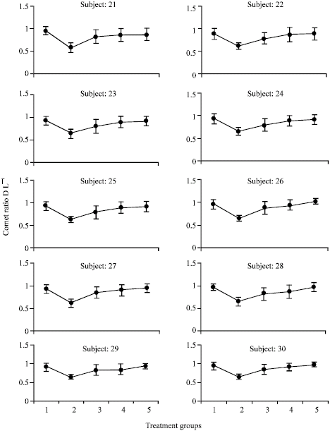
In all the three groups ginger showed a dose dependent inhibitory effect. Higher protective effect was observed in cells obtained from females compared to other groups. However maximum repair was noted in all the groups with 10 μg of ginger extract. The line graph of each individual of all the groups is being represented in Fig. 2-4.
GST μ Status
The subjects were categrorised as GST-Mu positive or negative. One individual (smoker) was negative for GST Mu and the rest of all the group subjects were positive for GST Mu expression. Mean comet ratios between treatment groups were analyzed by analysis of variance to ascertain the homogeneity. To test the difference between treatments, Duncan's multiple range test was used. It was observed that ginger showed similar protective effect in all the individuals irrespective of their GST-Mu status. The values of GST-μ expressed as P moles conjugated/mt/mg protein are shown in Table 1.
Assessment of Antioxidant Vitamin Status
The plasma vitamin levels were significantly different between smokers Vs non smokers (p<0.05). Females had low levels of vitamin E compared to smokers and non smokers, though statistically not significant (Table 1). However no differences were found in the plasma vitamin C levels in all the groups.
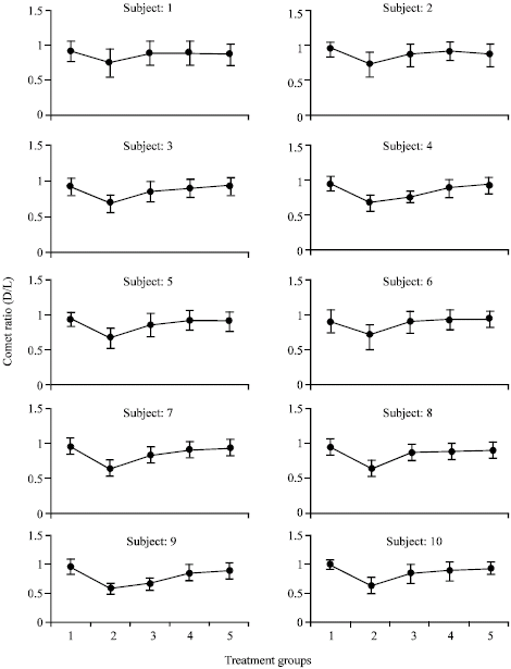 | |
| Fig. 2: | Line graph for each individual among male non-smokers. Values are Mean±SD. Treatment groups: 1)Control; 2) B(a)P; 3) B(a)P+2.5 μg GE; (4) B(a)P+5.0 μg GE; 5) B(a)P+10 μg GE B(a)P: 300 μM |
DISCUSSION
There is an increasing interest in reliable biological markers of exposure to genotoxicants. The damaging effects of environmentally occurring genotoxicants including diet and other life style factors result in the formation of reactive oxygen species which have damaging effects on DNA leading to genomic instabilities that are implicated in the etiology of cancer (Limoli et al., 1997). Epidemiological evidences suggest that higher consumption of fruits and vegetables lower the incidence of cancer (World Cancer Research Fund, 1997). These foods contain relatively high amounts of components with inherent antioxidant properties, such as vitamin C, carotenoids and flavonoids suggesting that dietary antioxidants may augment cellular antioxidant defenses and help to protect cellular components from oxidative damage.
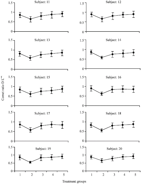 | |
| Fig. 3: | Line graph for each individual among male smokers. Values are Mean±SD. Treatment groups: 1) Control; 2) B(a)P; 3) B(a)P+2.5 μg GE; (4) B(a)P+5.0 μg GE; 5) B(a)P+10 μg GE B(a)P: 300 μM |
However experimental data confirming the physiological significance of this antioxidant effect is required.
Gingerol, the major pungent principle isolated from ginger oleoresin has been reported to possess strong antioxidant property whereas zingerone, a less pungent analog exhibits weaker antioxidant activity. Gingerol also exerts inhibitory effect on xanthine oxidase (Chang et al., 1994). In a study conducted in Insulin Dependent Diabetes Mellitus patients, higher level of DNA damage and decreases in the mean levels of vitamin A, glutathione peroxidase and uric acid was observed suggesting that alterations in antioxidant status may increase the likelihood of free radical induced changes within the tissues (Hannon et al., 1998).
 | |
| Fig. 4: | Line graph for each individual among females Values are Mean±SD. Treatment groups: 1) Control; 2) B(a)P; 3) B(a)P+2.5 μg GE; (4) B(a)P+5.0 μg GE; 5) B(a)P+10 μg GE B(a)P: 300 μM |
In a report by Sardas et al. (2001), it was shown that vitamin E supplementation for 12 weeks to diabetic individuals reduced the oxidative damage when evaluated by comet assay. The single gel electrophoresis assay commonly referred to as the ‘comet assay ‘ is used as biomarker of DNA damage. This test required fewer number of cells and the damage can be visualized in a single cell. This test can also be applied to determine the DNA damage in cells under a variety of experimental conditions. This technique has been employed to investigate the antioxidant properties of various compounds including carotenoids, in vivo and in vitro (Torbergsen and Collins, 2000; Collins et al., 1998; Astley et al., 1994). In a study reported by Astley et al. (2002), supplementation of carotenoids to Molt 17 lymphocyte cultures did not protect against the oxidative damage induced by hydrogen peroxide as measured by comet assay. However, there was a rapid decrease in single strand breaks in carotenoid supplemented cells possibly due to increase in DNA repair enzymes.
Aqueous ginger extract at three concentrations namely 2.5, 5 and 10 μg were tested for protective effect against B(a)P (300 μM) induced DNA damage in human peripheral blood lymphocytes obtained from 10 male smokers,10 non-smokers and 10 females. The damage was measured as comets under alkaline condition. B(a)P, a well known commonly present environmental mutagen/carcinogen was employed to induce DNA damage. This agent is present in tobacco smoke and has been implicated to play a major role in the epidemiology of lung as well as cancer at other sites and variety of chronic degenerative diseases (Deflora et al., 2003).
Previous studies on human lymphocytes have shown that in smokers increased DNA damage was observed. However some have reported contradictory observations (Frenzille et al., 1997). Smoking leads to large quantity of free radicals and carcinogenic products (Ly et al., 1995). It has been reported that DNA damage could be due to cumulative effect on exposure to genotoxicants and reduction in antioxidant status in smokers (Wojewodzska et al., 1999; Hininger et al., 1997).
In this study, it was observed that B(a)P induced significant DNA damage which was quantitated in terms of comet ratios. Significant reduction in comet ratios was seen in all the groups in the lymphocytes as a result of treatment with B(a)P. However, higher DNA damage was seen in the smokers followed by females and non-smokers. When the cells were exposed to carcinogen plus ginger extract the DNA damage was inhibited in a dose dependent manner as observed by increases in the comet ratio with increasing levels of ginger extracts.
It is well known that B(a)P and its diol epoxides are known to bind to cellular macromolecules (Phillips, 1983). This event is one of the key factor in the process of carcinogenesis (Dipple, 1983). In vitro exposure to B(a)P has been known to induce primary DNA damage in many tissues (Fairbairn et al., 1995). Many chemopreventers are known to modulate the outcome of B(a)P mutagenicity by preventing its binding to cell macromolecules or by binding covalently to activated carcinogens masking the DNA binding sites for carcinogens (Deflora et al., 2003). The levels of drug metabolism enzymes and their activity determine the ultimate carcinogenic metabolites that binds to cellular macromolecules. It was observed that ginger feeding resulted stimulation in levels of Glutathione S-transferase and Quinone reductase in liver, lung, kidney and intestine. Therefore under in vivo condition further protective effect may be achieved by consuming ginger.
Active principles in spices/herbs act as strong antioxidants and help in minimizing the oxidative damage occurring in tissues as a result of exposure to a variety of xenobiotics present in the environment (Aruoma, 2003). In the present investigation, plasma levels of micronutrients namely vitamin A, vitamin E and vitamin C at all subjects were estimated. There was some correlation between the DNA damage and the levels in different groups (p<0.01). The findings of this study suggest that ginger possess strong antigenotoxic potential and it may be related to its antioxidant and antimutagenic potential.
More recently, the Mu form of GST, which has a polymorphic expression in human leukocytes, is shown to modify cancer risk. A correlation between low GST Mu activity and DNA adduct in lymphocytes of smokers has been reported (Sony et al.,1998). The attenuation of cytogenetic damage was better expressed in N-Acetyl cysteine (NAC) treated human smokers who were either N-Acetyl transferase (NAT2) slow acetylators or GST M1 negative subject suggesting that pharmacogenomic approach can be adopted in chemoprevention research (Deflora et al., 2003). In this study, only one subject is GST Mu negative. Therefore, no conclusion could be made on relationship of GST Mu status and individual DNA damage.
Assessment of genotoxicity/antigenotoxicity can be performed at different steps of the interaction of the effects of the mutagen on DNA. The direct damage to DNA can be assessed by comet assay, but depending on the stage of cell cycle, repair capacity, genetic background of cells and type of mutagen, only a fraction of induced DNA damage will lead to fixed mutations.
In a study reported by Weidner and Sigwart (2001), it was observed that administration of ginger extract by oral gavage at 100, 333 and 1000 mg kg-1 to rats had no teratogenic and toxic effects and was reported to be pharmacologically safe.
In usual culinary practices, spices are subjected to thermal changes during cooking. Spices namely turmeric, curcumin, garlic, onion were tested for their antimutagenicity after subjecting them to boiling and frying to explore if their beneficial effects are destroyed by SOS chromotest using E. Coli PQ37. It was observed that all the spices tested exhibited antimutagenic property following heating procedures indicating that cooking conditions are not likely to destroy the antimutagenic potentials (Polasa et al., 1997; Polasa, 2000). Antioxidant activity of spices extract namely cloves, cinnamon, pepper, ginger, garlic, mint and onion were found to retain their antioxidant potential even after boiling for 30 min, at 100°C indicating that the spice constituents were resistant to thermal denaturation (Shobana and Naidu, 2000). Therefore intake of diet containing even low levels of different naturally occurring non-nutrients can be effective in exerting antigenotoxic effects and offset the oxidative stress occurring due to low level of antioxidant nutrients. Consumption of natural phytochemicals through balanced diet containing functional foods would be more cost effective than supplementation of individual antioxidants for protection against genotoxic stress.
Ginger has been proven to be possessing antigenotoxic potential using Comet Assay. However the minimum effective protective level in comet assay was 2.5 μg of ginger extract corresponding to 0.8 g of ginger. Therefore, comet assay appears to be a sensitive method of assaying the effective dose as has been reported (Goethem et al., 1997).
ACKNOWLEDGMENT
The authors wish to thank Spices Board, Cochin, India for supporting in part by grants.
REFERENCES
- Aruoma, O.I., 2003. Methodological considerations for characterizing potential antioxidant actions of bioactive components in plant foods. Mutat. Res./Fundam. Mol. Mech. Mutagen., 523-524: 9-20.
CrossRefDirect Link - Astley, S.B., R.H. Elliot, D.B. Archer and S. Southon, 2002. Increased cellular carotenoid levels reduce the persistence of DNA single stranded breaks after oxidative challenge. Nutr. Cancer, 43: 202-213.
Direct Link - Bieri, T.G., T.J. Tolliver and G.L. Catignani, 1979. Simultaneous determination of α-tocopherol and retinol in plasma or red cells by high pressure liquid chromatography. Am. J. Clin. Nutr., 32: 2143-2149.
Direct Link - Chang, W.S., Y.H. Chang, F.J. Lu and H.C. Chiang, 1994. Inhibitory effects of phenolics on xanthine oxidase. Anticancer Res., 14: 501-506.
Direct Link - Collins, A., B. Olmedilla, S. Southon, F. Granado and S.J. Duthie, 1998. Serum carotenoids and oxidative DNA damage in human lymphocytes. Carcinogenesis, 19: 2159-2162.
Direct Link - Deflora, S., F.D. Agostini, R. Balansky, A. Camorians, C. Bennicelli, M. Bagnasco, C. Cartiglia, E. Tampa, M.G. Longobardi, R.A. Lubet and A. Izzot, 2003. Modulation of cigarette smoke-related end points in mutagenesis and carcinogenesis. Mut. Res., 523-524: 237-252.
Direct Link - De, S. and C. Ganguly, 2003. Natural dietary agents can protect against DMBA genotoxicity in lymphocytes as revealed by single cell gel electrophoresis assay. Teratog. Carcinog. Mutagen., 23: 71-78.
Direct Link - Dipple, A., 1983. Formation, metabolism and mechanism of action of polycyclic aromatic hydrocarbons. Cancer Res., 43: 2422S-2425S.
PubMed - Fairbairn, D.W., P.C. Olive and K.L. O'Neill, 1995. The comet assay: A comprehensive review. Mut. Res./Rev. Genet. Toxicol., 339: 37-59.
CrossRefPubMedDirect Link - Ferguson, L.R., 2002. Natural and human made mutagens and carcinogens in the human diet. Toxicology, 181: 79-82.
Direct Link - Frenzilli, G., C. Betti, T. Davini, M. Desideri and E. Fornai et al., 1997. Evaluation of DNA damage in leukocytes of ex-smokers by single cell gel electrophoresis. Mutation Res./Fundam. Mol. Mech. Mutagen., 375: 117-123.
CrossRefPubMedDirect Link - Goethem, V.F., D. Lison and M. Kirsch-Volders, 1997. Comparative evaluation of the in vitro micronucleus test and the alkaline cell gel electrophoresis assay for the detection of DNA damaging agents: Genotoxic effects of cobalt power, tungsten carbide and cobalt-tungsten carbide. Mut. Res., 392: 31-43.
- Hannon, M.P.A., C. Hughes, M.J.O. Kana, K.W. Moles, C.R. Barnett and Y. Barnett, 1998. Antioxidant status and DNA damage in patients with insulin dependent Diabetes Mellitus. Biochem. Soc. Trans., 26: S57-S57.
Direct Link - Hininger, I., M. Chopra, D. Thurnam, F. Laporte, M.J. Richard, A. Favier and A.M. Roussel, 1997. Effect of increased fruit and vegetable intake on the susceptibility of lipoprotein in smokers. Eur. J. Clin. Nutr., 51: 601-606.
Direct Link - Katiyar, S.K., R. Agarwal and H. Mukhtar, 1996. Inhibition of tumor promotion in SENCAR mouse skin by ethanol extract of Zingiber officinale rhizome. Cancer Res., 56: 1023-1030.
Direct Link - Johanna, W.L., 2003. Spicing up a vegetarian diet: Chemopreventive effects of phytochemicals. Am. J. Clin. Nutr., 78: 579S-583S.
Direct Link - Lee, E. and Y.J. Surh, 1998. Induction of apoptosis in HL-60 cells by pungent vanilloids, [6]-gingerol and [6]-paradol. Cancer Lett., 134: 163-168.
CrossRefPubMedDirect Link - Limoli, C.L., M.L. Kaplan, J.W. Philips, G.M. Adair and W.F. Morgan, 1997. Differential induction of Chromosomal instability by DNA strand-breaking agents. Cancer Res., 57: 4048-4056.
Direct Link - Ly, Z., K. Stone and W.A. Pryor, 1995. Detection of free radicals in aqueous extracts of cigarette tar by electron spin resonance. Free Radic. Biol. Med., 19: 161-167.
Direct Link - Park, K.K., K.S. Chun, J.M. Lee, S.S. Lee and Y.J. Surh, 1998. Inhibitory effects of [6]-gingerol, a major pungent principle of ginger, on phorbol ester-induced inflammation, epidermal ornithine decarboxylase activity and skin tumor promotion in ICR mice. Cancer Lett., 129: 139-144.
CrossRefPubMedDirect Link - Park, E.J. and J.M. Pezzuto, 2002. Botanicals in cancer chemoprevention. Cancer Metastasis Rev., 21: 231-255.
CrossRefDirect Link - Sardas, S., M. Yilmaz, U. Oztok, N. Cakir and A.E. Karakaya, 2001. Assessment of DNA strand breakage by comet assay in diabetic patients and the role of antioxidant supplementation. Mut. Res., 490: 123-129.
Direct Link - Shobana, S. and K.A. Naidu, 2000. Antioxidant activity of selected Indian spices. Prostaglandins Leukot. Essent. Fatty Acids, 62: 107-110.
CrossRefPubMedDirect Link - Soni, M., M. Madurantakan and K. Krishnaswamy, 1998. Glutathione-s-transferase Mu (GST-Mu) deficiency and DNA adducts in lymphocytes of smokers. Toxicology, 126: 155-162.
Direct Link - Surh, Y.J., 1999. Molecular mechanisms of chemopreventive effects of selected dietary and medicinal phenolic substances. Mutat. Res., 428: 305-327.
Direct Link - Surh, Y.J., 2003. Cancer chemoprevention with dietary phytochemicals. Nat. Rev. Cancer, 3: 768-780.
CrossRefPubMedDirect Link - Torbergsen, A.C. and A.R. Collins, 2000. Recovery of human lymphocytes from oxidative DNA damage: The apparent enhancement of DNA repair by carotenoids is probably simply an antioxidant effect. Eur. J. Nutr., 39: 80-85.
Direct Link - Weidner, M.S. and K. Sigwart, 2001. Investigation of the teratogenic potential of a Zingiber extract in the rat. Reprod. Toxicol., 15: 75-79.
Direct Link - Wojewodzska, M., M. Kuszewski, T. Iwanenko, A.R. Collins and I. Szumiel, 1999. Lack of adverse effect on smoking habit on DNA strand breakage and base damage, as revealed by the alkaline comet assay. Mut. Res., 440: 19-25.
Direct Link - Zannoni, V., M. Lynch, S. Goldstein and P. Sato, 1974. A rapid micromethod for the determination of Ascorbic acid in plasma and tissues. Biochem. Med., 11: 41-48.
CrossRef - Singh, N.P., M.T. McCoy, R.R. Tice and E.L. Schneider, 1988. A simple technique for quantitation of low levels of DNA damage in individual cells. Exp. Cell Res., 175: 184-191.
CrossRefPubMedDirect Link