
Varsha Wankhade
Department of Zoology, University of Pune, Pune, MS, India
R.B. Andhale
Department of Zoology, University of Pune, Pune, MS, India
Sangita Lodha
Thalassemia Center, Nashik, India
Trends in Medical Research
Year: 2013 | Volume: 8 | Issue: 1 | Page No.: 27-31


ABSTRACT
In the present study, some cellular and crystalline components of urine of Sickle Cell Anemia (SCA) were studied. In total 67 samples were investigated. The normal and abnormal cellular components like presence of Red blood cells, White blood cells Epithelial cells, Renal tubules cells, Bacteria, Yeast and Protozoan were observed and counted. Noncellular components/crystals like Tyrosine, Cholesterol, A/T Phosphate, Leucine, B-Granule, Uric Acid, Oxalate and Cystine were counted. It was observed that RBCs, WBCs and Epithelial cells, Renal tubules cells, Bacteria, Yeast and Protozoan were in high number indicating various complications occurring in the body of sickle cell anemic patients. It was observed that RBCs were 13918.18/cumm±SE 4057.90 indicating tremendous hematourea, number of WBC were 224.59±50.002 SE, Sqaumous Epithelial cells were 35.68±10.989 SE, Renal cells were 110.68±74.15 SE. This indicates that SCA patients suffer from kidney damage, hematourea, Urinary Tract Inflammation, Interstitial nephritis, Glomerulonephritis and Pylonephritis.
PDF Abstract XML References Citation
Received: December 12, 2012;
Accepted: April 15, 2013;
Published: June 17, 2013
How to cite this article
Varsha Wankhade, R.B. Andhale and Sangita Lodha, 2013. Prediction of Physiological Status of Sickle Cell Anemic Patients by Quantitative Observations of Microscopic Components of Urine. Trends in Medical Research, 8: 27-31.
URL: https://scialert.net/abstract/?doi=tmr.2013.27.31
URL: https://scialert.net/abstract/?doi=tmr.2013.27.31
INTRODUCTION
Sickle Cell Disease (SCD) is one of the common genetic disorders affecting large population of the world. In sickle cell anemia RBCs become sickle shaped due to presence of abnormal Hemoglobin (HbS). SCD patients need regular blood transfusion. Based on the World Health Organization published global prevalence map of SCD and other data, it was estimated that about 20-25 million individuals worldwide have homozygous SCD; 12-15 million in sub-Saharan Africa, 5-10 million in India and about 3 million distributed in different parts of the world (Serjeant, 2006). It is estimated that each year over 300,000 babies with severe form of sickle cell disease are born worldwide. The majority of which is the middle and low income countries. About 5% of the population of world is carrier of gene for sickle cell disease, this percentage may vary and may be high up to 25% in some countries. A high concentration of the disease is in Asia, the Mediterranean basis, the Middle East and the Africa (http://www.who.int/genomics/public/Maphaemoglobin.pdf). Reports said that 10-30% tribal population of central India are affected by SCD. There may be 500000 and 1,000,000 cases of SCD in India (Adams et al., 1998; Alexy et al., 2005, 2006).
A variety of renal abnormalities have found to be associated with sickle cell (SS) disease (McCoy, 1969; Schlitt and Keitel, 1960; Sweeney et al., 1962). One of the common causes of death in adults from sickle cell disease are reported due to renal failure (Darbari et al., 2006). Kidney damage begins very early and progresses throughout life (Rossi-Espagnet et al., 1968; Eckman and Platt, 1991; Saborio and Scheinman, 1999).
Microscopic examination of urine is the cheapest and the easiest technique to determine the physiology of the kidney. By investigating urine’s components one can know the metabolic disturbances going on inside the various systems of the body. The objective of the present study was to know whether the quantitative analysis of urine of SCD generates information regarding the pathophysiological status of SCD. The purpose of the present study was to perform the quantitative analysis of normal and abnormal microscopic components of the urine samples of sickle cell anemic patients and correlate its association with the physiologic status of kidney in SCD. For this study we have selected the sickle cell population of district Amravati, Maharashtra, India. In the present study some cellular and crystalline components of urine of SCA were observed and counted.
MATERIALS AND METHODS
Study population: The study population consisted of 67 randomized Sickle cell diseased populations from district Amravati, Maharashtra, India. This study was approved by Institutional Human Ethics Committee. Written consent from each participant was taken.
Collection of sample: A clean catch urine sample was obtained. Urine sample was collected from the participants by the standard spontaneous voiding procedure as stated by Corwin (1996). Urine was collected at about noon to avoid contamination of urine by contents of urethra or vagina and therefore its constituents are more likely to reflect kidney origin. First void sample was also collected, because this is usually, a concentrated specimen and most informative. Urine was collected in a sterile, labeled bottle containing 4% formaldehyde. Urine was transported to the laboratory immediately in an ice bag. All the analysis were performed within a week (European Confederation of Laboratory Medicine, 2000; Skobe, 2004).
Preparation of urine sample: Ten milliliter of urine was centrifuged at 2,000 rpm for 5 min. Following aspiration of the supernatant to a marked level, the pellet was agitated into a homogeneous mixture followed by a sampling pipette provided by the manufacturer (Tarson). This was followed by microscopic analysis of urine using Magnus inclined trinocular microscope (Make-Olympus).
Microscopic analysis of urine: A drop of prepared urine sample was pipetted onto a Neubars chamber slide and a coverslip was placed. The slide was then examined. The urine sediment is routinely examined by bright field microscopy under both low and high power without staining. For the present study, prepared slide was observed under the Magnus inclined trinocular microscope (Make-Olympus). After that, RBCs, WBCs and ECs were examined using a High-power Field (HPF) and were counted.
The normal and abnormal cellular components like of RBCs, WBCs, Epithelial cells, Renal tubules cells, Bacteria, Yeast and Protozoan were counted and noted. Non-cellular components like Tyrosine, Cholesterol, A/T Phosphate, Leucine, B-Granule, Hyaline, Uric Acid, Oxalate and Cystine were also counted.
RESULTS AND DISCUSSION
Quantitative study of normal and abnormal cellular components and crystalline components of urine such as RBCs, WBCs, Epithelial cells, Renal tubules cells, Bacteria, Yeast and Protozoan was performed.
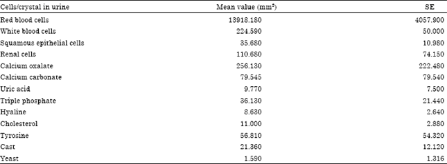
We observed that the number of cellular and crystalline component of urine is very high. It was observed that mean value of number of RBCs were 13918.18, WBCs were 224.59, Sqaumous Epithelial cells were 35.68182, Renal Cells were 110.6818 and Cholesterol were 11 per cumm. This indicates various complications in the kidney of sickle cell anemia. Quantitative analysis of Cellular and crystalline components of urine of SCA is shown in Table 1.
Crystalline components of urine in SCA: Non-cellular components like Tyrosine, Cholesterol, A/T Phosphate, Leucine, B-Granule, Hyaline, Uric Acid, Oxalate, Cystine were also counted in the urine sample of sickle cell anemic patients. The mean value of number of some crystals were very high in the urine. Uric acid crystals were 9.77, triple phosphate were 36.13, Tyrosine crystal 56.81, Casts were 21.36, Calcium oxalate were 256.13 and calcium carbonate were 79.54 per cumm area.
Microscopic analysis can help distinguish patients with Urinary Tract Infection or renal disease. In the present microscopic investigation on urine of SCD, we observed RBCs in high number indicating hematuria, may be because of the Glomerulonephritis, vasculitis and inflammation. White blood cells were also more in number which may appear in the urine in Interstial nephritis and pylonephritis. Epithelial cells are observed due to Acute tubular necrosis, interstitial nephritis and glomerulonephritis. Renal tubular cells were found due to renal tubular damage (Oser, 1976). Non-cellular/crystalline components in urine of SCD also express various abnormal conditions. Presence of large number of Uric acid, tyrosine, oxalate, A/T Phosphate, Hyaline etc. is the indication of acute nephropathy. Appearance of high cholesterol in urine indicates heavy protein urea due to improper filtration at Glomerulus (Oser, 1976). Presence of Cystine in the urine of SCD indicates the abnormal metabolic process in the body (Oser, 1976). On the basis of microscopic analysis of urine of SCA it could be concluded that the Kidney of such patients are under risk and suffering from various complications like nephrites, glomerular damage, hematourea etc.
| Table 1: | Quantitative analysis of microscopic components of urine of sickle cell anemic patients |
 | |
On the basis of cellular and non-cellular components of urine of SCA patients, probable physiological complications going on inside the important organs like kidney, liver, urinary bladder ect could be predicted. It was found that SCA patients suffered by various complications. Present study indicates that 69.7% patients of SCA suffers from Cystisis and Urinary Tract Inflammation, 96.9% patients suffer from Interstitial nephritis, Acute Tubular Necrosis and Glomerulonephritis. 66.7% SCA patients suffer from Pylonephritis which means inflammation of renal pelvis. 16% SCA patients suffer from Chyluria. In 15% SCA patients Atrophy and Cirrosis of liver could be seen while 53% patients suffers from liver dysfunction.
The renal concentrating defect in sickle cell anemia is apparently related to polymerization of sickle hemoglobin in the hyperosmolar renal medulla, which increases the concentration of hemoglobin within red cells. The ultimate result is almost complete destruction of the vasa recta and particularly disruption of the juxta medullary nephrons with long loops of Henle extending into the intramedullary zone (Miller et al., 2011),which results in an inability to super-concentrate urine in the presence of water deprivation (Van Eps et al., 1970). Studies examining the prevalence and severity of the renal concentrating defect in sickle cell disease were largely done in the 1950s by Kunz et al. (1954). Renal failure in SCD varies from 5-18% (Scheinman, 1994). Powars et al. (1991) reported 4.2% SCD patients affected with renal failure. Platt et al. (1994) observed 18% overall mortality in adult SCD patients with 40% of these due to renal failure. Renal thrombosis and intravascular hemolysis could be one cause of acute and renal insufficiency in SCD patients (Sklar et al., 1990). However, the renal abnormalities start at earlier ages (Tejani et al., 1985).
CONCLUSION
Quantitative study of Microscopic components of urine of SCD of district Amravati, MS, India exhibits high number of RBCs, WBCs, Sqaumous epithelial cells, renal tubular cells ect. Urine of the sickle cell anemic patients also shows high number of crystalline components in the urine. Present study shows that the SCA patients at district Amravati are under the threat of Glomerulonephritis, urinary tract infection, urinary tract inflammation, tubular necrosis, Interstitial nephritis, Pylonephritis etc. Thus we can conclude that the kidney of the SCA is in high risk, hence proper and timely precautions should be taken to lessen the damage to this important organ. SCA should be managed properly as soon as it is detected so that the vital organs could be protected and saved from deterioration.
ACKNOWLEDGMENTS
The authors are grateful to Board of College and University Development, University of Pune for financial support. First author is thankful to Centre for advanced studies and DST-PURSE for partial financial support. We are thankful to Thalassemia Center, Jankalyan Blood Bank, Nashik and Mr.Narendra Manwar, Department of Zoology, University of Pune for their co-operation throughout the research work.
REFERENCES
- Alexy, T., R.B. Wenby, E. Pais, L.J. Goldstein, W. Hogenauer and H.J. Meiselman, 2005. An automated tube-type blood viscometer: Validation studies. Biorheology, 42: 237-247.
PubMed - Darbari, D.S., P. Kple-Faget, J. Kwagyan, S. Rana, V.R. Gordeuk and O. Castro, 2006. Circumstances of death in adult sickle cell disease patients. Am. J. Hematol., 81: 858-863.
CrossRefDirect Link - Kunz, H.W., E.L. Pratt, G.W. Mellin and M.W. Cheung, 1954. Impairment of urinary concentration in sickle cell anemia. Pediatrics, 13: 352-356.
PubMed - McCoy, R.C., 1969. Ultrastructural alterations in the kidney of patients with sickle cell disease and the nephrotic syndrome. Lab. Invest., 21: 85-95.
PubMed - Platt, O.S., D.J. Brambilla, W.F. Rosse, P.F. Milner, O. Castro, M.H. Steinberg and P.P. Klug, 1994. Mortality in sickle cell disease-Life expectancy and risk factors for early death. N. Engl. J. Med., 330: 1639-1644.
Direct Link - Rossi-Espagnet, A., K.W. Newell, R. Maclennan, J.B. Mathison and S.P.H. Mandel, 1968. The relationship of sickle cell trait to variations in blood pressure. Am. J. Epidemiol., 88: 33-44.
PubMedDirect Link - Saborio, P. and J.I. Scheinman, 1999. Sickle cell nephropathy. J. Am. Soc. Nephrol., 10: 187-192.
Direct Link - European Confederation of Laboratory Medicine, 2000. European urinalysis guidelines. Scand. J. Clin. Lab. Invest. Suppl., 231: 1-86.
PubMedDirect Link - Schlitt, L.E. and H.G. Keitel, 1960. Renal manifestations of sickle cell disease: A review. Am. J. Med. Sci., 152: 773-778.
PubMedDirect Link - Serjeant, G.R., 2006. The case for dedicated sickle cell centres. Indian J. Hum. Genet., 12: 148-151.
CrossRefDirect Link - Sklar, A.H., J.C. Perez, R.J. Harp and R.J. Caruana, 1990. Acute renal failure in sickle cell anemia. Int. J. Artif. Organs, 13: 347-351.
PubMed - Skobe, C., 2004. The basics of specimen collection and handling of urine testing. LabNotes, Vol. 14, No. 2.
Direct Link - Sweeney, M.F., W.T. Dobbins and J.N. Etteldorf, 1962. Renal disease with elements of the nephrotic syndrome associated with sickle cell anemia. J. Pediatrics, 60: 42-51.
Direct Link - Tejani, A., K. Phadke, O. Adamson, A. Nicastri, C.K. Chen and D. Sen, 1985. Renal lesions in sickle cell nephropathy in children. Nephron, 39: 352-355.
PubMed