
C. K. Yap
Department of Biology, Faculty of Science, Universiti Putra Malaysia,
UPM 43400, Serdang, Selangor, Malaysia
F.B. Edward
Department of Biology, Faculty of Science, Universiti Putra Malaysia,
UPM 43400, Serdang, Selangor, Malaysia
S.G. Tan
Department of Biology, Faculty of Science, Universiti Putra Malaysia,
UPM 43400, Serdang, Selangor, Malaysia
Trends in Applied Sciences Research
Year: 2007 | Volume: 2 | Issue: 4 | Page No.: 284-294


ABSTRACT
In this study, the patterns of the distributions and redistributions of heavy metals in the different soft tissues of Perna viridis were determined. Crystalline style and muscle were found to be sensitive organs to pollution as evidenced by total disturbances in the patterns of metal occurrences. The present findings confirmed the use of different soft tissues as an effective way of monitoring the status of metal pollution in tropical coastal waters. Therefore, the distribution of metals in different parts of P. viridis is proposed as a good indicator of metal pollution in tropical coastal waters.
PDF Abstract XML References
How to cite this article
C. K. Yap, F.B. Edward and S.G. Tan, 2007. Determination of Heavy Metal Distributions in the Green-Lipped Mussel Perna viridis as Bioindicators of Heavy Metal Contamination in the Johore Straits and Senggarang, Peninsular Malaysia. Trends in Applied Sciences Research, 2: 284-294.
URL: https://scialert.net/abstract/?doi=tasr.2007.284.294
URL: https://scialert.net/abstract/?doi=tasr.2007.284.294
INTRODUCTION
Increasing anthropogenic activities nowadays have made heavy metal pollution in tropical coastal waters a major cause of concern from the ecotoxicological point of view. Efficient and practical approaches for assessing the problems caused by pollution are needed in order to find ways of overcoming them, therefore the use of biomonitors serves an important function due to the fact that they provide integrated measures of the bioavailable metals which are of ecotoxicological significance in a habitat (Philips and Rainbow, 1993; Rainbow, 1995; Rainbow et al., 2002, 2004; Yap et al., 2006a). The metals accumulated in the soft tissues of marine mussels can be a measure of the bioavailability of metals originating from both natural and anthropogenic sources (Rainbow et al., 2004; Yap et al., 2006a). However, the metal distribution in the different soft tissues is more informative in order to identify specific organs that may be particularly selective and sensitive to the accumulation of heavy metals (Pourang et al., 2004; Szefer et al., 1990). Information on the tissue distribution of metals also would allow insights into the factors governing trace metal concentrations in the different tissues of the molluscs (Depledge, 1989). There are a lot of factors including environmental, physiological and genetic ones which would directly or indirectly affect the distribution of heavy metal in the different tissues of molluscs (Yap et al., 2006a).
According to Couillard (1996), metallothionein in the soft tissues of mussels was associated with the distribution of Cd, Cu and Zn. Pourang and Amini (2001) also reported the roles of metallothionein in the distribution of trace metals in the shrimps Penaeus merguiensis and Metapenaues affinis. However, the influence of heavy metal pollution in the distribution of metals in the different soft tissues of bivalves has not been well studied. Therefore, the objective of this study was to determine the impact of heavy metal pollution in the distribution of Cu, Cd, Fe, Ni, Pb and Zn in the different soft tissues of P. viridis.
MATERIALS AND METHODS
Sample Preparation
Mussels were collected from the Straits of Johore and Senggarang during our sampling trips conducted from 10-12 August 2004. Descriptions of the sampling locations are presented in Table 1. The collected mussels were placed in plastic bags and stored in an ice compartment before being taken back to the Laboratory.
The mussels were carefully dissected into different soft tissues: gonad, gill, muscle, mantle, byssus, Crystalline Style (CS) and remainder (Yap et al., 2003a, 2003f, 2005a, 2004a, 2006a,b) Then, the pooled tissues were placed in aluminum foils and were dried in an oven for 72 h at 60°C to constant dry weights. The dried samples were then stored in clean plastics bags.
Digestion of Mussel Soft Tissues
About 0.5-0.7 g of dried tissues were weighed and placed in acid washed digestion tubes. Concentrated nitric acid (AnalaR grade, BDH 69%) was added to the digestion tube to digest the tissues (Yap et al., 2002, 2003g, 2004b,c, 2005d, 2006d). They were then placed in a digestion block at 40°C for 1 h and were then fully digested at 140°C for at least 2-3 h (Yap et al., 2002, 2003b, g, 2004b, c, 2005d, 2006d). After cooling, each sample was diluted to 40ml with double de-ionized water. The digested samples were then filtered through Whatman No. 1 (filter speed: medium) filter paper in a funnel into acid-washed pill boxes. Later, the pill boxes were stored in the refrigerator until used for metal determination.
Metal Determinations
All the samples stored in acid-washed pill boxes were determined for Cd, Cu, Fe, Ni, Pb and Zn by using an air-acetylene Perkin-ElmerTM flame atomic absorption spectrophotometer Model AAnalyst 800. Blank determinations were carried out for calibrations of the instrument. Standard solutions were prepared from 1000 ppm stock solutions provided by MERCK Titrisol for Cd, Cu, Fe, Ni, Pb and Zn and the data obtained from the AAS were presented in μg g-1 dry weight basis. Recoveries were done by using prepared standard solutions for each metal. The metal recoveries were satisfactory, being between 90-120%.
Quality Control and Quality Assurance
All the equipment and glassware were first acid washed in 10% nitric acid solution to avoid contamination. All the digested samples were stored in acid washed pill boxes and kept in a refrigerator until metal determination. The solutions were prepared by using double de-ionized water (USF Maxima, 18.2 MΏ cm-1). The Procedural blanks and quality control samples made from the standard solution for each metal were analyzed once in every ten samples to check for sample accuracy (Yap et al., 2002, 2003b, g, 2004b, c, 2005d, 2006d).
| Table 1: | Descriptions of the sampling locations |
 | |
Data Analysis
One-way ANOVA was applied to find the significance value of heavy metal concentrations between the different tissues; heavy metal concentrations between the five locations. The Statistical software, SPSS 12.0 for Windows was used for the data analysis.
RESULTS
CS, byssus and gill always exhibited high accumulations of heavy metal concentrations in all the five sampling locations (Table 2). Other tissues such as remainder, muscle and mantle also showed high concentrations of heavy metals but not in any consistent pattern as were shown by the CS and byssus. The Remainders were found to accumulate high concentrations of Cd in the Gelang Patah and Telok Jawa populations while high Pb was found in the Gelang Patah population. High concentrations of Cd and Pb were found in the muscle of P. viridis from the Senggarang and Telok Jawa populations, respectively. The Mantle accumulated high concentrations of Ni in Senibong.
| Table 2: | Heavy metals (Cu, Cd, Zn, Pb, Ni and Fe) concentrations (mean μg g-1 dw) in the different soft tissues of Perna viridis collected from Senggarang, Gelang Patah, Senibong, Telok Jawa and Kampung Masai |
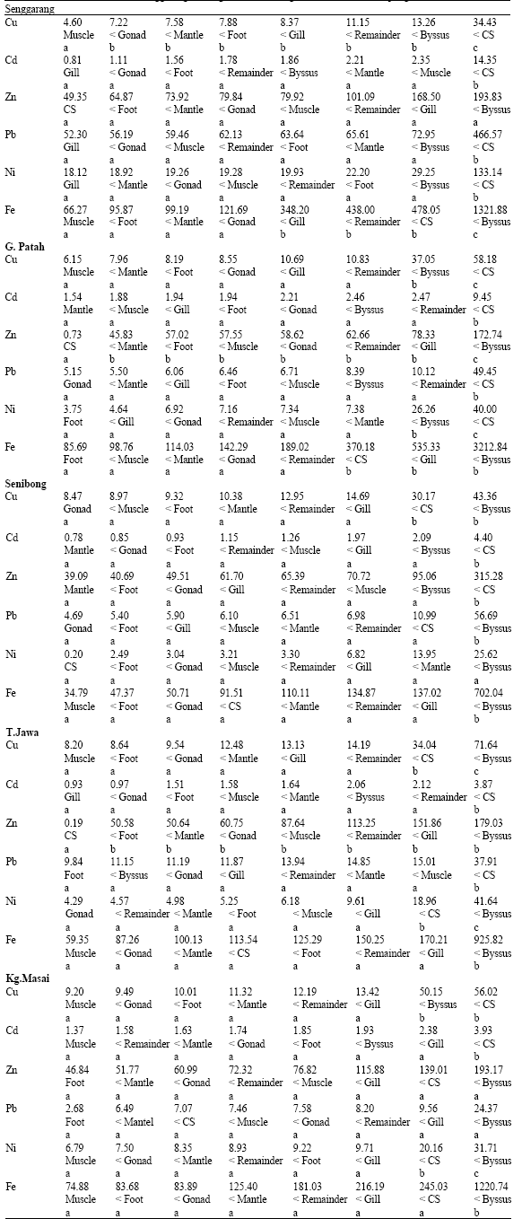 | |
| Objects indicated with different alphabet: p<0.05, Objects indicated with same alphabet: p>0.05 | |
Fe and Zn were the metals which were accumulated in the highest concentrations by all the different soft tissues while Cd was the least metal accumulated by the soft tissues (Table 3).
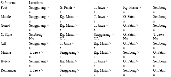
For Cu (Table 4), the highest concentrations were found in the mantle, gonad, byssus and remainder of P. viridis from Telok Jawa and in the foot and muscle of mussels from Kampung Masai; CS and gill of mussels from Gelang Patah and Senibong, respectively. The lowest Cu concentrations were found in the foot, mantle, gonad, gill, muscle and byssus of P. viridis from Senggarang; CS and remainder of those from Senibong and Gelang Patah, respectively.
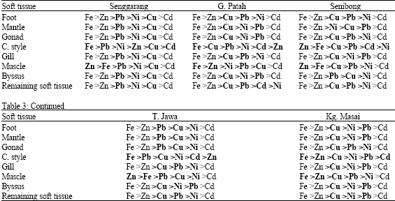
| Table 3: | Patterns of metal occurrence in the different soft tissues of P. viridis |
 | |
| Table 4: | Comparison of Cu concentration between different soft tissues of P. viridis |
 | |
| Objects indicated with different alphabet: p<0.05, Objects indicated with same alphabet: p>0.05, NA = Not Available | |
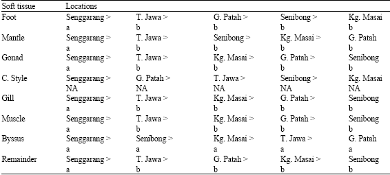
For Cd (Table 5), the highest concentrations were found in the foot, gonad, byssus and remainder of P. viridis from Gelang Patah; and the mantle, CS and muscle of mussels from Senggarang; while the highest Cd level was found in the gill of mussels from Kampung Masai. While foot, mantle, muscle and remainder of Senibong recorded the lowest Cd concentrations. The lowest concentrations of Cd were also found in the gill and byssus of P. viridis from Senggarang; and the CS of those from Telok Jawa.
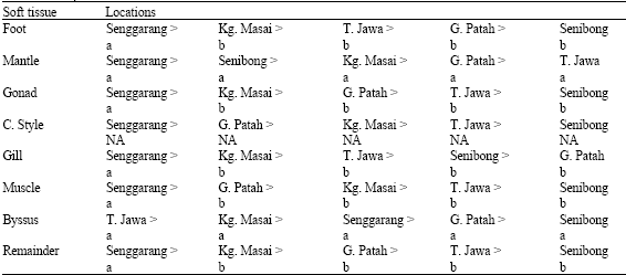
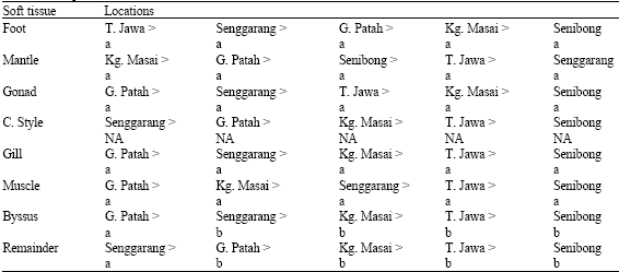
The foot, mantle, gonad, gill and byssus of mussels from Senggarang accumulated the highest concentrations of Zn (Table 6). High concentrations of Zn were also found in the CS of mussels from Senibong and the muscle and byssus of mussels from Telok Jawa. Meanwhile, the lowest concentrations of Zn were found in the foot, mantle, gonad, gill and byssus of mussels from Senibong. Low concentrations of Zn were also recorded in the CS of mussels from Telok Jawa. The highest Zn concentrations were found in the muscle and remainder of mussel from Gelang Patah. As for Pb (Table 7), highest concentrations were found in all soft tissues of P. viridis collected from Senggarang. The lowest concentrations of Pb were found in the foot and CS from Kampung Masai; the mantle and byssus from Gelang Patah; and the gonad, gill, muscle and remainder from Senibong.
| Table 5: | Comparison of Cd concentrations between different soft tissues of P. viridis |
 | |
| Objects indicated with different alphabet: p<0.05, Objects indicated with same alphabet: p>0.05, NA = Not Available | |
| Table 6: | Comparison of Zn concentrations between different soft tissues of P. viridis. |
 | |
| Objects indicated with different alphabet: p<0.05, Objects indicated with same alphabet: p>0.05, NA = Not Available | |
| Table 7: | Comparison of Pb concentrations between different soft tissues of P. viridis |
 | |
| Objects indicated with different alphabet: p<0.05, Objects indicated with same alphabet: p>0.05, NA = Not Available | |
| Table 8: | Comparison of Ni concentrations between different soft tissues of P. viridis |
 | |
| Objects indicated with different alphabet: p<0.05, Objects indicated with same alphabet: p>0.05, NA = Not Available | |
| Table 9: | Comparison of Fe concentrations between different soft tissues of P. viridis |
 | |
| Objects indicated with different alphabet (in bold fonts): p<0.05, Objects indicated with same alphabet: p>0.05, NA = Not Available | |
For Ni (Table 8), most of the soft tissues accumulated the highest concentrations in Senggarang except for byssus from Telok Jawa. Six of the soft tissues from Senibong recorded the lowest concentrations of Ni together with another two soft tissues namely the mantle and gill from Telok Jawa and Gelang Patah, respectively. Four soft tissues from Gelang Patah namely gonad, gill, muscle and byssus accumulated the highest concentrations of Fe (Table 9). Other soft tissues such as CS and remainder from Senggarang recorded the highest concentrations of Fe. High levels of Fe were also found in the foot and mantle from Telok Jawa and Kampung Masai, respectively.
DISCUSSION
Based on the above results, some important points can be noted. High levels of heavy metals were found in the CS and byssus. For the CS, the high levels of Cu found in this organ could be related to its function as the digestive tract with enzymatic activities which needed Cu in their physiological functions (Yap et al., 2006a). CS which plays a role in extracellular digestion where secreting digestive juices (Yap et al., 2006a) was found to accumulate the highest level of Cu. The byssus was reported as an important organ mainly involved in the excretion of pollutants or unwanted materials (Phillips and Rainbow, 1989; Phillips, 1995; Rainbow, 1995; Szefer et al., 2002; Yap et al., 2003c, d, e, 2005b, c, 2006c). Therefore, the elevated levels of heavy metals found in the byssus could be due to the metals regulated from the soft tissues. Other soft tissues like remainder, muscle and mantle which also accumulated high levels of heavy metals in some places could be influenced by the different metal bioavailabilities of the sampling sites.
In general, higher concentrations of Fe and Zn in all of the soft tissues indicated that the two essential metals which are associated with biochemical mechanisms tended to be present at fixed concentrations within a particular tissue regardless of the environmental exposures of the organisms to the metals (Turoczy et al., 2001). The distributions of heavy metals in the different tissues (highlighted in Table 2) were due to binding sites for metallothionein. This indicated that the pattern of metal occurrence for all the different soft tissues were changed (or disturbed) due to metal bioavailability or contamination of the mussel habitats. From the study conducted, it was found that the CS and muscle were very sensitive to contamination by the heavy metals since the most obvious change of the pattern of metal occurrence were found in these two tissues.
The different tissues (except for CS) of mussels from Kampung Masai, Telok Jawa and Senibong accumulated the highest level of Cu (Table 3) when compared to the other locations in the Johore Straits and Senggarang. This indicated the high bioavailability of Cu at the three sites which are situated on the east coast of the Johore Straits. This is in agreement with the report by Yap et al. (2006b), that the east coast is a marina and some petrochemical plants and port activity in the vicinity may be suspected as anthropogenic sources in this east part of the Straits. In the case of CS, the highest level of Cu was found in Gelang Patah. According to Yap et al. (2006b), Gelang Patah is a site where a boat garage, a jetty and an aquacultural area could be found. Since CS is a good biomonitoring organ to monitor Cu and Pb contamination (Yap et al., 2006a, c), therefore the high concentrations of Cu in the CS could be used as an indicator of the Cu bioavailability in that area. Cu leachate from the antifouling paints of the boats in the garage was believed to cause Cu contamination in Gelang Patah.
The high levels of Cd which were found in the different soft tissues of mussels collected from Gelang Patah, Senggarang and Kampung Masai were probably due to the marine bivalves being exposed to varying levels of Cd in their natural habitats in estuaries and in coastal areas (Sokolova et al., 2005). Since Gelang Patah and Kampung Masai are where the garages and a seaport located. These anthropogenic activities could possibly caused the Cd contamination of the sampling sites. However, the source for the Senggarang population was unknown since it was bought from the mussel stall. In Addition, the detoxification of Cd involving metallothionein binding could possibly result in high Cd concentrations in the different soft tissues. According to George et al. (1979), the major protein in M. edulis exposed to metal contaminant had a high-SH content, related to a metallothionein. The high tissue tolerance may be related to the mussel’s ability to isolate the metal internally via the granular vesicles (Fowler et al., 1975) or by protective chelating proteins such as metallothionein (Webb, 1987). Therefore, it is possible that the detoxification processes contributed to the high level of Cd in the soft tissues.
The Senggarang population was found to have high levels of Zn, Pb and Ni. This could be due to the high bioavailability of these metals in the mussels’ habitat. Meanwhile for Pb and Ni, the phenomena could be due to these non-essential metals being bound to metallothioneins (Viarengo et al., 1985). For Fe, the Johore Straits had high bioavailability of that metal as exhibited by six of the different soft tissue of mussels which originated from Telok Jawa (foot), Kampung Masai (mantle) and Gelang Patah (gonad, gill, muscle and byssus). However, the mussels from Senggarang recorded the highest Fe concentrations in the CS and remainder, which may also reveal the Fe’s bioavailability in the mussel’s habitat.
The widespread occurrence of metallothioneins in aquatic species is now firmly documented (Pourang et al., 2004). Over the recent decades, the roles and functions of metallothionein have been studied from various aspects and angles (detoxification, inducibility, homeostatic regulation of metals and etc.), but its possible role in the redistribution of heavy metals in the tissues of aquatic organisms has not yet been studied comprehensively. Therefore, further investigations are needed to study the biochemical processes of the distribution and redistribution of different trace elements in the different soft tissues of P. viridis.
CONCLUSIONS
As a conclusion, all the soft tissues of P. viridis are suitable to be used as biomonitoring tools for heavy metal contamination but the use of the CS is recommended since high concentrations of Cu, Cd, Zn, Pb, Ni and Fe were found in this organ. Therefore, it is suggested that the mussel watch approach should include metal distribution in the different soft tissues of P. viridis in order to increase the accuracy of the data interpretation besides using the total soft tissues.
ACKNOWLEDGMENT
This research was partly supported by Fundamental Research Grant 2006 from University Putra Malaysia vot: 5523266.
REFERENCES
- Depledge, M.H., 1989. Re evaluation of metabolic requirements for copper and zinc in decapod crustaceans. Mar. Environ. Res., 27: 115-126.
CrossRef - Fowler, B.A., D.A. Wolfe and W.F. Kettler, 1975. Mercury and iron uptake by cytosomes in mantle epithelial cells of quahog clams (Mercenaria mercenaria) exposed to mercury. J. Fish. Res. Board Can., 32: 1767-1775.
CrossRefDirect Link - George, S.G., E. Carpene, T.L. Coombs, J. Overnell and A. Youngson, 1979. Characterisation of cadmium binding proteins from mussel Mytilus edulis (L.) exposed to cadmium. Biochim. Biophys. Acta, 580: 225-233.
PubMed - Phillips, D.J.H. and P.S. Rainbow, 1989. Strategies of trace metals sequestration in aquatic organisms. Mar. Environ. Res., 28: 207-210.
Direct Link - Phillips, D.J.H., 1995. The chemistries and environmental fates of trace metals and organochlorines in aquatic ecosystems. Mar. Pollut. Bull., 31: 193-200.
CrossRefDirect Link - Pourang, N. and G. Amini, 2001. Distribution of trace elements in tissues of two shrimp species from persian gulf and effects of storage temperature on elements transportation. Water, Air Soil Pollut., 129: 229-243.
CrossRefDirect Link - Pourang, N., J.H. Dennis and H. Ghourchian, 2004. Tissue distribution and redistribution of trace elements in shrimp species with the emphasis on the roles of metallothionein. Ecotoxicology, 13: 519-533.
PubMed - Rainbow, P.S., 1995. Biomonitoring of heavy metal availability in the marine environment. Mar. Pollut. Bull., 31: 183-192.
CrossRefDirect Link - Rainbow, P.S., B.D. Smith and S.S.S. Lau, 2002. Biomonitoring of trace metal availabilities in the thames estuary using a suite of littoral biomonitors. J. Mar. Biol. Assoc. U.K., 82: 793-799.
CrossRefDirect Link - Rainbow, P.S., W. Fialkowski, A. Sokolowski, B.D. Smith and M. Wolowicz, 2004. Geographical and seasonal variation of trace metal bioavailabilities in the Gulf of Gdansk, Baltic Sea using mussels (Mytilus trossulus) and barnacles (Balanus improvises) as biomonitors. Mar. Biol., 144: 271-286.
CrossRefDirect Link - Turoczy, N.J., B.D. Mitchell, A.H. Levings and V.S. Rajendram, 2001. Cadmium copper mercury and zinc concentrations in tissues of king crab (Pseudocarcinus gigas) from southeast australia waters. Environ. Int., 27: 327-334.
Direct Link - Webb, M., 1987. Toxicological significance of metallothionein. Experientia Supplementum, 52: 109-134.
CrossRef - Yap, C.K., A. Ismail, S.G. Tan and H. Omar, 2002. Correlations between speciation of Cd Cu Pb and Zn in sediment and their concentrations in total soft tissue of green lipped mussel Perna viridis from the west coast of peninsular malaysia. Environ. Int., 28: 117-126.
PubMed - Yap, C.K., A. Ismail, H. Omar and S.G. Tan, 2003. Accumulation depuration and distribution of cadmium and zinc in the green lipped mussel Perna viridis (Linnaeus) under laboratory conditions. Hydrobiologia, 498: 151-160.
Direct Link - Yap, C.K., A. Ismail and S.G. Tan, 2003. Can the byssus of green-lipped mussel Perna veridis (Linnaeus) from the west coast of Peninsular Malaysia be a biomonitoring organ for Cd, Pb and Zn?Field and laboratory studies. Environ. Int., 29: 521-528.
CrossRefDirect Link - Yap, C.K., A. Ismail and S.G. Tan, 2003. Concentrations of Cd, Cu, Pb and zn in different parts of the byssus of green-lipped mussel Perna viridis (linnaeus). Pak. J. Biol. Sci., 6: 789-792.
CrossRefDirect Link - Yap, C.K., A. Ismail and S.G. Tan, 2003. Effects of body size (total soft tissue) and shell thickness on the accumulation of heavy metals (cd cu pb and zn) in the green lipped mussel Perna viridis (L.). Russian J. Mar. Biol., 29: 323-327.
CrossRef - Yap, C.K., A. Ismail, S.G. Tan and A.R. Ismail, 2004. Assessment of different soft tissues of green lipped mussel Perna viridis (Linnaeus) as biomonitoring agents of pb field and laboratory studies. Water Air Soil Pollut., 153: 253-268.
CrossRef - Yap, C.K., A. Ismail and S.G. Tan, 2004. Heavy metal (Cd Cu Pb and Zn) concentrations in the green-lipped mussel Perna viridis (Linnaeus) collected from some wild and aquacultural sites in the west coast of Peninsular Malaysia. Food Chem., 84: 569-575.
CrossRefDirect Link - Yap, C.K., W.H. Cheng, A. Ismail and S.G. Tan, 2005. Tissue distribution of heavy metals (Cd, Cu, Pb and Zn) in the green lipped mussel Perna viridis from nenasi and kuala pontian east coast of peninsular Malaysia. Pertanika J. Trop. Agric. Sci., 28: 13-22.
Direct Link - Yap, C.K., A. Ismail and S.G. Tan, 2005. Byssus of the green-lipped mussel Perna viridis (Linnaeus) as a biomonitoring material for Zn. Russian J. Mar. Biol., 31: 102-108.
CrossRefDirect Link - Yap, C.K., A. Ismail and S.G. Tan, 2005. Cadmium copper lead and zinc levels in the green-lipped mussel Perna viridis (L.) from the west coast of peninsular Malaysia: Safe as food. Pertanika J. Trop. Agric. Sci., 28: 41-47.
Direct Link - Yap, C.K., A. Ismail, W.H. Cheng and S.G. Tan, 2006. Crystalline style and tissue redistribution in Perna viridis as indicators of Cu and Pb bioavailabilities and contamination in coastal waters. Ecotoxicol. Environ. Saf., 63: 413-423.
CrossRef - Yap, C.K., A. Ismail, F.B. Edward, S.G. Tan and S.S. Siraj, 2006. Use of different soft tissues of Perna viridis as biomonitors of bioavailability and contamination by heavy metals (Cd Cu Fe, Pb, Ni and Zn) in a semi enclosed intertidal water the johore straits. Toxicol. Environ. Chem., 88: 683-695.
CrossRefDirect Link