
R.K. Sharma
Department of Zoology, Reproductive Physiology Laboratory, Kurukshetra University, Kurukshetra-136119, India
P.K. Chauhan
Department of Zoology, Reproductive Physiology Laboratory, Kurukshetra University, Kurukshetra-136119, India
A. Fulia
Department of Zoology, Reproductive Physiology Laboratory, Kurukshetra University, Kurukshetra-136119, India
Research Journal of Veterinary Sciences
Year: 2012 | Volume: 5 | Issue: 3 | Page No.: 59-68


ABSTRACT
Atrazine, an agricultural pesticide widely used all over the world, has now been recognized as an endocrine disrupting chemical which affects the reproductive system of mammals. The objective of the present study was to analyze the effect of single dose of atrazine on spermatids of Capra hircus in vitro. Small pieces (approximately 1 mm3) of testicular tissue were divided into experimental and control group. The experimental group was treated with atrazine concentration (1.0 nmol mL-1) and exposed for 1, 4 and 8 h. Atrazine, 1.0 nmol mL-1 revealed increasing morphological alterations in spermatids. After 1 h exposure, pycnosis and chromatolysis were observed in few spermatids. Atrazine exposure induced alterations in fine morphology of spermatids, degenerated nuclear membrane and pinching off of the nuclear material was clearly visible. Nanomolar concentration of atrazine has deleterious effects on testicular structure including fine morphology and severely impaired the spermatozoa formation and finally affects the reproductive potential of goat.
PDF Abstract XML References Citation
Received: December 20, 2011;
Accepted: June 07, 2012;
Published: August 10, 2012
How to cite this article
R.K. Sharma, P.K. Chauhan and A. Fulia, 2012. Histopathological Effects Induced by Single Dose of Atrazine in Spermatids of Goat in vitro. Research Journal of Veterinary Sciences, 5: 59-68.
URL: https://scialert.net/abstract/?doi=rjvs.2012.59.68
URL: https://scialert.net/abstract/?doi=rjvs.2012.59.68
INTRODUCTION
The endocrine disrupting chemicals present in the environment are found in low doses in thousands of products of daily use. Most of the pesticides used in agriculture are endocrine disruptors and are associated with the abnormal sexual development, lowered male fertility, breast, testicular and prostate cancers, damage to the thyroid and pituitary glands, lowered immunity, developmental and behavioral problems (Devesa et al., 1995; Moharram et al., 2011). Diazinon is an Organophosphate Insecticide used to control pests. Diazinon disturbs the Leydig cell steroidogenesis hence the synthesis of testosterone and glucose release from liver into blood and it led to anemia (Alahyary et al., 2008). Endosulfan induced fine morphological alterations in the goat Capra hircus in vitro (Sharma et al., 2010). Atrazine, an agricultural pesticide widely used all over the world, has now been recognized as an endocrine disrupting chemical which affects the reproductive system of mammals (Ashby et al., 2002; Rayner et al., 2004; Kniewald et al., 2000; Friedmann, 2002; Trentacoste et al., 2001). Decline in male fertility has been observed in recent years in human and some laboratory animals by several researchers (Carlsen et al., 1992; Auger et al., 1995; Irvine et al., 1996; Fisch et al., 1996; Swan et al., 1997; Andersen et al., 2000). Increase in testicular weight, degenerative changes in seminiferous tubules and abnormality in reproductive function in rats have been recorded due to exposure of pesticides (Lee et al., 1978; Mishra et al., 1993). Chlorpyrifos-methyl, diazinon and profenofos induced decline in sperm count associated with increased number of abnormal spermatozoa (Zidan, 2009). Earlier studies revealed that atrazine enhanced cancer risks as well as delayed reproductive development in laboratory animals (Laws et al., 2003; Stoker et al., 2000, 2002). There is only paper on goat testis wherein atrazine induced histomorohological alterations has been elaborated (Sharma and Chauhan, 2009). It is in the light of this information on the hazardous effect of agricultural pesticide atrazine, the present study has been focussed on the effect of atrazine on spermatids of goat (Capra hircus) in vitro.
MATERIALS AND METHODS
The mature goat (Capra hircus) testis was collected from the slaughter houses around Kurukshetra (29°6’N, 76°5’E) and Delhi (28°38’N, 77°12’E). The testis was brought to the Reproductive Physiology Laboratory, Department of Zoology, Kurukshetra University, Kurukshetra at 4°C in normal saline during November 2008.
Testicular tissue culture: After washing with the normal saline the testis was decapsulated and cut into small pieces (approximately 1 mm3) for culture. After washing with TCM-199, small pieces of testicular tissue were immediately placed on nucleopore filter and floated on medium. The culture medium was prepared by mixing TCM-199 and antibiotics (200 unit penicillin 100 IU mL-1 and streptomycin 100 g mL-1). The small testicular tissues were divided into two groups (one experimental group and one control group). Experimental group was treated with atrazine (1.0 nmol mL-1) and was further divided into three subgroups (i), (ii) and (iii) exposed for 1, 4 and 8 h, respectively at temperature 39°C, humidity 95 and 5% CO2 concentration and the control was run simultaneously.
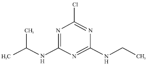 |
| Atrazine: | |
| IUPAC name: | 6-chloro-N-ethyl-N’-isopropyl-1,3,5-triazine-2,4-diamine |
| Chemical formula: | C8H14ClN5 |
| Molecular mass: | 215.7 |
Harvesting of testicular tissues was carried out after the specified duration and tissues from all the experimental and control groups were processed for histomorphological studies using the standard techniques given in Pearse (1968) and the results were statistically analyzed according to Zar (1984) with the help of mean, standard deviation, standard error, student “t” test and chi-square test.
Transmission electron microscopy (Zamboni, 1976): Harvested testicular tissues simultaneously were fixed in 2.5% glutaraldehyde in 0.2 M phosphate buffer saline (pH 7.2-7.4) at 4°C for 24 h. After trimming, samples were fixed and dehydrated with graded series of acetone. Dehydrated samples were cleared with epoxy propane and blocks were cut into “thick” sections (1 μm) and select the required portion. Specific portions were finally trimmed and the serial 60-90 nm thick sections were cut and stained with uranyl acetate followed by lead citrate (Hertig and Adams, 1967). The sections were examined and photographed under electron microscope model installed CM-10 Philips at All India Institute of Medical Sciences, New Delhi.
RESULTS
Histomorphological studies: During the present investigation, time dependant histopathological effect of atrazine (1.0 nmol mL-1), a chlorotriazine herbicide on the testis of goat (Capra hircus) has been evaluated. Histological preparations of the testicular tissue and also the plastic embedded sections prepared for electron microscopy were analyzed in experimental as well as control groups. Histologically, the testis of sexually mature goat was comprised of seminiferous tubules and interstitium. In the control group, a large number of seminiferous tubules were packed in the loose connective tissue. Seminiferous tubule was characterized by the presence of a series of well defined cellular associations and stages. The spermatids were present next to the layer of spermatocytes toward the lumen of seminiferous epithelium (Fig. 1).
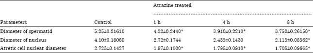
Atrazine, revealed morphological alterations in spermatids. Pycnosis and chromatolysis in the spermatids were observed after one hour. Hyalinization, dislodging, condensation and vacuolization were frequently noticed after 4 h of exposure duration (Fig. 2). Fragmentation was also recorded after 8 h of exposure duration. Atretic spermatid percentage was increased and condensation was also increased. These changes have a strong positive correlation with the duration of exposure from 4-8 h (Fig. 3). Spermatid cell diameter was declined from 5.25±0.2161 μm in control to 4.22±0.244 μm after 1 h, 3.91±0.221 μm after 4 h and 3.75±0.2615 μm after 8 h of exposure (Table 1). All the values were statistically significant at (p≤0.05). Spermatid nuclear diameter was declined from 4.1±0.1806 μm in control to 2.115±0.08562 μm after 8 h of exposure durations (Table 1).
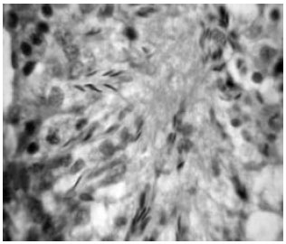 | |
| Fig. 1: | Light micrograph of control testicular tissue displaying sertoli cells laid on the basal lamina in between the spermatogonia and tapered towards the lumen. Spermatocytes, spermatids and spermatozoa were placed in layer next to spermatogonia. (x1000) |
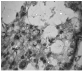 | |
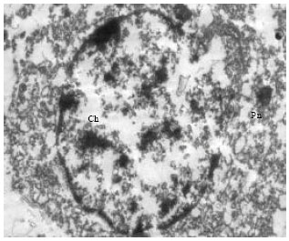
| Fig. 2: | A portion of testicular section of goat exposed to atrazine (1.0 nmol mL-1) for 4 h showing increased vacuolization (V) of germ cells. Fragmentation (F), pycnosis (P) and chromatolysis (Ch) was observed after the exposure of atrazine. (x1000) |
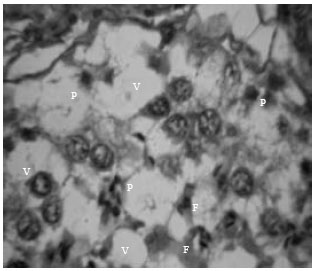 | |
| Fig. 3: | A section of goat exposed to atrazine (1.0 nmol mL-1 ) for 4 h showing increase in the hyaline cells and pycnotic nuclei (P) in spermatids. Vacuolization (V) in seminiferous tubule is clearly visible. (x1000) |
| Table 1: | Effects of atrazine on spermatid after different exposure durations |
 | |
| *Values are statistically significant at p≤0.05 | |
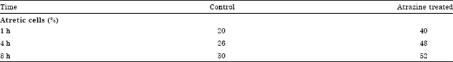
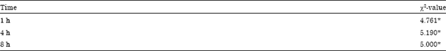
All the values were statistically significant at p≤0.05. Atretic spermatid at advanced stage of atresia in control group were 20, 26 and 30% after 1, 4 and 8 h, respectively. However, this percentage was increased up to 40, 48 and 52% after 1, 4 and 8 h of exposure durations, respectively due to the exposure of 1.0 nmol mL-1 dose of atrazine (Table 2). Chi-square values were 4.761, 5.190 and 5.00 after 1, 4 and 8 h, respectively and all the values were statistically significant at p = 0.05 (Table 3).
Transmission electron microscopic studies: Transmission electron microscopic studies in control group showed that nucleus was very large and round in shape and occupied most of the space in the cell. Chromatin material was highly condensed and occupies the central position in the nucleus of the spermatid. There was intact nuclear membrane and clearly visible cap present in the cap phase spermatid (Fig. 4). After exposure of atrazine (1.0 nmol mL-1) induced alterations in fine morphology spermatid.
| Table 2: | Percentage of atretic spermatid observed after exposure of atrazine, harvested after different exposure durations |
 | |
| Table 3: | The chi-square values comparison of No. of atretic cells spermatid of control and atrazine treated tests (1 .0 nmol mL-1) |
 | |
| *Statistically significant at p = 0.05 | |
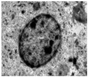 | |
| Fig. 4: | Transmission electron micrograph of control testicular tissue showing a magnified view of early cap phase spermatid (rSpd) with an arc shaped head cap (hc) at one end of the nucleus. Nuclei of cap phase spermatids occur either in spherical or ovoidal forms and occupy most of the space in the cell. (x5000) |
 | |
| Fig. 5: | Transmission electron micrograph of testicular tissue culture treated with atrazine (1.0 nmol mL-1) for 1 h exhibiting disrupted nuclear membrane and pinching off (Pn) of nuclear material in spermatid. Chromatolysis (Ch) and clumps of chromatin material were clearly visible with heterogeneous cytoplasm. (x5500) |
Atrazine induced degenerating changes in the cytoplasm and nucleus at all the stages of spermatid. After 1 h of exposure duration of atrazine pinching off of nuclear material was clearly visible. Degenerating spermatid was noticed at 1 h of exposure duration. Swollen and ruptured mitochondria, Golgi vesicles were seen and degenerated nuclear membrane were noticed. Cytoplasm became loose and detached. Abnormal and vacuolated organelles were increased due to the treatment. Vacuolization is clearly visible. Degenerated Golgi phase spermatid was evident. Nuclear membrane was degenerated at few places due to the atrazine treatment for 1 h. Slight margination of nuclear material was observed in the Golgi phase spermatid (Fig. 5).
As the exposure duration of atrazine was enhanced from 1-4 h at the same dose level, number and size of vacuoles were enhanced. Degenerated nuclear membrane was observed in Golgi phase spermatid. In cap phase spermatid nuclear membrane was also disrupted. Small clumps of chromatin material were observed. Abnormal vesicles were also noticed in the cap phase spermatid. A very clear vacuole in the nucleoplasm of cap phase spermatid was visible. Chromatin condensation and margination of chromatin material was increased (Fig. 6).
As the exposure duration was further enhanced from 4-8 h after treatment of atrazine at the dose level 1.0 nmol mL-1, spermatids showed higher degree of degeneration. Vacuoles of various sized and shaped were present in the cytoplasm of Golgi phase spermatid. Degenerated cap phase spermatid with vesicles of various shapes was noticed. Nuclear membrane is almost degenerated. Swollen round mitochondria were present in cytoplasm. Round mitochondria with ruptured membranes were frequently encountered. Chromatolysis in the cap phase spermatids were noticed. Large vacuoles were visible in nucleoplasm. Vacuole has almost covered the nucleus leading to disruption of spermatid. Cytoplasm is filled with debris of disrupted organelles due to the effect of atrazine treatment. Chromatolysis and multinuclear bodies were observed in spermatid.
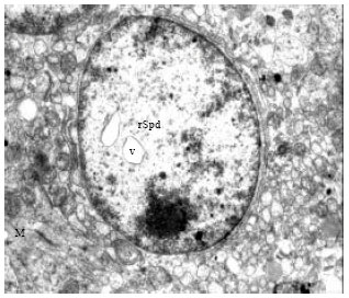 | |
| Fig. 6: | Transmission electron micrograph of testicular tissue culture treated with atrazine (1.0 nmol mL-1) for 4 h exhibiting Golgi phase spermatid (rSpd) with increased degeneration. Interanuclear vacuoles (v) and degenerating mitochondria (M) were observed. (x5000) |
DISCUSSION
During the present investigation effect of atrazine at dose level 1.0 nmol mL-1 on spermatids of goat Capra hircus have been analyzed. The results of the present investigations demonstrated that degenerative changes in spermatid were induced by nanomolar concentration of atrazine in vitro. Histologically, pycnotic nuclei, fragmented nuclei, chromatolysis, hyalinization and condensation in spermatids were noticed after atrazine exposure. These degenerative changes were increased as the exposure duration enhanced. The results of the present findings are in agreement with the findings of Dunnick et al. (1984) who observed the dose dependent effect of Dimethyl Methylphosphonate (DMMP). Histological studies revealed that the characteristic changes induced by DMMP were lack of spermatogenesis or by degeneration, vacuolization and necrosis of cells in the spermatogenic tubules. Results of the present study demonstrated that there was increase in atretic percentage of spermatids as the exposure duration increased. Atretic spermatid at advanced stage of atresia showed 40, 48 and 52% after 1, 4 and 8 h of exposure durations, respectively at 1.0 nmol mL-1 dose of atrazine. Effect of Sumithio NP 25/2.5 EC insecticide was investigated by Al-Jahdali and Bisher (2007) who have observed that Sumithio NP 25/2.5 EC induced damage to the seminiferous tubules leading to the separation of spermatogenic cells from the germinal epithelial membrane. Decreasing number of the spermatogenic cells leading to hypospermatogenesis due to the exposure of Sumithio® NP 25/2.5 EC. All these observations are in consistent with the present study that atrazine affect the spermatogenesis. The observations of present study that atrazine induced chromatolysis, fragmentation and other morphological alterations in spermatids strongly supports the findings of Khan and Sinha (1994) who have reported that in the primary spermatocytes of mice, the pesticides induced various types of structural alterations in chromosomes, pairing impairments among homologues and division-disruptive changes. During the present investigation atrazine depicted similar degenerative changes in testis to as induced by lead toxicity. Almansour (2009) observed the pycnosis, desquamation, chromatolysis and spermatid giant cell were observed due to the exposure of lead in testis of adult quail. The results of the present study are in consistent with the findings of Masouleh et al. (2011) who have been observed that exposure of diazinon induced decline in number of spermatocytes and spermatids in fish in vitro. The observations of present investigation strongly advocate the findings of Moustafa et al. (2007) where in they have studied the effect of pesticide profenofos, on the nuclei of the spermatogonia. Some sections exhibited azoospermia and spermatid abnormalities, besides cytoplasmic inclusion bodies in a few spermatogonial cells. The elongation of Leydig cells indicative of hyper-activity was also recorded due to the exposure of profenofos. The results of present transmission electron microscopic analysis revealed alterations in fine morphology of spermatids after the exposure of 1.0 nmol mL-1 atrazine and exhibited intracytoplasmic vacuolization. As the exposure duration was increased, extensive accumulation of intracytoplasmic vacuoles was observed in the spermatid. Sizes of these vacuoles were enlarged with the increase in exposure duration from 1-4 and 4-8 h. Degree of cytoplasmic and nuclear degeneration was also enhanced with the increase in exposure durations. Clumps of chromatin material were observed during the present study at 4 and 8 h exposure of atrazine at dose level 1.0 nmol mL-1 in spermatids. These observations are in agreement with the findings of Kniewald et al. (2000) where in histological analysis of testicular tissue from treated rats depicted the cellular disorganization such as cluster formation, vacuolated cytoplasm and reduced collagen fiber using electron microscopy.
CONCLUSION
From the present study, it becomes clear that nanomolar concentration of atrazine has deleterious effects on testicular structure including fine morphology and severely impaired the spermatozoa formation and finally affects the reproductive potential of goat. The data generated will be of great value in assessing the health hazards of pesticides to domestic animals and can provide important clues about the damage induced by the exposure of pesticides in reproductive potential of wild life and humans.
ACKNOWLEDGMENTS
Authors are thankful to University Grants Commission (New Delhi) for financial assistance in the form of Rajiv Gandhi National Fellowship and Department of Zoology, Kurukshetra University, Kurukshetra for providing all the facilities throughout the study.
REFERENCES
- Devesa, S.S., W.J. Blot, B.J. Stone, B.A. Miller, R.E. Tarone and J.F. Jr. Fraumeni, 1995. Recent cancer trends in the United States. J. Natl. Cancer Inst., 87: 175-182.
PubMed - Moharram, S.G., O.M. Wahbi and Z.A. El-Greisy, 2011. Effect of polluted water from the Egyptian Eastern Mediterranean coast on reproductive, toxicological and hematological characteristics of Siganus rivulatus. Pak. J. Biol. Sci., 14: 668-681.
CrossRefDirect Link - Alahyary, P., M.I. Poor, F.F. Azarbaijani and V. Nejati, 2008. The potential toxicity of diazinon on physiological factors in male rat. Pak. J. Biol. Sci., 11: 127-130.
CrossRefPubMedDirect Link - Ashby, J., H. Tinwell, J. Stevens, T. Pastoor and C.B. Breckenridge, 2002. The effects of atrazine on the sexual maturation of female rats. Regul. Toxicol. Pharmacol., 35: 468-473.
PubMed - Rayner, J.L., C. Wood and S.E. Fenton, 2004. Exposure parameters necessary for delayed puberty and mammary gland development in long-evans rats exposed in utero to atrazine. Toxicol. Applied Pharmacol., 195: 23-34.
PubMed - Kniewald, J., M. Jakominic, A. Tomljenovic, B. Simic, P. Romac, D. Vranesic and Z. Kniewald, 2000. Disorders of male rat reproductive tract under the influence of atrazine. J. Applied Toxicol., 20: 61-68.
PubMed - Friedmann, A.S., 2002. Atrazine inhibition of testosterone production in rat males following peripubertal exposure. Reprod. Toxicol., 16: 275-279.
PubMed - Trentacoste, S.V., A.S. Friedmann, R.T. Youker, C.B. Breckenridge and B.R. Zirkin, 2001. Atrazine effects on testosterone levels and androgen-dependent reproductive organs in peripubertal male rats. J. Androl., 22: 142-148.
PubMed - Carlsen, E., A. Giwercman, N. Keiding and N.E. Skakkebaek, 1992. Evidence for decreasing quality of semen during past 50 years. Br. Med. J., 305: 609-613.
PubMedDirect Link - Auger, J., J.M. Kunstmann, F. Czyglik and P. Jouannet, 1995. Decline in semen quality among fertile men in Paris durin the past 20 years. N. Engl. J. Med., 332: 281-285.
PubMedDirect Link - Irvine, S., E. Cawood, D. Richardson, E. MacDonald and J. Aitken, 1996. Evidence of deteriorating semen quality in the United Kingdom: Birth cohort study in 577 men in Scotland over 11 years. Br. Med. J., 312: 467-471.
PubMedDirect Link - Fisch, H., E.T. Goluboff, J.H. Olson, J. Feldshuh, S.J. Broder and D.H. Barad, 1996. Semen analysis in 1,283 men from the United States over a 25-year period: No decline in quality. Fertility Sterility, 65: 1009-1014.
PubMed - Swan, S.H., E.P. Elkin and L. Fenster, 1997. Have sperm densities declined? A reanalysis of global trend data. Environ. Health Perspect., 105: 1228-1232.
PubMedDirect Link - Andersen, A.G., T.K. Jensen, E. Carlsen, N. Jorgensen and A.M. Anderson et al., 2000. High frequency of sub-optimal semen quality in an unselected population of young men. Hum. Reprod., 15: 366-372.
Direct Link - Lee, C.C., J.Q. Russell and J.L. Minor, 1978. Oral toxicity of ferric dimethyl-dithiocarbamate (ferbam) and tetramethylthiuram disulfide (thiram) in rodents. J. Toxicol. Environ. Health, 4: 93-106.
PubMed - Mishra, V.K., M.K. Srivastava and R.B. Raizada, 1993. Testicular toxicity of thiram in rat. Morphological and biochemical evaluations. Ind. Health, 31: 59-67.
PubMed - Zidan, N.E.H.A., 2009. Evaluation of the reproductive toxicity of chlorpyrifos methyl, diazinon and profenofos pesticides in male rats. Int. J. Pharmacol., 5: 51-57.
CrossRefDirect Link - Sharma, R.K., P.K. Chauhan and A. Fulia, 2010. Endosulphan induced changes in fine morphology of goat spermatogonia in vitro. Res. J. Environ. Toxicol., 4: 214-222.
CrossRefDirect Link - Laws, S.C., J.M. Ferrell, T.E. Stoker and R.L. Cooper, 2003. Pubertal development in female Wister rats following exposure to propazin and atrazine biotransformation by-products, diamino-S-chlorotriazine and hydroxyatrazine. Toxicol. Sci., 76: 190-200.
PubMed - Stoker, T.E., S.C. Laws, D.L. Guidici and R.L. Cooper, 2000. The effect of atrazine on puberty in male Wistar rats: An evaluation in the protocol for the assessment of pubertal development and thyroid function. Toxicol. Sci., 58: 50-59.
PubMed - Stoker, T.E., D.L. Guidici, S.C. Laws and R.L. Cooper, 2002. The effects of atrazine metabolites on puberty and thyroid function in the male wistar rat. Toxicol. Sci., 67: 198-206.
CrossRef - Hertig, A.T. and E.C. Adams, 1967. Studies on human oocyte and its follicle, ultrastructure and its cytochemical observation on the pre ovulatory follicles. J. Cell. Biol., 34: 647-675.
Direct Link - Dunnick, J.K., B.N. Gupta, M.W. Harris and J.C. Lamb, 1984. Reproductive toxicity of dimethyl methyl phosphonate (DMMP) in the male Fischer 344 rat. Toxicol. Applied Pharmacol., 72: 379-387.
CrossRef - Al-Jahdali, M.O. and A.S.B. Bisher, 2007. Testicular histopathological alterations in rats treated with Sumithion&ref; NP 25/2.5 EC, insecticide. J. Boil. Sci., 7: 520-525.
CrossRefDirect Link - Khan, P.K. and S.P. Sinha, 1994. Impact of higher doses of vitamin C in modulating pesticide genotoxicity. Teratog. Carcinog. Mutagen., 14: 175-181.
PubMedDirect Link - Masouleh, F.F., B.M. Amiri, A.R. Mirvaghefi and M.A. Nemtollahi, 2011. In vitro effects of diazinon on male reproductive tissue and sperm motility of caspian kutum (Rutilus frisii kutum). Res. J. Environ. Toxicol., 5: 108-116.
CrossRefDirect Link - Almansour, M.I., 2009. Histological alterations induced by lead in the testes of the quail Coturnix coturnix. Res. J. Environ. Toxicol., 3: 24-30.
CrossRefDirect Link - Moustafa, G.G., Z.S. Ibrahim, Y. Hashimoto, A.M. Alkelch, K.Q. Sakamoto, M. Ishizuka and S. Fujita, 2007. Testicular toxicity of profenofos in matured male rats. Arch. Toxicol., 81: 875-881.
CrossRefDirect Link