
R.A. Adisa
Laboratories for Biomembrane Research and Biotechnology, Department of Biochemistry, College of Medicine, University of Ibadan, Ibadan, Nigeria
A. Abass Khan
H.E.J. Research Institute of Chemistry, University of Karachi, Karachi, Pakistan
I. Oladosu
Department of Chemistry, University of Ibadan, Ibadan, Nigeria
A. Ajaz
H.E.J. Research Institute of Chemistry, University of Karachi, Karachi, Pakistan
M.I. Choudhary
H.E.J. Research Institute of Chemistry, University of Karachi, Karachi, Pakistan
O.O. Olorunsogo
Laboratories for Biomembrane Research and Biotechnology, Department of Biochemistry, College of Medicine, University of Ibadan, Ibadan, Nigeria
A. Ur Rahman
H.E.J. Research Institute of Chemistry, University of Karachi, Karachi, Pakistan
Research Journal of Phytochemistry
Year: 2011 | Volume: 5 | Issue: 4 | Page No.: 177-189


ABSTRACT
The presence of yet to be explored radical scavengers in the leaf extracts of Cnestis ferruginea (De Candolle) have been reported. Thus, the present study is focused on isolating and characterizing these radical scavengers for possible therapeutic use as lead drug candidates against oxidatively induced pathological conditions. The defatted methanol extracts (MECF) of the leaves of Cnestis ferruginea was fractionated into Chloroform (CF), Ethylacetate (EF), Butanol (BF) and Water (WF) fractions. Total antioxidant property, superoxide anion, 1,1-diphenyl-2-picrylhydrazyl (DPPH) radical scavenging activities and inhibition of xanthine oxidase activity by the fractions were determined. The extent of inhibition of Fe2+/ascorbate stimulated lipid peroxidation in mitochondrial membranes was also evaluated. All fractions and crude extracts exhibited antioxidant properties at different degrees. The pattern of DPPH radical scavenging activity and inhibition of mitochondrial membrane lipid peroxidation by MECF and EF were similar. β-sitosterol, para-hydroxyphenol and six other sub-fractions were isolated from EF. The DPPH radical scavenging activities of the sub-fractions were not significantly (p>0.05) different from one another. Robustaside B or 6'-(3", 4"-dihydroxycinnamoyl) arbutin was also isolated from sub-fraction III from EF using sephadex LH 20. Structural elucidation of all compounds was determined by spectroscopy (IR, UV, 1D and 2D 1H and 13C NMR). The present study reported for the first time the isolation and radical scavenging activity of Robustaside B-an arbutin derivative and para-hydroxyphenol from leaves of Cnestis ferruginea. Leaf extracts of Cnestis ferruginea was demonstrated to be a rich source of antioxidants.
PDF Abstract XML References Citation
Received: May 05, 2011;
Accepted: September 28, 2011;
Published: October 29, 2011
How to cite this article
R.A. Adisa, A. Abass Khan, I. Oladosu, A. Ajaz, M.I. Choudhary, O.O. Olorunsogo and A. Ur Rahman, 2011. Purification and Characterization of Phenolic Compounds from the Leaves of Cnestis ferruginea (De Candolle): Investigation of Antioxidant Property. Research Journal of Phytochemistry, 5: 177-189.
URL: https://scialert.net/abstract/?doi=rjphyto.2011.177.189
URL: https://scialert.net/abstract/?doi=rjphyto.2011.177.189
INTRODUCTION
Cnestis ferruginea (De Candolle) (Capparaceae) (Irvine, 1961) is a climber of genus Cnestis ferruginea DC (Connaracea). In Nigeria, it is locally referred to as Gboyin-gboyin, Akara-aje, Omu-aje by the Yorubas; Ukpe, Ibi-eka by Binis, Amunketa, Okpe-nketa, or Okpe-isi-uketa by Igbos and Utinabua, Usiere-ebua by the Efiks. It has a wide distribution in Africa and is specifically found in deciduous and secondary scrub such as Udi-Plateau. The fruits are orange-red with velvety hairs on the follicle and have leaves ten inches long with eight pairs of leaflets. All leaf parts being ferruginously pubescent and the flowers are white with ferruginous sepals. The local uses of the plant include as laxative, remedy for dysentery and gonorrhoea (Dalziel, 1937). In West-Africa the bitter fruit juice is widely used for the treatment of tooth-ache, mouth and skin infections (Boakye-Yiadom and Konning, 1975) while the bark is rubbed on the gums for pyorrhea (Okwu and Iroabuchi, 2004).
The first reported chemistry of C. ferruginea was by Olugbade et al. (1982) when phytochemistry revealed the presence of coumarin, flavonoid, squalene, β-sitosterol and triacontal-1-ol in its roots. Five years later, the petroleum ether fraction of C. ferruginea fruit was demonstrated to contain octacosanyl stearate, 1-myristo-2-stearo-3-palmitin (Ogbechie et al., 1987). At exactly one decade after the first report, a novel isoflavone glycoside, afrormosin-7-O-beta-D-galactoside with antimicrobial activity against Staphylococcus aureus and Escherichia coli was isolated in the fruit testa (Parvez and Rahman, 1992). The compound also possess antifungal effects against Candida albicans but was unable to inhibit the growth of Aspergillus niger. Other compounds such as squalene, myricyl alcohol, β-sitosterol, cyanidin, delphinidin and apigenidin (Ogbede et al., 1986) have also been isolated from the plant. Extracts of roots, stems and leaves of Cnestis ferruginea have been shown to possess antibacterial and anticonvulsant (Declume et al., 1984) activities. Recently, Atere and Ajao (2009) reported the adverse effects of the crude alkaloid fraction of the roots on hepatic functions. The methanol extracts of leaves of C. ferruginea have been reported to inhibit haemoglobin glycosylation in vitro (Adisa et al., 2004). More recently, methanol and ethylacetate fraction of C. ferruginea were confirmed to contain highly potent antihyperglycaemic principles capable of attenuating secondary complications of diabetes such as atherosclerosis, liver and renal dysfunction (Adisa et al., 2010).
Polyphenolic compounds including flavonoids are established to have antioxidant properties and have been isolated from many natural sources (Nijveldt et al., 2001; Heijnen et al., 2002; Arya and Yadav, 2011; Butkhup and Samappito, 2011). Several reports have shown that these substances are effective against myriads of oxidative stress-induced disease conditions including cancer, neurodegenerative disorders, ageing and diabetes (Ames et al., 1993; Jain et al., 2011). Meanwhile, there is renewed interest in the investigation of natural products for the discovery of new bioactive substances with better pharmacological activities (antioxidant and anticancer). These drug candidates could serve as substitutes to synthetic drug’s adverse effects (Kim et al., 2003). Therefore, the present study was designed to investigate the phytochemistry of leaves of C. ferruginea. Bioactive compounds with antioxidant property would be isolated, purified and characterized using bioassay guided isolation.
MATERIALS AND METHODS
Spectroscopic measurements: One dimensional (1D) NMR (1H-NMR, 13C-NMR, DEPT 135 and DEPT 90) and two dimensional (2D) NMR spectra were recorded in deuterated solvents (CDCl3 or MeOD or DMSO) on Bruker AM-400, or 500 MHZ spectrometers. Chemical shifts were measured in ppm (δ) and coupling constants (J) are given in Hz. Electron Impact Mass spectra (EIMS) was recorded on Varian MAT 312 double focusing spectrometer or on a Finnigan MAT 311 with MASS SPEC data system. Peak matching and Field Desorption (FD) experiments were performed on Finnigan MAT 312X mass spectrometer. Fast atom bombardment mass spectra (FAB-MS) were recorded on Jeol HX 600 mass spectrometer. Exact molecular formulae were determined by High Resolution Mass Spectrometry (HRMS). Glycerol or thioglycerol were used as reference compounds for FAB (+ve). Melting points of compounds were determined on the Yanaco micromelting point apparatus and were uncorrected. UV and IR spectra were recorded on Hitachi-UV-3200 and Jasco 320-A spectrophotometers, respectively. Column chromatography was carried out using silica gel 60 Merck; 70-230 mesh; pore diameter (0.06-0.20 mm) and Sephadex LH 20 (Reverse Phase 18F254s (Merck KGa A) 64271 Darmstadt Germany). Thin layer chromatography (TLC) was done on silica gel 60 pre-coated plates, F-254 (Merck) and Reverse Phase 18F254s (Merck KGa A) 64271 Darmstadt Germany) pre-coated plates.
Plant material: Fresh leaves of C. ferruginea were collected at the Forest Reserve Area of International Institute of Tropical Agriculture Ibadan in the month of October, 2004. The leaves were immediately rinsed of debris and shade-dried for one week on laboratory trays. The dried leaves were powdered and weighed. Leaves were authenticated and identified at the Herbarium, Forestry Research Institute of Nigeria (FRIN) Ibadan, Oyo State where specimens (Voucher No. 106524) were deposited. The laboratory experiments were continuously on-going up to year 2008.
Animals: Male wistar strain albino rats (180-200 g, 16 weeks old) used in this study were bred and housed at the animal house of HEJ Research Institute of Chemistry, University of Karachi, Karachi under a 12 h light/dark condition. The experimental design was approved by the Animal Care Ethics Committee of H.E.J Research Institute of Chemistry, University of Karachi, Karachi and the protocols conformed to the National Institute of Health (NIH) guidelines. Animals had free access to food (rat chow) and water throughout the experimental period.
Preparation of rat liver mitochondria: Mitochondrial fraction were isolated from liver homogenate of male Wistar strain albino rats (180-200 g) as described by Johnson and Lardy (1967). The weighed livers were washed in ice-cold 1.15% KCl solution and then chopped before homogenizing in 0.25 M sucrose (ice-cold). The homogenate was centrifuged twice at 3000 rpm for 5 min. The supernatant was further centrifuged at 10,000 rpm for 20 min to pellet the mitochondria. The mitochondria were washed twice in 0.25 M sucrose at 10,000 rpm, dispensed into pre-cooled eppendorf tubes and used fresh.
Protein estimation: The protein content was determined using the method of Lowry et al. (1951).
Extraction procedures: The air-dried, powdered (1 kg) leaves of C. ferruginea (D.C) were exhaustively extracted separately with n-hexane (100%) and methanol (95% v/v) (Sigma Aldrich Chemical Co. St Louis USA), in a giant size Soxhlet apparatus at 60°C continuously for 12 h in each case. The crude extracts were collected and concentrated with a rotary evaporator at 40°C. The methanol extract concentrate of C. ferruginea (MECF) obtained was a greenish brown substance. MECF (234.9 g) was dissolved in distilled water and partitioned with equal volume of chloroform, ethylacetate and butanol in succession to yield the Chloroform (13%), (CF), Ethylacetate (EF) (18%), Butanol (BF) (7.2%) and Water (WF) (7.2%) fractions. The water fraction was lyophilized in a freeze dryer. The ethylacetate fraction (EF, 20 g) was subjected to column chromatography on silica gel (Merck; 70-230 mesh; pore diameter 0.06-0.20 mm) and eluted with gradients of dichloromethane-hexane (15:85, 2L), dichloromethane-methanol (98:2, 2.5 L; 85:15, 1 L; 85:15, 0.75 L; 85:15, 0.5 L; 80: 20, 1 L; 75:25, 1.75 L; 75:25, 2.5 L) at a flow rate of 24 mL min-1. The fractions were subjected to Thin Layer Chromatography (TLC) in solvent systems (40:60; H2O: MeOH) on Reverse Phase 18F254s (Merck KGa A) 64271 Darmstadt Germany) chromatographic plates. The fractions were combined, concentrated on a rotary evaporator and weighed.
The subfractions obtained were further screened qualitatively on TLC plates by spraying with DPPH, AlCl3 and FeCl3.
Determination of antioxidant activities: Antioxidant activities of MECF, HF, EF, CF, BF and WF were determined using in vitro free radical generating systems such as horse radish peroxidase catalysed oxidation of 2,2-azinobis-(3-ethylbenzothiazoline-6-sulfonic acid) (ABTS), Fe2+/ascorbate induced-mitochondrial membrane lipid peroxidation, PMS-NADH-NBT reduction system and inhibition of xanthine oxidase activity assay.
Total antioxidant activity: The total antioxidant activity of extracts of C. ferruginea was measured using horse radish peroxidase catalysed oxidation of ABTS with a slight modification of the method described by Cano et al. (2002). The reaction mixture (200 μL) contained 1 mm ABTS, 30 μM H2O2 and 6 μM horse radish peroxidase in acidified ethanol (phosphoric acid 0.7% w/v). The test sample, control or standard (Trolox) was added to the reaction medium in a microtitre plate and the decrease in absorbance at 730 nm was determined after 5 min at 27°C in a spectramax 384 USA (Molecular devices).
Superoxide anion radical scavenging activity: The superoxide anion radical scavenging activity of MECF, HF, EF, CF, BF and WF was assessed by a slight modification of the method described by Nishikimi et al. (1972) and De Gaulejac et al. (1999) in the Phenazine Methosulphate (PMS)-NADH coupling reaction system that generates superoxide anion radical which reduces nitroblue tetrazolium (NBT). The observed decrease in absorbance at 560 nm in the presence of antioxidants indicates the consumption of superoxide anion in the reaction mixture (Oktay et al., 2003) at a temperature of 27°C. The continuous formation of the blue colour of formazan dye was monitored for 5 min immediately after the addition of PMS. The percent radical scavenging activity of samples was determined in comparison with a DMSO treated control group and propyl gallate group (positive control).
Inhibition of xanthine oxidase activity assay: The inhibition of xanthine oxidase activity by MECF and fractions-HF, CF, EF, BF and WF of C. ferruginea was evaluated according to the method described by Lee et al. (1998).
Fe2+/ascorbate induced-mitochondrial membrane lipid peroxidation: Mitochondrial membrane lipid peroxidation was stimulated in Fe2+/ascorbate system spectrophotometrically using the Thiobarbituric Acid Reactive Substances (TBARS) index as described by Varshney and Kale (1990). The absorbance of the clear pink supernatant was measured against a reference blank of distilled water at 532 nm using a Camspec 106 spectrophotometer.
DPPH radical scavenging activity: DPPH radical scavenging activity of MECF and fractions-HF, CF, EF, BF and WF was evaluated by a slight modification of the method described by Tomohiro et al. (1994). Sub-fractions III-VII and pure compounds (1 mM) obtained from column chromatography of EF on silica gel and sephadex LH 20 were also evaluated for DPPH radical scavenging activity at varying concentrations (12.5-200 μg mL-1) and IC50 values were determined using the EZ-Fit software (Parrella Scientific Inc. USA). Propyl gallate was used as positive control.
Statistical analysis: All experiments were run in triplicates and the data were expressed as Mean±SD for each group and the analysis of variance was used to calculate the statistical significance. Values with p<0.05 were considered significant.
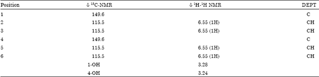
Spectral information: Compound B: FAB-MS = 110.036, Calculated MS for C6H6O2 =110.03678; IR-C-H = 2925, O-H = 3258.5/3624, 1601.2, C = C-1514 and 1365.5, C-O = 1216.1/1096.4, aromatic = 827/758.8, UV = 189 nm, 388.8 nm. Synonyms: para-hydroxyphenol or quininol.
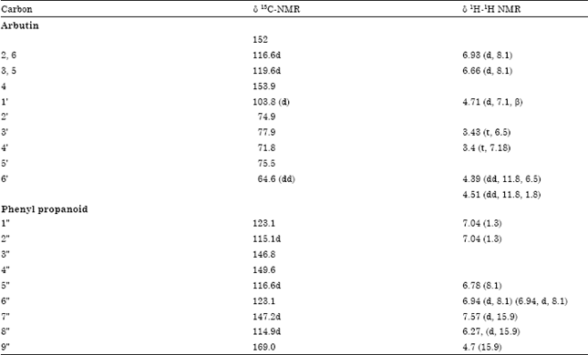
Compound C: Amorphous powder, 1H-NMR spectral data (CD3OD) δ: 3.40 (1H, t, J = 7.18 Hz, H-4'), 3.43 (1H, t, J = 6.5 Hz, H-3'), 4.39 (1H, dd, J = 11.8, 6.5 Hz, H-6'), 4.51 (1H, dd, J =11.8, 1.8 Hz, H-6'), 4.71 (1H, d, J = 7.1 Hz, H-1'), 6.27 (1H, d, J = 15.9, Hz, H-8"), 6.66 (2H, d, J = 8.1 Hz, H-3, H-5), 6.78 (2H, J = 8.1, H-5'), 6.94 (1H, d, J = 8.1 Hz, H-5'), 6.94 (1H, d, J = 8.1 Hz, H-6'), 7.04 (1H, J = 1.3, H-2'), 6.93 (2H, d, J = 8.1 Hz, H-2, H-6), 7.98 (1H, d, J = 15.9 Hz, H-7"). For 13C-NMR spectral data (CD3OD) melting point = 143; FAB MS (positive ion mode) m/z: HR-FAB MS negative ion mode) m/z: 433, calculated for C21H23O10: 435.1219.
Synonym (s): 6-O-Caffeoylarbutin, Robustaside B.
RESULTS
The Total Antioxidant (TA) activity of MECF presented in Fig. 1 is greatest (85.5%, 200 μg mL-1) compared to in CF (44.4%), WF (37.7%), HF (33.3%), BF (23.6%) and EF (5.3%), respectively. TA activity of MECF was not significantly different (p>0.05) from that of allopurinol (96.6%, 1 mM) (standard antioxidant).
As clearly presented in Fig. 1, MECF at 200 μg mL-1 significantly (p<0.05) scavenged superoxide anion radical by 80.4% while similar concentrations of BF, CF, WF, EF and HF elicited 78.6, 68, 57.5, 54 and 30% radical scavenging activities, respectively. There was no significant difference (p<0.05) between the superoxide anion radical scavenging activity of MECF and BF compared to that by allopurinol (85.7%), the standard antioxidant. Furthermore, radical scavenging activity of CF, EF and WF were not significantly different from one another.
Similarly, 200 μg mL-1 of MECF and EF significantly (p<0.05) inhibited the activity of xanthine oxidase by 80 and 87.1% compared to control (Fig. 1). There was no significant difference (p<0.05) in the inhibitions by MECF, EF and allopurinol-the standard antioxidant. Inhibitions by HF, CF, BF and WF of 43.3, 44.2, 39.5 and 48.5%, respectively were also not significantly different from one another (p<0.05) but significantly different from MECF, EF and allopurinol (Fig. 1).
MECF and EF demonstrated similar pattern of inhibition of mitochondrial lipid peroxidation in Fe2+/ascorbate system (Fig. 1). Maximum inhibition of mitochondrial lipid peroxidation (93%) was obtained at 0.2 mg mL-1 of EF and MECF, respectively compared to inhibitions by other fractions. Increasing the concentrations to over 0.3 mg mL-1, showed no significant difference (p<0.05) in the inhibitions by EF, MECF and CF.
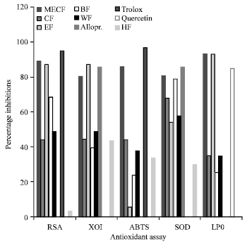 | |
| Fig. 1: | Comparison of antioxidant properties of 200 μg mL-1 of methanolic crude extracts and purified fractions of Cnestis ferruginea. The 200 μg mL -1 each of HF-hexane fraction; CF-chloroform fraction, EF-ethylacetate fraction, BF-Butanol fraction, WF-water fraction were tested for TA-Total antioxidant (ABTS), radical (DPPH, superoxide anion) scavenging activities (RSA), inhibition of Fe2+/ascorbate-induced lipid peroxidation (LPO) and xanthine oxidase activity (IXO) spectrophotometrically at different wavelengths. Values are Mean±SD of three independent experiments. One-way ANOVA was used for comparisons of multiple groups |
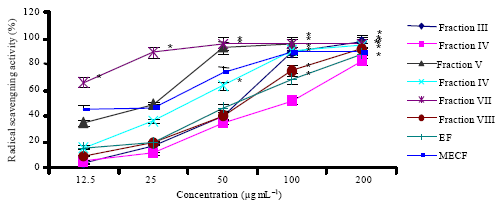 | |
| Fig. 2: | DPPH radical scavenging activities of varying concentrations of subfractions of ethylacetate and methanol fractions of Cnestis ferruginea |
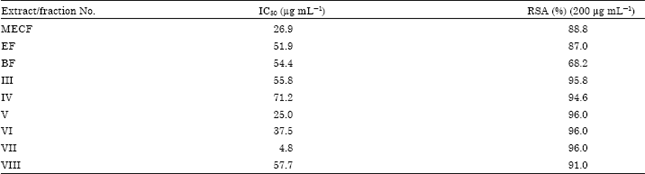
Similarly, MECF and EF (200 μg mL-1) showed very strong DPPH radical scavenging activity (RSA) of 88.8 and 87%, respectively (Fig. 1). But BF showed moderate DPPH RSA of 68.2% while CF and WF exhibited 40 and 48.5% activity, respectively. HF did not show any significant (p>0.05) RSA against DPPH radical at this concentration. Increasing concentrations (12.5-200 μg mL-1) of MECF and EF, scavenged DPPH radicals in a concentration-dependent manner (Fig. 2) and the IC50 values obtained for MECF, EF and BF thereafter, were 26.9, 51.9 and 54.4 μg mL-1, respectively (Table 1).
EF was further purified on silica gel column chromatography because it exhibited similar RSA and anti-lipid peroxidation activity with MECF. Six Sub-fractions (III-VIII) and two pure compounds (A and B) were obtained from this separation process. All these fractions also exhibited very strong DPPH radical scavenging activity of not less than 90% at 200 μg mL-1 which were not significantly different (p<0.05) from each other. Increasing concentrations of these sub-fractions indicated an IC50 value in the order VII> V>VI>III>VIII>IV (Table 1). Although, sub-fraction III showed similar radical scavenging activity (96%) with others but the yield was highest (2.4 g, 0.12%). The chromatograms of these fractions were separately sprayed with DPPH radical solution (1 mg mL-1), FeCl3 and AlCl3. A particular band in the fractions was observed to change the purple colour of DPPH to yellow, FeCl3 to blue and AlCl3 to yellow. Fraction III appeared to contain the highest quantity of the constituents of this band compared to other fractions. Based on this, fraction III was purified further on sephadex LH 20 column and compound C was isolated. The isolated compounds were screened quantitatively for DPPH radical scavenging activity. Compounds B and C at 1 mM exhibited very strong DPPH radical scavenging activities of 86.4 and 85.3%, respectively compared to 92% radical scavenging activity of propylgallate (Fig. 3).
The structure of each compound A, B, C was elucidated by UV, IR, MS. 1H- and 13C-NMR and 2D-NMR methods as β-sitosterol, para-hydroxyphenol and Robustaside B (6'-3", 4"-dihydroxycinnamoyl arbutin. These structures were also confirmed by comparing spectral with previously reported spectral data.
| Table 1: | DPPH free radical scavenging activity of varying concentrations of extract and fractions from Cnestis ferruginea |
 | |
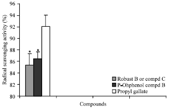 | |
| Fig. 3: | Each compound-Robustaside B, p-hydroxyphenol and propylgallate (1 mM) were incubated with DPPH radical solution (300 μM) for 30 min. Absorbances were read on microplate reader. Values are Mean±SD of three independent experiments. One-way ANOVA was used for comparisons of multiple groups. * Significantly different (p<0.05) from control |
| Table 2: | 13C-NMR and 1H-1H spectral data (CD3OD, 400 MHZ) for Compound B |
 | |
| Table 3: | 13C-NMR and 1H-1H spectral data (500 MHZ) for compound C |
 | |
The spectral data for para-hydroxyphenol and Robustaside B or 6'-(3", 4"-dihydroxycinnamoyl) arbutin are as shown on Table 2 and 3.
DISCUSSION
The measurement of antioxidant activity involves an assessment of the capacity of the substance to inhibit peroxidation of membranes such as erythrocytes, tissue homogenates, mitochondria, microsomes or liposomes (Halliwell, 1990). The phospholipids bilayers of cellular and subcellular membranes are undoubtedly major targets for free radicals. Extracts or compounds inhibit membrane phospholipids peroxidation probably by exerting a pharmacological effect in the prevention of radical-induced oxidative pathological processes (Halliwell, 1991). The peroxidation of PUFAs on the cardiolipin of mitochondrial inner membranes has been suggested to contribute to age-related decline of mitochondrial function. Therefore, mitochondria are among cell-free systems usually chosen to evaluate antioxidant effects on lipid peroxidation (Hsiao et al., 1996). Exposure of mitochondria to Fe2+ and ascorbate elicits lipid peroxidation by a mechanism whose induction step may primarily involve site-bound iron-mediated decomposition of lipid hydroperoxides to yield alkoxy or peroxyl radicals, leading to the chain reaction of lipid peroxidation (Hsiao et al., 1996). DPPH radical is known to abstract labile hydrogen (Constantin et al., 1990) and therefore, the DPPH radical scavenging activity is not unrelated to the inhibition of lipid peroxidation (Ratty et al., 1988; Rekka and Kourounakis, 1991).
In the present study, MECF and its purified fractions scavenged the radicals generated in in vitro systems to different extents. Among the six samples/extracts tested, MECF showed the highest radical scavenging activity, total antioxidant activity, inhibition of xanthine oxidase activity and mitochondrial lipid peroxidation. Thus, it is a rich source of compounds possessing strong antioxidant properties useful for chemotherapy of oxidant-induced pathological conditions in consonance with the reports of Verghese et al. (2008). However, the variation in the radical scavenging activity observed in other fractions supports the claim that different extracts elicit their antioxidant activity by different indices. This finding correlates with previous reports that a single plant extract may not possess the activity of all the different measurements of antioxidant activity (Cakir et al., 2003; Annegowda et al., 2010). Fraction EF and MECF showed similar and strong DPPH radical scavenging activity at 200 μg mL-1 probably by reducing DPPH radicals (violet) to DPPH-H (yellow). They also quenched the radical chain reactions usually propagated by the OH. generated by the Fe2+/ascorbate system to initiate lipid peroxidation to the same extent. These data suggest that the same type of polyhydroxyl phenolic containing compounds may be the major constituents of MECF and EF. This finding confirms and proves that MECF and EF act as direct free radical scavenger. Thus, explaining their strong radical scavenging activity and the low levels of TBARS observed in the present study.
On this basis, the Ethylacetate Fraction (EF) was subjected to further purification on silica gel column using chromatographic techniques. Phytochemical screening of chromatograms of the sub-fractions with DPPH radical solution indicated that Fraction III contained high concentration of a band which markedly reduced DPPH radicals. Again, this band changed FeCl3 to blue and AlCl3 to yellow confirming the presence of trihydroxyl phenols and flavones, respectively. These observations were substantiated by a quantitative DPPH radical scavenging activity assay which indicated the presence of the radical scavenger in all the sub-fractions (Table 1). However, their DPPH RSA is concentration dependent and the strength varies at similar concentrations (Fig. 2). The IC50 values of these sub-fractions revealed their DPPH radical scavenging potential to be in the order VII>V>VI>III>VIII>IV (Fig. 2, Table 1). Sub-fraction III was further purified on sephadex LH 20 because of its high yield and the presence of large quantity of trihydroxylphenol and flavones compared to others. Compound C with RF value 0.43 was obtained from this separation.
The structure of compound B was elucidated by subjecting it to spectroscopic analyses. The HR-FAB MS spectrum of compound B (MZ-111) M++H+ was used to establish the molecular formula C6H6O2. The UV spectrum (λmax-388.3 and 189.2 nm) implied the presence of conjugated chromophore. The IR spectra indicated the presence of conjugated system C = C, signals at 2925 cm-1 represents C-H stretching vibrations. The characteristic signals of OH, C-O and aromatic system are revealed at 3258, 1216.1 and 827/758.8 cm-1, respectively. The standard 13C-NMR, DEPT as well as the polarization transfer experiment of compound B depicted 6 carbon atoms. The DEPT experiment showed resonance for two quarternary carbon, four methines, absence of methylene and methyl groups giving an attached proton formula of C6H4. This molecular formula possesses a double bond equivalent of 4 which was consistent with the exact mass measurement.
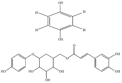 | |
| Fig. 4: | The structure of compound B (para-hydroxyphenol) and C (Robustaside B) |
The clustering of signals between 3.24 and 3.28 is an indication of the presence of hydroxyl group moiety buttressing the IR signals at 3258 cm-1. The structure of compound B (Fig. 4) was proposed to be para-Hydroxyphenol based on comparison with reported spectral data (Table 2).
Also, the structure of the compound C (Fig. 4) was elucidated by subjecting it to spectroscopic analyses. The HR-FAB MS spectrum of compound C (m/z 453) M+H+ established the molecular formula C21H22O10. The UV spectrum (λmax 328 and 199.2 nm) implied the presence of conjugated chromophore. The 1H-NMR spectrum of compound C and 1H-1H COSY, showed signals for an ABC-system [a broad singlet at δ 7.04 (δc 115.1 C-2") and a multiplet at δ6.94 (δc 117.9, 123.1, C-5" and C-6"). The carbon signals are 149.6 and 146.8 at C-3" and C-4", respectively. The 13C-NMR (Table 3) and HMQC spectra of this compound showed 17 signals assigned for 21 carbon atoms in the molecule. These signals also included signals for a sugar moiety, 12 aromatic olefinic carbons and an ester carbonyl. The sugar moiety was identified as glucose by comparing 13C-NMR spectral data with those reported for methyl O-glucosides and by considering the glycosidation effect (Agrawal et al., 1985). These spectral data (Table 3) suggested an arbutin residue acylated at C-6" of the sugar moiety (δ4.39 and 4.51, 64.7) with a p-hydroxycinnamate (Occolowitz, 1964). Fragments ions at m/z 324.3 and 162.2 (cinnamoyl) supports this partial structure. The coupling constant of H-1' (J = 7.1 Hz) indicated a β configuration of the sugar moiety. Compound C was named 6'-(3", 4"-dihydroxycinnamoyl) arbutin or Robustaside B based on comparison with previously reported spectral data and authentic standards (Dommisse et al., 1986). This compound has an existing chemical structure previously isolated in Grevillea robusta by Ahmed et al. (2000) along with its several similar analogues. However, the present study was the first chemical investigation of C. ferruginea leaves which led to the isolation of Robustaside B and para-Hydroxyphenol from this source. Robustaside B belongs to the derivatives originating from Arbutin (Ahmed et al., 2000; He et al., 2006). Arbutins are known for their diuretic and urinary anti-infective activities for centuries either as a plant extract or in purified form (Robertson and Howard, 1987). Furthermore, arbutin is known to be an inhibitor of melanin biosynthesis (Akiu et al., 1988) and is used as a skin-whitening agent in cosmetics. In addition, the present study revealed the radical scavenging activity of arbutin-derivative-Robustaside B for the first time.
In conclusion, the present study isolated Robustaside B and para-Hydroxyphenol which are phenolic compounds from the leaves of C. ferruginea for the first time. Robustaside B and para-Hydroxyphenol were established to be potent free radical scavengers. C. ferruginea was also identified from this study to be a rich source of potent antioxidant principles which are direct free radical scavengers. These bioactive agents would find therapeutic uses in militating against age related disorders; cancer, diabetes, gout, liver and kidney dysfunction; and other radical-induced pathological conditions.
ACKNOWLEDGMENT
This study was supported by the Third World Organization for Women in Science through their Postgraduate Training Fellowship received by Dr. Rahmat Adetutu Adisa in 2004 and utilized at H.E.J Research Institute of Chemistry, University of Karachi, Karachi.
REFERENCES
- Adisa, R., M. Choudhary, E. Adewoye and O. Olorunsogo, 2010. Hypoglycaemic and biochemical properties of Cnestis ferruginea. Afr. J. Tradit. Complement Altern Med., 7: 185-194.
PubMed - Adisa, R.A., J.M. Oke, S.A. Olomu and O.O. Olorunsogo, 2004. Inhibition of human haemoglobin glycosylation by flavonoid-containing leaf extracts of Cnestis ferruginea. J. Cameroon Acad. Sci., 4: 351-359.
Direct Link - Agrawal, P.K., D.C. Jain, R.K. Gupta and R.S. Thakur, 1985. Carbon-13 NMR spectroscopy of steroidal sapogenins and steroidal saponins. Phytochemistry, 24: 2479-2496.
CrossRef - Ahmed, A.S., N. Nakamura, M.R. Meselhy, M.A. Makhboul, N. El-Emary and M. Hattori, 2000. Phenolic constituents from Grevillea robusta. Phytochemistry, 53: 149-154.
CrossRef - Ames, B.N., M.K. Shigenaga and T.M. Hagen, 1993. Oxidants, antioxidants and the degenerative diseases of aging. Proc. Nat. Acad. Sci. USA, 90: 7915-7922.
CrossRefDirect Link - Annegowda, H.V., C.W. Nee, M.N. Mordi, S. Ramanathan and S.M. Mansor, 2010. Evaluation of phenolic content and antioxidant property of hydrolysed extracts of Terminalia catappa L. leaf. Asian J. Plant Sci., 9: 479-485.
CrossRefDirect Link - Arya, V. and J.P. Yadav, 2011. Antioxidant properties of the methanol extracts of the leaves, seeds and stem of Cassia occidentalis. Res. J. Med. Plant, 5: 547-556.
CrossRef - Atere, T.G. and A.T. Ajao, 2009. Toxicological implications of crude alkaloidal fraction from Cnestis ferruginea D.C root on liver function indices of male Wistar rats. Int. J. Biom. Health Sci., 5 : 145-156.
Direct Link - Boakye-Yiadom, K. and G.H. Konning, 1975. Incidence of antibacterial activity in the Connaraceae. Planta Med., 28: 397-400.
CrossRef - Butkhup, L. and S. Samappito, 2011. In vitro free radical scavenging and antimicrobial activity of some selected Thai medicinal plants. Res. J. Med. Plant, 5: 254-265.
CrossRefDirect Link - Cakir, A., A. Mavi, A. Yildirim, M.E. Duru, M. Harmandar and C. Kazaz, 2003. Isolation and characterization of antioxidant phenolic compounds from the aerial parts of Hypericum hyssopifolium L. by activity-guided fractionation. J. Ethnopharmcol., 87: 73-83.
PubMed - Cano, A., O. Alcaraz, M. Acosta and M.B. Arnao, 2002. On-line antioxidant activity determination: Comparison of hydrophilic and lipophilic antioxidant activity using the ABTS.+ assay. Redox Rep., 7: 103-109.
CrossRef - Constantin, M., C. Bromont, R. Fickat and R. Massingham, 1990. Studies on the activity of bepridil as a scavenger of free radicals. Biochem. Pharmacol., 40: 1615-1622.
CrossRef - De Gaulejac, N.S.C., C. Provost and N. Vivas, 1999. Comparative study of polyphenol scavenging activities assessed by different methods. J. Agric. Food Chem., 47: 425-431.
CrossRefPubMedDirect Link - Dommisse, R.A., L. van Hoof and A.J. Vlietinck, 1986. Structural analysis of phenolic glucosides from Salicaceae by NMR spectroscopy. Phytochemistry, 25: 1201-1204.
CrossRef - Halliwell, B., 1991. Drug antioxidant effects. A basis for drug selection. Drugs, 42: 569-605.
CrossRefPubMedDirect Link - Halliwell, B., 1990. How to characterize a biological antioxidant. Free Raical Res. Commun., 9: 1-32.
PubMedDirect Link - Heijnen, C.G., G.R. Haenen, R.M. Oostveen, E.M. Stalpers and A. Bast, 2002. Protection of flavonoids against lipid peroxidation: The structure activity relationship revisited. Free Radic. Res., 36: 575-581.
PubMed - Jain, V., S.K. Verma, S.S. Katewa, S. Anandjiwala and B. Singh, 2011. Free radical scavenging property of Bombax ceiba Linn. root. Res. J. Med. Plant, 5: 462-470.
CrossRefDirect Link - Johnson, D. and H. Lardy, 1967. Isolation of liver or kidney mitochondria. Methods Enzymol, 10: 94-96.
CrossRefDirect Link - Kim, D.O., O.K. Chun, Y.J. Kim, H.Y. Moon and C.Y. Lee, 2003. Quantification of polyphenolics and their antioxidant capacity in fresh plums. J. Agric. Food Chem., 51: 6509-6515.
CrossRefDirect Link - Lee, S.K., Z.H. Mbwambo, H. Chung, L. Luyengi and E.J. Gamez et al., 1998. Evaluation of antioxidant potential of natural products. Comb. Chem. High Throughput Screen., 1: 35-46.
PubMed - Lowry, O.H., N.J. Rosebrough, A.L. Farr and R.J. Randall, 1951. Protein measurement with the folin phenol reagent. J. Biol. Chem., 193: 265-275.
CrossRefPubMedDirect Link - Nijveldt, R.J., E. van Nood, D.E.C. van Hoorn, P.G. Boelens, K. van Norren and P.A.M. van Leeuwen, 2001. Flavonoids: A review of probable mechanisms of action and potential applications. Am. J. Clin. Nutr., 74: 418-425.
CrossRefDirect Link - Nishikimi, M., N.A. Rao and K. Yagi, 1972. The occurrence of superoxide anion in the reaction of reduced phenazine methosulfate and molecular oxygen. Biochem. Biophys. Res. Commun., 46: 849-854.
CrossRefPubMedDirect Link - Occolowitz, J.L., 1964. Mass spectrometry of naturally occurring alkenyl phenols and their derivatives Anal. Chem., 36: 2177-2181.
CrossRef - Oktay, M., I. Gulcin and O.I. Kufrevioglu, 2003. Determination of in vitro antioxidant activity of fennel (Foeniculum vulgare) seed extracts. LWT-Food Sci. Technol., 36: 263-271.
CrossRefDirect Link - Olugbade, T.A., J.O. Oluwadiya and W.A. Yisak, 1982. Chemical constituents of Cnestis ferruginea DC. I. Petroleum ether fraction. J. Ethnopharmacol., 6: 365-370.
Direct Link - Ratty, A.K., J. Sunamoto and N.P. Das, 1998. Interaction of flavonoids with 1,1-diphenyl-2-picrylhydrazyl free radical, liposomal membranes and soybean lipoxygenase-1. Biochem. Pharmacol., 37: 989-995.
CrossRefDirect Link - Rekka, E. and P.N. Kourounakis, 1991. Effect of hydroxyethyl rutosides and related compounds on lipid peroxidation and free radical scavenging activity. Some structural aspects. J. Pharm. Pharmacol., 43: 486-491.
CrossRefPubMedDirect Link - Robertson, J.A. and L.A. Howard, 1987. Effect of carbohydrates on growth of Ureaplasma urealyticum and Mycoplasma hominis. J. Clin. Microbiol., 25: 160-161.
Direct Link - Tomohiro, T., K. Futoshi, W. Naoharu, Y. Akihito and S. Kanzo, 1994. A simple screening method for antioxidants and isolation of several antioxidants produced by marine bacteria from fish and shellfish. Biosci. Biotechnol. Biochem., 58: 1780-1783.
Direct Link - Varshney, R. and R.K. Kale, 1990. Effects of calmodulin antagonists on radiation-induced lipid peroxidation in microsomes. Int. J. Radiat. Biol., 58: 733-743.
CrossRefDirect Link - Verghese, M., M.S.C. Fullerton, L.T. Walker, L.A. Shackelford and E. Cebert et al., 2008. Determination of antioxidant contents in red sorrel and its anticarcinogenic potential in azoxymethane-induced colonic aberrant crypt foci. Res. J. Phytochem., 2: 69-76.
CrossRefDirect Link