
C.U. Nwaigwe
Department of Veterinary Physiology and Pharmacology, Faculty of Veterinary Medicine, University of Nigeria, Nsukka, Nigeria
I.I. Madubunyi
Department of Veterinary Physiology and Pharmacology, Faculty of Veterinary Medicine, University of Nigeria, Nsukka, Nigeria
S.C. Udem
Department of Veterinary Physiology and Pharmacology, Faculty of Veterinary Medicine, University of Nigeria, Nsukka, Nigeria
C.O. Nwaigwe
Department of Animal Health and Production, Faculty of Veterinary Medicine, University of Nigeria, Nsukka, Nigeria
Research Journal of Medicinal Plants
Year: 2012 | Volume: 6 | Issue: 5 | Page No.: 395-405


ABSTRACT
The hepatoprotective activity of Olax viridis Oliv. (Olacaceae Juss.) methanol root extracts were tested in vivo and in vitro. The plant material was defatted with n-hexane and extracted with 70% methanol, respectively. The methanol extract was recovered in 11.73% w/w yield. An oral 100 mg kg-1 administration of the methanol extract significantly reduced pentobarbitone-induced sleep in rats poisoned with acetaminophen (p<0.05). In this model, a protection of 87% against the cytotoxicity of acetaminophen was obtained by pretreatment with the methanol extract as compared to a protection of 91% when the animals were pretreated with silymarin. The n-hexane extract was without a significant hepatoprotective effect in this model. Intraperitoneal injection of the methanol extract into rats showed no significant effect on pentobarbitone-induced hypnosis (p<0.05). The elevation of serum glutamate oxaloacetate transaminase (GOT), glutamate pyruvate transaminase (GPT), alkaline phosphatase (ALP) and urea induced by paracetamol intoxication in rats was also significantly attenuated by the methanolic extract (p<0.05). These findings demonstrated that O. viridis root extract has protective effects on experimental model of liver diseases in rats and validates its use in Nigerian traditional medicine for the prevention of liver diseases. Even in its crude form, the effects were comparable to that of silymarin, a flavanolignan with proven antihepatotoxic activity. This suggests that the extract could be a potential source of a novel antihepatotoxic agent.
PDF Abstract XML References Citation
Received: August 02, 2011;
Accepted: December 07, 2011;
Published: January 31, 2012
How to cite this article
C.U. Nwaigwe, I.I. Madubunyi, S.C. Udem and C.O. Nwaigwe, 2012. Methanolic Root Extract of Olax viridis Protects the Liver against Acetaminophen-induced Liver Damage. Research Journal of Medicinal Plants, 6: 395-405.
URL: https://scialert.net/abstract/?doi=rjmp.2012.395.405
URL: https://scialert.net/abstract/?doi=rjmp.2012.395.405
INTRODUCTION
In recent decades, there are many claims on the use of medicinal plants in the treatment of many diseases (Agrawal et al., 2011; Gill et al., 2012). Despite the traditional claims, it is very important to have sufficient knowledge regarding their therapeutic uses through detailed scientific investigation (Serdevi et al., 2007; Dalirsani et al., 2011). The use of the medicinal herbs for treating ailments has been documented in history of all civilizations (Paliwal et al., 2011; Danquah et al., 2011). From World Health Organization records, plant-based remedies are now being used by 80% of the world population as their primary form of healthcare (Hemabarathy et al., 2009; Sarwar et al., 2011; Shafaei et al., 2011). Consequently, herbal remedies are being investigated for their efficacy in the treatment of many conditions including hepatic injuries caused by Carbon tetra chloride (Alisi et al., 2011), Paracetamol (PCM) (El-Gohary et al., 2009) etc.
One of such traditionally acclaimed hepatoprotective plants is Olax viridis Oliv. (Olacaceae Juss.), known as “Atu ndi umoha” in Nsukka native language of Nigeria. It is a shrub commonly found in the Tropics and grows well in the Forest and in Savannah regions (Burkill, 1997).
In our studies on Nigerian medicinal plants, we now report the hepatoprotective effects of O. viridis root in an attempt to establish the scientific basis for its use in Nigerian traditional medicine for the management of liver ailments.
MATERIALS AND METHODS
The experiment was performed as approved by the Ethics Committee of the University of Nigeria, Nsukka, in accordance with the guide to the care and use of laboratory animals in research and teaching in the University. Freshly prepared solutions of drugs and physiological solutions were used in all experiments.
Collection and identification of plant material: The fresh root materials used in this investigation were collected in December, 2010 from Obollo-Eke in Udenu Local Government Area of Enugu State, Nigeria and identified as Olax viridis Oliv. (Olacaceae Juss.), in the Department of Botany, University of Nigeria, Nsukka. A voucher specimen was deposited in the departmental Herbarium.
Collection of the experimental animals: Male Wistar albino rats weighing between 150-200 g, were procured from the Federal University of Agriculture, Makurdi, Nigeria. They were kept in clean stainless steel cages and were fed ad libitum with standard laboratory animal feed (Guinea Feed®) and with access to clean drinking water. They were maintained in accordance with the recommendation in the Guide for the Care and Use of Laboratory Animal (DDHS, 1985). They were allowed two weeks to acclimatize before the commencement of the experiments.
Preparation of extracts from roots of O. viridis: The fresh roots of O. viridis were dried at room temperature and reduced to a powder by grinding using a Hammer-mill with a 2 mm filter. One kilogram of the dried powder was defatted with 3 L absolute n-hexane in a Soxhlet apparatus for 24 h and then extracted with 70% methanol for 72 h. Removal of solvents in vacuo at 40°C afforded 17 g of n-hexane extract (HE) and 117.3 g of methanol extract. The extracts were stored at 4°C until used.
Acute toxicity test: The acute toxicity test of the ethyl acetate portion of the methanol root extract of O. viridis was determined. Thirty five rats were randomly selected into five groups of six animals each and were allowed free access to feed and water. Groups 2-4 were injected with varying doses (i.e., 0.2, 0.4, 0.8, 1.2, 2.0 mg kg-1 b.wt., i.p.) of the 70% methanol extract in normal saline, while group 1 which served as the control group received the same volume of normal saline only by the same route. The volumes of normal saline and extract administered were maintained at 3 mL kg-1. After treatment the rats were fed diet containing 16% protein, with drinking water given ad libitum. They were accommodated in metal cages and observed for clinical signs over a period of 24 h. Deaths within this period were recorded and the LD50 determined following the method of Lorke (1983).
Effect of the extract on pentobarbitone sleeping time: This experiment was carried out using the method of MacLeod (1970). Five groups of five rats (100-150 g) of both sexes per group were used for the experiment. Group 1 which served as the control was injected with pentobarbitone-Na (35 mg kg-1 b.wt., i.p.). Groups 2 to 3 were similarly treated 30 min after they were treated with increasing doses of 100 and 200 mg kg-1, b.wt., p.o. of the extract, respectively. The time of injection, time of sleep (when writhing reflex was lost) and the time of awakening (when writhing reflex was regained) were recorded and the mean sleeping times calculated and subjected to one-way analysis of variance (ANOVA).
Effect on acetaminophen-induced hepatotoxicity in rats: The extract was dissolved in normal saline and administered orally to groups of randomly selected rats (5 per group). In testing for protective activity of the extracts, treatment was repeated every 12 h for five days. one hour after the last dose, each animal was challenged with a single oral dose of acetaminophen (2000 mg kg-1 b.wt.). Eighteen hours later, each group was given a single dose of pentobarbitone-Sodium (35 mg kg-1 b.wt., i.p.) and the duration of sleep for each animal recorded. These experiments were conducted parallel with silymarin (100 mg kg-1 b.wt.) as a reference drug. The time of injection, time of sleep (when writhing reflex was lost) and the time of awakening (when writhing reflex was regained) were recorded. The mean sleeping times calculated and subjected to one-way analysis of variance (ANOVA).
Effects of Olax viridis methanol root extract on acetaminophen-induced elevation of serum enzymes and bilirubin level in rats: Five groups of five rats (100-150 g) were used for this experiment. The extract was reconstituted in normal saline and administered orally to the experimental groups. The control group (group 1) received only normal saline twice daily for five days. Groups 2 and 3 were administered 100 and 200 mg kg-1 of the extract, respectively and group 4 was of silymarin (100 mg kg-1, b.wt., p.o.). Treatment was repeated every 12 h for five consecutive days.
One hour after the last dose, each animal was challenged with a single oral dose of acetaminophen (200 mg kg-1 b.wt., p.o.). Another control group received acetaminophen in normal saline only. Eighteen hours later, the animals were anaesthetized with ether and blood samples obtained using the orbital technique into clean glass test tubes. The blood samples were kept at room temperature for 30 min to clot after which they were centrifuged at 3,000 revolutions per minute for ten minutes using a table centrifuge. The clear supernatant was then carefully aspirated with a syringe, into a clean sample bottle for the enzymes and bilirubin determination using Quimica Clinica Applicada (QCA) test kits. Serum levels of aspartate amino-transferase (GOT) and alanine amino-transferase (GPT) were measured in the serum by the method of Reitman and Frankel (1957). The serum alkaline phosphate level was determined using the phenolphthalein monophosphate method as described by Babson et al. (1966) while the total bilirubin and direct bilirubin levels in the serum were determined using the Jendrassik-Grof method as described by Doumas et al. (1973).
Histopathological studies: After the blood (2 mL) collection for serum enzyme assay, the animals were sacrificed by cervical decapitation and sections of the liver were removed for histopathology. The liver tissues collected were washed in normal saline and fixed in 10% solution of formalin. They were then dehydrated in graded alcohol, cleared in xylene and embedded in paraffin wax. Cut sections (5 μm thick) were stained with haematoxylin and eosin (H and E) stain for histopathological examination using light microscope.
Statistical analyses: Data were analyzed using one-way analysis of variance (ANOVA) followed by post hoc Duncan’s test. The p-values less than 0.05 were considered statistically significant.
RESULTS
Preparation of the extract: The weight of the methanol extract of O. viridis root recovered was 117.3 g. The extract was reddish brown in color, possessed a mild aromatic flavor and gave a percentage yield of 11.73% w/w. The results of the phytochemical analysis revealed the presence of saponins, alkaloids, flavonoids and tannins in the methanol extract of O. viridis roots.
Acute toxicity test: At the doses of 200 and 400 mg kg-1 b.wt., the animals did not show any sign of toxicity. The animals given 800 mg kg-1 b.wt., exhibited loss of appetite, dullness and clumping together but no deaths were recorded. At 1200 and 2000 mg kg-1 b.wt., the animals had loss of appetite and hyper-apnea. One mortality was recorded at the dose of 1200 mg kg-1 b.wt., while only one survivor was recorded in the group administered 2000 mg kg-1 b.wt. of the methanol extract. The LD50 was calculated to be 1,585 mg kg-1 b.wt.
Effect of extracts on pentobarbitone-induced hypnosis in rats intoxicated with acetaminophen: A prophylactic application of the methanol extract (100 and 200 mg kg-1 b.wt.) of O. viridis root significantly (p<0.05) reduced the prolongation of pentobarbitone-induced hypnosis in rats challenged with acetaminophen (2000 mg kg-1 b.wt.), as compared to animals that received acetaminophen alone (Table 1). The effect of the methanol extract was comparable to that of silymarin. The petroleum ether extract did not show any form of hepatoprotective effect in this model.
Effect of methanol extract on pentobarbitone-induced hypnosis in rats: The methanol extract of O. viridis root showed no significant effect on the duration of sleep in pentobarbitone anesthesia in rats at the dose range of 50-100 mg kg-1 (Table 2).
| Table 1: | Effect of prophylactic doses of Olax viridis root extract on pentobarbital-induced hypnosis in acetaminophen poisoned rats |
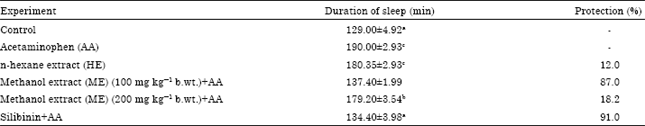 | |
| Values are Mean±SEM; n = 6. Means with different superscripts are significant at p<0.05 | |
| Table 2: | Effect of the methanol root extract of Olax viridis on pentobarbital-induced sleep in rats |
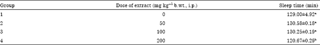 | |
| Values are Means±SEM; n = 5. Means with different superscripts are significant at p<0.05 | |
Effect on acetaminophen-induced elevation of serum enzymes and total bilirubin: Acetaminophen caused liver damage as manifested by the remarkable increase in the serum levels of glutamate oxaloacetate transaminase (GOT), glutamate pyruvate transaminase (GPT) and alkaline phosphate (ALP) as compared to control. At the probability level of (p<0.05), the serum SGOT, SGPT, ALP, total and direct bilirubin levels of the groups given 100 mg kg-1 of the extract and the group that received 100 mg kg-1 silymarin did not differ significantly from each other or the control group, but had a significantly lower (p<0.05) SGOT and SGPT levels when compared to the group that received 200 mg kg-1 of the extract and the group that received only acetaminophen. The group that received 200 mg kg-1 of the extract had a significantly lower (p<0.05) level of SGPT, but showed no significant difference (p<0.05) in the levels of SGOT, ALP, total bilirubin and direct bilirubin levels when compared to the group that received only acetaminophen. There was no significant difference (p<0.05) in the ALP, total bilirubin and direct bilirubin levels of the group treated with 100 mg kg-1 of the extract when compared to the group that received 200 mg kg-1 of the extract. The serum enzymes and bilirubin levels of the group that received acetaminophen was significantly higher (p<0.05) when compared to the control group (Table 3). These results were comparable to that of silymarin.
Histopathological studies: The control group had a normal liver architecture (Fig. 1). The group that received (100 mg kg-1 b.wt.) had a normal liver architecture comparable to the control group (Fig. 2). Mild glycogen degeneration around the central vein was observed in the animals treated with O. viridis root extract (100 mg kg-1 b.wt., p.o.).
| Table 3: | Effect of the methanol root extract of Olax viridis on acetaminophen-induced elevation of serum enzymes and bilirubin in rats |
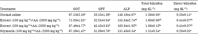 | |
| GOT: Glutamate oxaloacetate transaminase, GPT: Glutamate pyruvate transaminase, ALP: Alkaline phosphatase (ALP), AA: Acetaminophen. Values are Means±SEM; n = 5. Means with different superscripts down a column are significantly different at p<0.05 | |
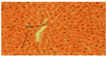 | |
| Fig. 1: | Liver of rats in the control group administered normal saline showing normal hepatocytes (NH) (H and E x400) |
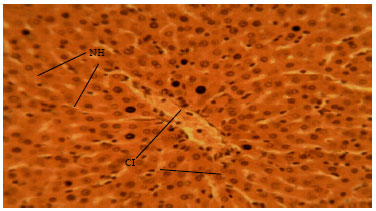 | |
| Fig. 2: | Liver of rats treated with 100 mg kg¯1 of silymarin and exposed to acetaminophen showing normal hepatocytes (NH), with mild cellular infiltration (CI) of the hepatocytes (H and E x400) |
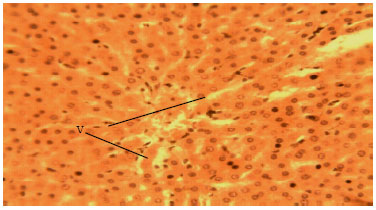 | |
| Fig. 3: | Liver of rats treated with 100 mg kg¯1 of methanol extract of Olax viridis and exposed to Acetaminophen showing mild vacuolation (V) (H and E x400) |
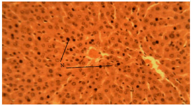 | |
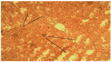
| Fig. 4: | Liver of rats treated with methanol root extract of Olax viridis at 200 mg kg¯1 and exposed to acetaminophen showing mild necrosis (N) of hepatocytes (H and E x400) |
 | |
| Fig. 5: | Liver of rats exposed to acetaminophen only showing severe cellular degeneration (SD) and necrosis (N) (H and E x100) |
The cells were radially arranged and the vacuolation was presented similar to that of the normal liver (Fig. 3). Glycogen degeneration and hepatic necrosis were seen in the liver histopathology of the group that received 200 mg kg-1 b.wt. of the extract (Fig. 4, 5).
DISCUSSION
The hepatoprotective activity of the extracts of O. viridis roots was first of all ascertained by determining a functional parameter, that is, pentobarbitone-induced hypnosis in rats poisoned with acetaminophen. The Methanol Extract (ME) at a dose of 100 mg kg-1 b.wt. remarkably protected the liver from acetaminophen-induced damage. ME, at a dose of 200 mg kg-1 b.wt. was less effective in this model. The n-hexane (HE) extract was without effect in this model (Table 1). This finding indicated that the hepatoprotective component(s) are present in the methanol extract. Therefore all further investigations were done with the methanol extract only.
To insure that the hepatoprotective effect of the ME is not due to its action on the Central Nervous System (CNS), the influence of the ME extract on the duration of barbiturate-induced hypnosis in normal albino rats was tested at a dose range (50-200 mg kg-1) used for the experiments. The ME extract (at a dose of 100 mg kg-1 b.wt., p.o.) did not show any effect on barbiturate-induced hypnosis in rats (Table 2), suggesting that it has no influence on the central nervous system (CNS). The liver protective effect of the ME extract was therefore not due to its action on the CNS since at a dose range of 50-100 mg kg-1, it did not influence the duration of barbiturate-induced hypnosis in normal animals (Table 2).
The length of barbiturate-induced sleep is related to the rate of detoxification of barbiturate by the P-450 cytochromes in the liver (Lutz and Krafft, 1997). Therefore, the ability of the extract to reduce the prolongation of the barbiturate-induced sleep in rats challenged with acetaminophen is indicative of the antihepatotoxic potential of the extract. Barbiturates are almost exclusively metabolized in the liver. Therefore, the sleeping time after a given dose can serve as a measure of hepatic metabolism (Vogel, 1977). If there was any pre-existing liver damage as in the case of acetaminophen-poisoning, the sleeping time after a given dose of the barbiturate will be prolonged because the amount of the hypnotic broken down per unit time will be less (Gupta and Dixit, 2009).
Result presented in Table 1 indicate that the duration of sleep of the animals that received 100 mg kg-1 b.wt. of the extract did not differ significantly from that of the control group and the group that received the reference drug, silymarin. This observation further proves the efficacy of the extract as a hepatoprotective agent. The significantly longer duration of sleep of the group treated 200 mg kg-1 b.wt. and the group that received acetaminophen only is an indication of hepatic impairment. This agrees with the work of Gupta and Dixit (2009).
The development of liver necrosis is acetaminophen poison is associated with the release of hepatic enzymes into the serum with the consequent increase in the level of these enzymes in the serum. The hepatoprotective activity of the methanol extract of O. viridis roots was therefore further evaluated by determining its effect s on the enzyme levels in rats challenged with acetaminophen. The lowering of enzyme level is a definite indication of the hepatoprotective action of the drug. Our results were further substantiated by the finding that pretreatment with the methanol extract remarkably reduced the increases in the serum level of serum transaminases in the liver of rats challenged with acetaminophen (Table 3). Elevated levels of serum enzymes are indicative of cellular leakage and loss of functional integrity of cell membrane of the liver (Drotman and Lawhan, 1978). High level of SGOT can be due to liver damage, viral hepatitis as well as cardiac infarction and muscle injury. SGPT is more specific to the liver and is thus a better parameter for detecting liver injury. Even though there was a significant increase in the SGPT level in the group treated with methanol extract (200 mg kg-1 b.wt., p.o.) when compared to the control, its significant lower level when compared to the group that received acetaminophen only is an indication that this dose offered a mild protection to the liver. While acute hepatocellular necrosis results in dramatic increases in serum SGOT and SGPT levels, minimal increases in serum ALP levels are observed in the same disease condition. Increase in serum ALP as observed in the group treated with acetaminophen only and in the group treated with 200 mg kg-1 of the extract is likely due to increased synthesis of this enzyme in the presence of increasing biliary pressure (Muriel et al., 1992).
In hepatocellular damage, free bilirubin combined with albumin might not be separated by the liver and continues to circulate at high concentrations (Reece, 2004). According to Coles (1986), if more than 25% of the total serum bilirubin is of the conjugated type, hepatocellular damage should be suspected and if conjugated bilirubin is greater than 30% of total serum bilirubin, cholestasis should be suspected. Based on this formula, form the results in Table 3, it can be seen that the damage caused by acetaminophen is hepatocellular and not cholestatic in nature and that all doses of the extract offered protection against acetaminophen- induced liver damage, even though this was not apparent in the group that received 200 mg kg-1 of the extract. The extract and silymarin, at a dose of 100 mg kg-1, respectively, protected the liver against hepatic damage caused by acetaminophen as observed by the decrease in serum level of SGOT, SGPT. ALP, total and conjugated bilirubin when compared to groups that received the m ethanol extract (200 mg kg-1 b.wt., p.o.).
The hepatoprotective activity of O. viridis methanol extract was further confirmed through histopathological studies on the liver rats. The administration of acetaminophen alone produced extensive vascular degenerative changes and necrosis of hepatocytes. These findings agree with the works of Olaleye et al. (2006), who reported that disorientation of the parenchyma tissue of the liver, vacuolar formation in the parenchyma and necrosis of hepatocytes were observed in the histopathological studies of liver of rats treated with acetaminophen (2000 mg kg-1 b.wt., p.o.). Pretreatment with ME before inducing liver damage with acetaminophen produced a lesser degree of damage to the liver cells as compared to the animals treated with acetaminophen alone. The sections of the liver of animals treated with of ME (100 mg kg-1 b.wt., p.o.) and then acetaminophen showed a better hepato-protective activity having only mild degenerative changes and the absence of pyknotic or necrotic hepatocytes. These observations further pointed out the efficacy of ME as a source of a novel hepatoprotective agent.
The hepatotoxicity of acetaminophen is thought to occur in two stages: (1) metabolic phase and (2) oxidative phase (James et al., 2003). The metabolic consists of formation of toxic metabolites when part of it is oxidized by hepatic cytochrome P-450-dependant mixed-function oxidase system (Savides and Oehme, 1983) to a highly reactive cytotoxic, electrophilic metabolite N-acetyl-para-benzoquinoneimine (NAPQI), which under normal conditions is rapidly detoxified by conjugation with reduced hepatic glutathione (GSH)-glutathione transferase (GST) systems to yield a harmless product called mercapturic acid that is renally excreted (Mitchell et al., 1973). Although remarkably safe at therapeutic doses, excessive amounts of acetaminophen can produce centrilobular hepatic necrosis and acute renal tubular necrosis which are the leading causes of acute liver failure (Savides and Oehme, 1983).
Phytochemical studies show that the roots contain flavonoids. Flavonoids are known to scavenge on free radicals (Pathak et al., 1991; Al-Humaid et al., 2010). This suggests the possibility that the active component(s) in the methanol extract of O. viridis root may be scavenging on the radicals. Further investigations for the isolation, identification and structure determination of the active principles responsible for the hepatoprotective activity of O. viridis root methanol extract are in progress.
CONCLUSION
These findings demonstrate that O. viridis root has protective effects on experimental model of liver disease in rats and validates its use in Nigerian traditional medicine for the prevention of liver diseases. Even in its crude form, the effects were comparable to that of silymarin, a flavanolignan with proven antihepatotoxic activity which suggests that the extract could be a potential source of a novel antihepatotoxic agent.
REFERENCES
- Agrawal, B., S. Das and A. Pandey, 2011. Boerhaavia diffusa Linn: A review on its phytochemical and pharmacological profile. Asian J. Applied Sci., 4: 663-684.
CrossRef - Al-Humaid, A.I., H.M. Mousa, R.A. El-Mergawi and A.M. Abdel-Salam, 2010. Chemical composition and antioxidant activity of dates and dates-camel-milk mixtures as a protective meal against lipid peroxidation in rats. Am. J. Food Technol., 5: 22-30.
CrossRefDirect Link - Danquah, C.A., E. Woode, E.B. Gyasi, M. Duwiejua and C. Ansah, 2011. Anti-inflammatory and antipyretic effects of an ethanolic extract of Capparis erythrocarpos isert roots. Res. J. Med. Plant, 5: 158-168.
CrossRefDirect Link - Babson, A.L., S.J. Greeley, C.M. Coleman and G.E. Philips, 1966. Phenolphthalein monophosphate as a substrate for serum alkaline phosphatase. Clin. Chem., 12: 482-490.
Direct Link - Dalirsani, Z., M. Aghazadeh, M. Adibpour, M. Amirchaghmaghi and A. Pakfetrat et al., 2011. In vitro comparison of the antimicrobial activity of ten herbal extracts against Streptococcus mutans with chlorhexidine. J. Applied Sci., 11: 878-882.
Direct Link - Doumas, B.T., B.W. Perry, E.A. Sasse and J.V. Straumfjord, Jr., 1973. Standardization in bilirubin assays: Evaluation of selected methods and stability of bilirubin solutions. Clin. Chem., 19: 984-993.
Direct Link - Drotman, R.B. and G.T. Lawhorn, 1978. Serum enzymes are indications of chemical induced Liver damage. Drug Chem. Toxicol., 1: 163-171.
PubMed - Gill, N.S., R. Kaur, R. Arora and M. Bali, 2012. Phytochemical investigation of Caesalpinia crista seed extract for their therapeutic potential. Res. J. Med. Plant, 6: 100-107.
CrossRef - El-Gohary, M., M. Gamil, K. Girgis and S. Nabil, 2009. Scalp nerve block in children undergoing a supratentorial craniotomy: A randomized controlled study. Asian J. Scientific Res., 2: 105-112.
CrossRefDirect Link - Hemabarathy, B., S.B. Budin and V. Feizal, 2009. Paracetamol hepatotoxicity in rats treated with crude extract of Alpinia galanga. J. Biol. Sci., 9: 57-62.
CrossRefDirect Link - James, L.P., P.R. Mayeux and J.A. Hinson, 2003. Acetaminophen-induced hepatotoxicity. Drug Metab. Dispos., 31: 1499-1506.
CrossRefPubMedDirect Link - Lorke, D., 1983. A new approach to practical acute toxicity testing. Arch. Toxicol., 54: 275-287.
CrossRefPubMedDirect Link - Lutz, J. and M.P. Krafft, 1997. Longitudinal studies on the interaction of perfluorochemicals with liver cytochromes p-450 by means of testing the rate of detoxification of pentobarbital. Adv. Exp. Med. Boil., 411: 391-394.
PubMed - Mitchell, J.R., D.J. Jollow, W.Z. Potter, D.C. Davis, J.R. Gillette and B.B. Brodie, 1973. Acetaminophen-induced hepatic necrosis. I. Role of drug metabolism. J. Pharmacol. Exp. Ther., 187: 185-194.
PubMedDirect Link - Muriel, P., T. Garciapina, V. Perez-Alvarez and M. Mourelle, 1992. Silymarin protects against paracetamol-induced lipid peroxidation and liver damage. J. Applied Toxicol., 12: 439-442.
CrossRefPubMedDirect Link - Olaleye, M.T., O.O. Adegboye and A.A. Akindahunsi, 2006. Alchornea cordifolia extract protects wistar albino rats against acetaminophen-induced liver damage. Afr. J. Biotechnol., 5: 2439-2445.
Direct Link - Paliwal, R., V. Sharma and Pracheta, 2011. A review on horse radish tree (Moringa oleifera): A multipurpose tree with high economic and commercial importance. Asian J. Biotechnol., 3: 317-328.
CrossRefDirect Link - Pathak, D., K. Pathak and A.K. Singla, 1991. Flavonoids as medicinal agents-recent advances. Fitoterapia, 62: 371-389.
Direct Link - Reitman, S. and S. Frankel, 1957. A colorimetric method for the determination of serum glutamic oxalacetic and glutamic pyruvic transaminases. Am. J. Clin. Pathol., 28: 56-63.
CrossRefPubMedDirect Link - Sarwar, M., I.H. Attitalla and M. Abdollahi, 2011. A review on the recent advances in pharmacological studies on medicinal plants: Animal studies are done but clinical studies needs completing. Asian J. Anim. Vet. Adv., 6: 867-883.
CrossRef - Savides, M.C. and F.W. Oehme, 1983. Acetaminophen and its toxicity. J. Applied Toxicol., 3: 96-111.
CrossRefDirect Link - Sridevi, M., G. Chandramohan and K.V. Pugalendi, 2007. Protective effect of Solanum surattense Leaf-extract on blood glucose, oxidative stress and hepatic marker enzymes in STZ-diabetic rats. Asian J. Biochem., 2: 247-255.
CrossRefDirect Link - Shafaei, A., E. Farsi, B.M.K. Ahamed, M.J.A. Siddiqui, I.H. Attitalla, I. Zhari and M.Z. Asmawi, 2011. Evaluation of toxicological and standardization parameters and phytochemical investigation of Ficus deltoidea leaves. Am. J. Biochem. Mol. Biol., 1: 237-243.
CrossRefDirect Link - Alisi, C.S., A.O. Ojiako, G.O.C. Onyeze and G.C. Osuagwu, 2011. Normalisation of lipoprotein phenotypes by Chromolaena odorata-linn. in carbon tetrachloride hepatotoxicity-induced dyslipidaemia. Am. J. Drug Discov. Dev., 1: 209-219.
CrossRefDirect Link