
Naresh Singh Gill
Rayat School of Pharmacy, SBS Nagar, Punjab Technical University, Ropar-144533, India
Manoj Bali
Quest Group of Institutions, Jhanjeri, Punjab Technical University, Mohali, India
Research Journal of Medicinal Plants
Year: 2012 | Volume: 6 | Issue: 4 | Page No.: 309-317


ABSTRACT
The 9-beta-methyl-19-norlanosta-5-ene cucurbitane glycoside from Cucumis sativus seeds was evaluated for its antioxidant, antiulcer activity. Isolation was done by simple chromatographic technique. The evaluation of antioxidant activity by 1, 1-diphenyl-1-picrylhydrazyl and hydrogen peroxide method further more it was evaluated for antiulcer activity using pyloric ligation, water immersion and non steroidal anti-inflammatory drug (indomethacin) induced gastric ulcer model. The triterpenoid glycosides showed maximum antioxidant activity i.e., 73.21±3.11 and 71.63±2.51% by 1,1-diphenyl-1-picrylhydrazyl, hydrogen peroxide method at 300 μg mL-1, respectively as compared to ascorbic acid. Further, it was evaluated for anti ulcerogenic activity, the compound showed optimum percentage inhibition i.e., 56.6, 68.5 and 62.6% by pyloric ligation, water immersion stress and non steroidal anti-inflammatory drug (indomethacin) induced ulcer modal at 300 μg mL-1 against ranitidine in rats. Thus, it can be concluded that the isolated triterpenoid glycosides may be responsible for the antioxidant and antiulcer activity.
PDF Abstract XML References Citation
Received: October 31, 2011;
Accepted: December 29, 2011;
Published: January 12, 2012
How to cite this article
Naresh Singh Gill and Manoj Bali, 2012. Evaluation of Antioxidant, Antiulcer Activity of 9-beta-methyl-19-norlanosta-5-ene Type Glycosides from Cucumis sativus Seeds. Research Journal of Medicinal Plants, 6: 309-317.
URL: https://scialert.net/abstract/?doi=rjmp.2012.309.317
URL: https://scialert.net/abstract/?doi=rjmp.2012.309.317
INTRODUCTION
Traditional system of medicines has still reliable remedies for the prevention of various diseases (Makhija et al., 2011). The world’s one fourth population are dependent on traditional medicines for the cure and ailments (Divya et al., 2011). It is well accepted that the plant products are cheaper, natural and harmless to the human body compare to synthetic medicines.
Cucurbitaceae is an important plants family which consists of 118-119 genera and 700-800 species and is distributed in tropical and subtropical regions of the world (Kocyan et al., 2007). The major constituent of this family is cucurbitacins, it has been used as purgative, emetic and as insect antifeedants (Sook et al., 2009; Miro et al., 1995).
Research has confirmed that many of the common disease and ailments are associated with tissue deficiency and low dietary level of compound (Kundan et al., 2011). Polyphenols are present in abundance in fruits, vegetables and plants which are associated with the risk of certain cancers, cardiovascular disease and atherosclerosis (Annegowda et al., 2010). The production of free radical is more due to less consumption of fruits and vegetables (Khanahmadi et al., 2010).
The antioxidant constituents in fruits and vegetables play important role in chelation of pro-oxidant metal ions (Chipurura et al., 2010). Gastric hyperacidity and ulceration of the stomach mucosa are due to various factors which include increased acid pepsin, inefficient neutralization of bicarbonate, inheritance, cigarette smoking and diet habits and is serious health problems of global concern (Desai et al., 1997; Gill et al., 2011a). It is a imbalance between gastro-protective and aggressive factors (Gill et al., 2011b). Antioxidant had been an important part in protecting the gastric mucosa against various noxious stimuli and prevents initiation of lipid peroxidation and by scavenging free radicals as free radicals have been responsible for many ailments including gastro duodenal ulcers (Etuk et al., 2009). It has been well explanted that the free radicals is involved in the production of various types of ulcer (Oluwole et al., 2007; Pandey et al., 2011).
Decrease in gastric mucosal damage can be observed with the antioxidants and synthetic drugs such as H+ K+ ATPase pump inhibitors, histamine H2-receptor blockers (Salim, 1994; Waldum et al., 2005). But the synthetic drugs have various adverse effects such as diarrhea, headache, drowsiness, fatigue and muscular pain (Zimmerman, 1984). Thus, researchers are paying more attention towards natural antioxidant these days. Natural compounds are in demand so that they could replace synthetic drugs (Rahman et al., 2011). This study reported the isolation, characterization and antiulcer activity of a triterpenoid type glycosides extracted from C. sativus.
MATERIALS AND METHODS
Chemicals and drugs: All solvents used in this investigation wear of analytical grade. Ranitidine was obtained as a free sample from (Jackson Laboratories Amritsar). Phenobarbitone (Neon pharmaceuticals), methanol, hexane, hydrogen peroxide and sodium hydroxide (Merk, Ranbaxy, SD fine chemicals and Loba chemicals). 1, 1-diphenyl-1-picrylhydrazyl was purchased from (HIMEDIA chemicals).
Animals: Wistar albino rats (200-250 g) of either sex were obtained from NIPER S.A.S. Nagar. They were kept at standard laboratory diet, environmental temperature and humidity. A Twelve hour light and dark cycle was maintained throughout the experimental protocol. The experimental protocol was duly approved by Institutional Animal Ethics Committee (IAEC) and care of the animals was carried out as per the guidelines of Committee for the Purpose of Control and Supervision of Experiments on Animals (CPCSEA), Ministry of Environment and Forest, Government of India (Registration No. 874/ac/05/CPCSEA).
Plant material and extraction procedure: C. sativus seeds were purchased from local market of Palampur (Himachal Pradesh) in 2007. It was authenticated from Department of Vegetable Science and Floriculture CSK Himachal Pradesh Krishi Vishvavidyalaya Palampur and Botanical and Environmental Science Department, Guru Nanak Dev University, Amritsar, (Punjab). The voucher specimen has been deposited their. The seeds were cleaned, washed, dried under shade and reduced to a fine powder. The powdered material (250 g) was extracted successively with methanol for 72 h. Extract was concentrated under reduced pressure and partitioned between aqueous layer and hexane. The aqueous layer was concentrate to dryness and used for further studies.
Preliminary phytochemical screening: The phytochemical screening of the extract was carried out to know the presence various constituents as per the standard procedures (Harborne, 1973).
Isolation by column chromatography and characterization of compound: Phytochemical study showed maximum presence of constituent’ in methanolic extract. It was further subjected to isolate the active constituent which are responsible for activities. The stationary phase was mix with methanolic extract and dry. The dried mixture was packed in column as stationary phase, hexane and ethyl acetate was used as mobile phase by increasing polarity to obtain pure fractions. Fractions with similar Rf values were pooled, concentrated and subjected to thin layer chromatography profiling in randomly selected solvent system (Hexane: Ethyl acetate) (8:2). The pure fraction was used for characterization by IR, 1H NMR and 13C NMR.
Antioxidant activity by 1, 1-diphenyl-1-picrylhydrazyl: Antioxidant activity of isolated compound was determined by 1,1-diphenyl-2 picryl-hydrazyl method Sreejayan and Rao (1996). Briefly, prepared 0.05 mM solution of DPPH in methanol and add 1.5 mL of this solution to 0.5 mL of isolated compound solution in methanol at different concentrations (100-300). Shake the mixture and allowed to stand for 30 min at room temperature. Absorbance was measured at 517 nm using a spectrophotometer. A blank reading without DPPH was used to remove the influence of the colour of the samples. A methanolic solution of DPPH was used as negative control. Ascorbic acid was used as a reference drug. All measures were carried out in triplicate. The DPPH radical scavenging activity was calculated using the formula:
where, Ao is absorbance of the negative control and As is the absorbance of the sample.
Antioxidant activity by hydrogen peroxide radical scavenging activity: Hydrogen peroxide radical scavenging activity method (Sood et al., 2009). In brief, the isolated compound 1 mL (50-250 μg mL-1) solution was mixed with 2.4 mL, phosphate buffer (0.1 M, pH 7.4) and 0.6 mL of hydrogen peroxide solution (43 mM). After 10 min the absorbance was measured at 230 nm using spectrophotometer against a blank solution. The percentage inhibition was calculated. Each reading was performed in triplicate.
Antiulcer activity by pyloric ligation induced gastric ulcer: The assay was performed according to Mahendran with a few modifications (Mahendran et al., 2002). In brief, animals were marked in six groups, each group comprising six rats. Methanolic extract of C. sativus was administered as dose range of 150 and 300 mg kg-1 for eight days. On eighth day normal saline, ranitidine and methanolic extract of Cucumis sativus were administered 1 h before a prior to pyloric ligation. Animals were anaesthetized using pentobarbitone (35 mg kg-1, i.p.) and the abdomen was opened and pylorus was ligated without causing any damage to its blood vessels. The stomach was replaced carefully and the abdominal wall was closed with interrupted sutures. After 4 h of ligation, the animals were sacrificed by cervical dislocation. The abdomen was opened and a ligature was placed around the cardiac sphincter. The stomach was removed. Gastric volume, free and total acid content of gastric juices were determined. Mean ulcer score for each animal was expressed as ulcerative index and the percentage ulcer protection was calculated.
Antiulcer activity by water immersion stress induced gastric ulcer: The assay was performed using according to Alphine and Word (1969) with a few modifications. In brief, the animals were divided into six groups comprising of 6 rats. Gastric ulcer was induced by water immersion stress method. The animals were immersed vertically up to the level of xiphoid in plastic containers containing water maintained at 23°C for 4 h. Animals were fasted for 24 h prior to the experiment. All drugs were administered by oral route. After 4 h animals were removed, they were then sacrificed and stomach was opened along the greatest curvature, washed with normal saline (0.9% w/v NaCl). Then ulcerative index and percentage ulcer protection were calculated.
Experimental design for indomethacin induced ulcer model: Animals were divided into five groups each comprising of 6 animals. Group I served as normal control; group II served as ulcer control. Group III served as standard and IV and V treated with isolated compound at the dose of 150 and 300 mg kg-1, p.o., respectively and standard drug ranitidine (50 mg kg-1, p.o.). Indomethacin was administered after 30 min. of administration of dose of test compound. The animals were sacrificed by cervical dislocation after 6 h after the dose of Indomethacin (Parmar et al., 1993). The stomachs were isolated, washed gently under tap water and cut opened along the greater curvature (Ubaka et al., 2010). It was examined under a dissecting microscope, having square-grid eyepiece to observe the formation of ulcers. The ulcerated and total areas were counted and were measured as mm. The ulcer indexes were calculated for each stomach.
Estimation of gastric volume and free and total activity changes in Pl model
Gastric volume: Stomachs of all animals were dissected out after 4 h of ligation and their contents were collected to measure the volume of gastric contents.
Free and total acidity: Gastric content collected from pylorus ligated rats was centrifuged. Then gastric content was subjected to titration to determine free and total acidity. 1 mL of gastric juice was pipette into a 100 mL conical flask and diluted with 10 mL of distilled water 2 or 3 drops of Topfer’s reagent was added and titrated with 0.01N sodium hydroxide until all traces of red colour disappears and the colour of the solution turns to orange. The volume of alkali added was noted. This volume corresponds to free acidity. Then 2 or 3 drops of phenolphthalein solution was added and titration was continued until pink colour appeared. Again the total volume of alkali added was noted and the volume obtained corresponds to total acidity (Venkat et al., 2011).
Acidity was calculated by using the formula:
Estimation of gastric ulcerative index changes in Pl, WIS and non steroidal anti-inflammatory drug induced ulcer model: Stomach was opened along the greater curvature. The tracing of the stomach boundary and the ulcerated area on the transparent film was placed on top of a graph paper. The total surface area of the stomach and the lesions was determined in mm2 from the graph paper. The ratio of total surface area and the total ulcerated area was determined and scoring of the ulcer index was done. Percentage protection was calculated in the drug treated groups against control (Ganguly, 1969).
where, X = Total mucosal area/Total ulcerated area
where: Uc is ulcer index of treated group, and Ut is ulcer index of disease control group.
Statistical analysis: All results are expressed as Mean±standard deviation. Mean values between the groups were considered statistically significant p<0.05 after analyzed by one way ANOVA and compared by using Tukey-Kramer multiple comparison tests.
RESULTS AND DISCUSSION
Preliminary phytochemical screening: The phytochemical screening of the extract showed maximum presence of triterpenoids.
Isolation and characterization: Isolation of the extract was done by column chromatography. Four fractions was collected i.e., A1, A2, A3 and A4 and subjected to Liebermann-Buchard’s test. A3 fraction gives the positive test for the presence of triterpenoid and A3 fraction was characterization by IR, 1H NMR and 13C NMR.
Identification and characterization of compound
IR (liquid): It showed characteristic peaks at 3317.24, 2944.82, 2833.33, 1713, 1448 and 1019 cm-1 indicating the presence of ketone (C = O) and alcoholic (R-OH) group.
1HNMR (400 MHZ, CDCL3): δ 0.67-1.18 (m, 27H, -CH3), δ 1.21-1.50 (m, 4H, H6, H7, H8, H9),δ 1.51-1.59 (m, 6H, H2, H10, H17, H18, H19, H20), δ 1.83-1.86 (m, 2H, H3, H4),δ 1.99-2.27 (m, 5H, H11, H12, H14, H15, H16),δ 3.48 (m, 1H, H13),δ 4.98-5.18 (brs, 3H, -OH),δ 5.34-5.35 (s, 2H, H1, H5).
13C NMR (400 MHZ, CDCl3): δ 24.38, 25.43, 26.03, 28.27, 28.95, 30.27, 31.66, 31.90, 32.43, 33.71, 33.94, 36.16, 36.51, 37.25, 38.83, 39.77, 40.54, 42.30, 42.32, 45.82, 50.12, 51.25, 55.94, 56.09, 56.87, 71.82, 121.75, 121.84 (C = C), 129.26, 129.76 (C = C), 138.35 (C = O), 140.75 (C = O).
Therefore, from the above data the structure of the compound was characterized as 9-beta-methyl-19-norlanosta-5-ene type glycosides. This was resembled with the basic skeleton of cucurbitane Glycoside. C. sativus has not been explored for its isolation. But the other plant of this family was studied i.e., isolation of α-spinasterol, α-7-avenasterol, stigmastadien-3 β-ol and α-7-ergostenol from C. pepo (Bombardelli and Morrazzoni, 1995).
The isolated cucurbitane glycoside was evaluated for its antioxidant and antiulcer potential. The percentage scavenging activity by 1, 1-diphenyl-1-picrylhydrazyl method was 73.21±3.11% at 300 μg mL-1 as shown in Table 1 and by hydrogen peroxide method it was found to be 71.63±2.51% at 300 μg mL-1 as compare to standard ascorbic acid as shown in Table 2. Plants containing antioxidants interaction with DPPH and neutralizing its free radical, by transfer an electron. Previously C. sativus and its parts has not been explored only the extract of C. sativus seeds was evaluated for its antioxidant activity (Gill et al., 2009, 2010).
It was further evaluated for the antiulcer activity by pyloric ligation, water immersion stress and non steroidal anti-inflammatory drug induced ulcer model in various rat.
| Table 1: | Percentage scavenging of isolated glycosides by 1, 1-diphenyl-1-picrylhydrazyl radical |
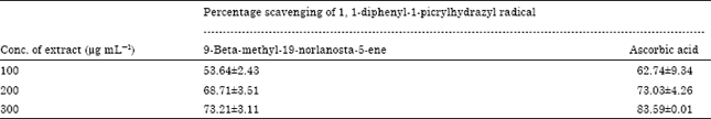 | |
| Values are the average of triplicate experiments and represented as Mean±SEM and p<0.05 as compared to standard ascorbic acid | |
| Table 2: | Percentage scavenging of isolated glycosides by hydrogen peroxide radical |
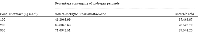 | |
| Values are the average of triplicate experiments and are represented as Mean±SEM and p<0.01 as compared to standard ascorbic acid | |
| Table 3: | Effect of triterpenoid type glycosides on gastric secretion, free acidity and total acidity in pylorus ligation induced gastric ulcer in rats |
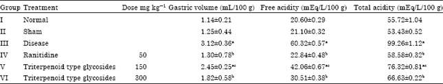 | |
| Values are Mean±SEM, n = 6 animals in each group; ap<0.05 as compared with sham control group, bp<0.05 compared with disease control groups, cp<0.05 compared with ranitidine treated group | |
| Table 4: | Effect of isolated glycosides on ulcerative index and percentage inhibition in Pl, WIS and non steroidal anti-inflammatory drug induced gastric ulcer in rats |
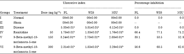 | |
| Values are Mean±SEM, n = 6 animals in each group; ap< 0.05 compared with sham control group, bp< 0.05 compared with PL and WIS groups respective columns, cp< 0.05 | |
The causes of gastric ulcer pyloric ligation are believed to be due to stress induced increase in gastric hydrochloric acid secretion and/or stasis of acid and the volume of secretion is also an important factor in the formation of ulcer due to exposure of the unprotected lumen of the stomach to the accumulating acid (Dhuley, 1999). Ulcer formation induced by indomethacin is known to be related with inhibition of cyclooxygenase that prevents prostaglandin biosynthesis which in turn inhibits the release of mucus, a defensive factor against gastrointestinal damage. Cucurbitane type glycosides showed reduction in gastric secretion, free and total acidity and ulcerative index at highest dose i.e., 300 μg mL-1 as compare to the standard ranitidine as shown in Table 3 (Bandyopadhyay et al., 2000). The % age inhibition in PL, WIS and NIU models was found to be 56.6, 68.5 and 62.6%, respectively at higher dose as Table 4. Previously C. sativus and its parts has not been explored only the extract was evaluated for its antiulcer activity (Gill et al., 2009). This indicated that the isolated comp of MECS has significant anti-ulcer activity.
CONCLUSION
Thus, from the above study it may be concluded that 9-beta-methyl-19-norlanosta-5-ene a cucurbitane type glycosides isolated from the C. sativus has tetra cyclic nucleus which may be responsible for antiulcer and antioxidant activity. These seeds can be used for the health benefit.
ACKNOWLEDGMENT
Thanks to Professor A. C. Rana and all faculty members of Rayat School of Pharmacy for their encouragement and support. We are also grateful to Rayat and Bahra Educational and Research Trust for their unconditional helps to carry out this project.
REFERENCES
- Annegowda, H.V., C.W. Nee, M.N. Mordi, S. Ramanathan and S.M. Mansor, 2010. Evaluation of phenolic content and antioxidant property of hydrolysed extracts of Terminalia catappa L. leaf. Asian J. Plant Sci., 9: 479-485.
CrossRefDirect Link - Alphine, R.S. and J.W. Word, 1969. Antihistaminic activity and ulceration. Eur. J. Pharmacol., 6: 61-66.
PubMed - Bandyopadhyay, S.K., S.C. Pakrashi and A. Pakrashi, 2000. The role of antioxidant activity of Phyllanthus emblica fruits on prevention from indomethacin induced gastric ulcer. J. Ethnopharmacol., 70: 171-176.
CrossRefPubMedDirect Link - Chipurura, B., M. Muchuweti and F. Manditseraa, 2010. Effects of thermal treatment on the phenolic content and antioxidant activity of some vegetables. Asian J. Clin. Nutr., 2: 93-100.
CrossRefDirect Link - Divya, B., K. Mruthunjaya and S.N. Manjula, 2011. Parkinsonia aculeata: A phytopharmacological review. Asian J. Plant Sci., 10: 175-181.
CrossRefDirect Link - Dhuley, J.N., 1999. Protective effect of Rhinax, a herbal formulation against physical and chemical factors induced gastric and duodenal ulcers in rats. Indian J. Pharmacol., 31: 128-132.
Direct Link - Desai, J.K., R.K. Goal and N.S. Parmar, 1997. Pathogenesis of peptic ulcer disease and current trends in therapy. Indian J. Physiol. Pharmacol., 41: 3-15.
PubMed - Etuk, E.U., V. Igbokwe, O.P. Ajagbonna and M.O. Egua, 2009. Toxicological studies of a nigerian commercial polyherbal product in albino rats. Res. J. Med. Plant, 3: 52-60.
CrossRefDirect Link - Ganguly, A.K., 1969. A method for quantitative assessment of experimentally produced ulcers in the stomach of albino rats. Experientia, 25: 1224-1224.
CrossRefPubMedDirect Link - Gill, N.S., A. Sharma, R. Arora and M. Bali, 2011. Evaluation of Cassia tora seeds for their antioxidant and antiulcer activity. J. Med. Sci., 11: 96-101.
CrossRef - Gill, N.S., K. Dhiman, P. Sharma, J. Bajwa and S. Sood et al., 2011. Evaluation of free radical scavenging and antiulcer potential of methanolic extract of Benincasa hispida seeds. Res. J. Med. Plant, 5: 596-604.
CrossRefDirect Link - Gill, N.S., M. Garg, R. Bansal, S. Sood, A. Muthuraman, M. Bali and P.D. Sharma, 2009. Evaluation of antioxidant and antiulcer potential of Cucumis sativum L. seed extract in rats. Asian J. Clin. Nutr., 1: 131-138.
CrossRef - Rahman, M.H., M.B. Alam, N.S. Chowdhury, M.K. Jha and M. Hasan et al., 2011. Antioxidant, analgesic and toxic potentiality of Stephania japonica (Thunb.) Miers. Leaf. Int. J. Pharmacol., 7: 257-262.
CrossRefDirect Link - Miro, M., 1995. Cucurbitacins and their pharmacological effects. Phytother. Res., 9: 159-168.
CrossRefDirect Link - Kocyan, A., L.B. Zhang, H. Schaefer and S.S. Renner, 2007. A multi-locus chloroplast phylogeny for the Cucurbitaceae and its implications for character evolution and classification. Mol. Phylogenet. Evol., 44: 553-557.
CrossRefPubMedDirect Link - Kundan, P. and B. Ganga, 2011. Evaluation of nutritive minerals and antioxidants values of Euphorbia thymifolia Linn. Curr. Res. Chem., 3: 98-105.
CrossRef - Khanahmadi, M., S.H. Rezazadeh and M. Taran, 2010. In vitro antimicrobial and antioxidant properties of Smyrnium cordifolium Boiss. (Umbelliferae) extract. Asian J. Plant Sci., 9: 99-103.
CrossRef - Mahendran, P., A.J. Vanisree and C.S.S. Devi, 2002. The antiulcer activity of Garcinia cambogia extract against indomethacin induced gastric ulcer in rats. Phytother. Res., 16: 80-83.
PubMed - Makhija, I.K., L. Richard, S.P. Kirti, K. Saleemullah, M. Jessy and S. Annie, 2011. Sphaeranthus indicus: A review of its chemical, pharmacological and ethnomedicinal properties. Int. J. Pharmacol., 7: 171-179.
CrossRef - Oluwole, F.S., B.O. Omolaso and J.A. Ayo, 2007. Methanolic extract of Entandrophragma angolense induces gastric mucus cell counts and gastric mucus secretion. J. Boil. Sci., 7: 1531-1534.
CrossRefDirect Link - Pandey, A.K., V. Ojha, S. Yadav and S.K. Sahu, 2011. Phytochemical evaluation and radical scavenging activity of Bauhinia variegata, Saraca asoka and Terminalia arjuna barks. Res. J. Phytochem., 2: 89-97.
CrossRef - Parmar, N.S. and J.K. Desai, 1993. A review of the current methodology for the evaluation of gastric and duodenal anti-ulcer agents. Indian J. Pharmacol., 25: 120-135.
Direct Link - Salim, A.S., 1994. Scavenging free radicals to prevent stress induced gastric mucosal injury. J. Surg. Res., 56: 45-52.
PubMed - Sood, S., S. Bansal, A. Muthuraman, N.S. Gill and M. Bali, 2009. Therapeutic potential of Citrus medica L. peel extract in carrageenan induced inflammatory pain in rat. Res. J. Med. Plant, 3: 123-133.
CrossRefDirect Link - Lee, S.Y., S.H. Eom, Y.K. Kim, N.I. Park and S.U. Park, 2009. Cucurbitane-type triterpenoids in Momordica charantia Linn. J. Med. Plants Res., 3: 1264-1269.
Direct Link - Sreejayan, N. and M.N. Rao, 1996. Free radical scavenging activity of curcuminoids. Arzneimittelforschung, 46: 169-171.
PubMedDirect Link - Ubaka, M.C., V.C. Ukwe, C.T. Okoye and O.M. Adibe, 2010. Investigation into anti-ulcer activity of aqueous leaf extract of Asilia Africana C.D. Adams. Asian J. Med. Sci., 2: 40-43.
Direct Link - Waldum, H.L., B. Gustafsson, R. Fossmark and G. Qvigstad, 2005. Antiulcer drugs and gastric cancer. Dig. Dis. Sci., 50: S39-S44.
PubMed - Zimmerman, T.W., 1984. Problems associated with medical treatment of peptic ulcer disease. Am. J. Med., 77: 51-56.
PubMed