
Pierre C. Djemgou
Department of Chemistry, Faculty of Science, University of Dschang,
P.O. Box 67, Dschang, Cameroon
Donatien Gatsing
Department of Biochemistry, Faculty of Science, University of Dschang,
P.O. Box 67, Dschang, Cameroon
Marguerite Kenmogne
Department of Chemistry, Faculty of Science, University of Dschang,
P.O. Box 67, Dschang, Cameroon
Dieudonne Ngamga
Department of Chemistry, Faculty of Science, University of Dschang,
P.O. Box 67, Dschang, Cameroon
Roseline Aliyu
Department of Biochemistry, Faculty of Medical Sciences, University of Jos,
PMB 2084 Jos, Nigeria
Abiodun H. Adebayo
Department of Biological Sciences, College of Science and Technology, Covenant University, PMB 1023, Canaanland, Ota, Ogun State, Nigeria
Pierre Tane
Department of Chemistry, Faculty of Science, University of Dschang,
P.O. Box 67, Dschang, Cameroon
Bonaventure T. Ngadjui
Department of Organic Chemistry, Faculty of Science, University of Yaounde I,
P.O. Box 812, Yaounde, Cameroon
Elisabeth Seguin
Laboratoire de Pharmacognosie, UFR de M6decine-Pharrnacie,
76183 Rouen Cedex, France
Godwin I. Adoga
Department of Biochemistry, Faculty of Medical Sciences, University of Jos,
PMB 2084 Jos, Nigeria
Research Journal of Medicinal Plants
Year: 2007 | Volume: 1 | Issue: 2 | Page No.: 65-71


ABSTRACT
Phytochemical and biological investigation of the leaves of Cassia petersiana afforded four compounds including a new dihydroanthracenone (1), two known chromones (2,3), in addition to stigmasterol glucoside (4). The work was guided by the antisalmonellal activity of the extract and fractions. Compound 4 was found to be the active principle. The structures of the compounds were determined by combination of spectroscopic techniques, including 1H, 13C, DEPT, COSY, HMQC, HMBC, MS and IR.
PDF Abstract XML References
How to cite this article
Pierre C. Djemgou, Donatien Gatsing, Marguerite Kenmogne, Dieudonne Ngamga, Roseline Aliyu, Abiodun H. Adebayo, Pierre Tane, Bonaventure T. Ngadjui, Elisabeth Seguin and Godwin I. Adoga, 2007. An Antisalmonellal Agent and a New Dihydroanthracenone from Cassia petersiana. Research Journal of Medicinal Plants, 1: 65-71.
URL: https://scialert.net/abstract/?doi=rjmp.2007.65.71
URL: https://scialert.net/abstract/?doi=rjmp.2007.65.71
INTRODUCTION
Cassia is the major genus of the family Caesalpiniaceae and possesses about 600 species (Viegas et al., 2004). Aromatic compounds are mostly published from phytochemical investigation of the genus (Rao et al., 1999; Ingkaninan et al., 2000). Some authors have also isolated terpenes and alkaloids (Ingkaninan et al., 2000). Significant biological activities are reported from members of this genus (Moriyama et al., 2003). Cassia petersiana Bolle is a tree generally distributed in equatorial countries from Sierra Leone to D. R. Congo. This plant species is commonly found growing on sandy soils up to 12 m high and at altitude of up to 1050 m above sea level. It has pinnate leaves and yellow flowers. In Cameroon the leaves are widely used for the treatment of typhoid fever. The compressed, hairy pods are eaten either raw or cooked as gruel. The roots of the plant are used as a treatment for coughs, colds, syphilis and stomachache. It is also used as an anthelmentic. The roots are mixed with those of Fagara nitens (Rutaceae) and Stegmanotaenia araliacea (Umbellifereae) and burnt to charcoal, which when pulverized, is rubbed on incisions cut in the ankles and between the forefinger against snake bite. In Southern Africa, the leaves are used as a febrifuge and as a cure for skin diseases (Msonti, 1984).
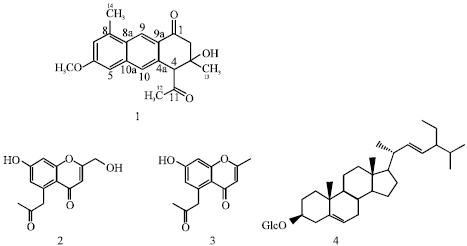 | |
| Fig. 1: | Structures of the isolated compounds |
Typhoid fever is caused by Salmonella typhi, whereas paratyphoid fevers are caused by Salmonella paratyphi A and Salmonella paratyphi B (Cheesbrough, 1991). Typhoid fever continues to be a marked public health problem in developing countries in general and in Sub-Saharan Africa in particular, where it is endemic (Gatsing et al., 2003). The greater prevalence of resistance to all three first line antimicrobial (ampicillin, chloramphenicol and co-trimoxazole) has been established (WHO, 1981; Gatsing et al., 2003). Previous phytochemical investigation on this plant species led to the isolation of diterpene (Msonti, 1984) and flavonoids (Coetze et al., 1999). As part of our contribution to the phytochemical and chemotaxonomic survey of the genus Cassia and in a continuation of our search for therapeutic agents from natural sources with potential for the treatment of typhoid and paratyphoid fevers (Gatsing et al., 2003; Gatsing et al., 2006), we carried out the investigation of the CH2Cl2-MeOH (1:1) extract of the leaves of C. petersiana, a species from Cameroon. We report herein the isolation and structural elucidation of four compounds including one new and three known (Fig. 1). The extract and the pure compounds were assessed for their antisalmonellal activity. The compounds were characterised from comprehensive 1D and 2D NMR interpretation and comparison with literature data.
MATERIALS AND METHODS
General
1H, 13C-NMR and 2D spectra were recorded at 400 MHZ using a Bruker DPX 400 spectrometer; trimethylsilane (TMS) was used as the internal standard. EIMS spectra were recorded on TSQ-70-Triple Stage Quadruple mass spectrometer (70 ev). The IR spectra (CHCl3) were recorded on a Perkin-Elmer FT-IR-spectrometer.
Plant Material
The leaves of Cassia petersiana Bolle were collected in Bafia, Centre province of Cameroon in July 2003. Plant material was identified by Dr. Onana at the Cameroon National Herbarium, Yaoundé, where a voucher specimen (N° 6494/SFR/Cam) was deposited.
Test Bacteria and Culture Media
The test microorganisms, Salmonella typhi, Salmonella paratyphi A and Salmonella paratyphi B, were obtained from the Medical Bacteriology Laboratory of the Pasteur Centre, Yaoundé, Cameroon. The culture media used namely Salmonella-Shigella agar (SS agar) and Selenite Broth, were supplied by International Diagnostics Group PLC, Topley House, 52 Wash Lane, Bury, Lancashire BL96AU, UK.
Extraction and Isolation
The air-dried and pulverized leaves of C. petersiana (800 g) were extracted by maceration with CH2Cl2-MeOH (1:1), (11 L, 72 h) and evaporated under reduced pressure to give 80 g of crude extract. Part of this extract (30 g) was fractionated by silica gel column chromatography (CC) eluted successively with n-hexane-EtOAc and EtOAc-MeOH in a step gradient by using different ratios. Three fractions A (8 g), B (11 g) and C (3 g) were recorded. Fraction A was purified on Sephadex LH-20 column (n-hexane-CH2Cl2-MeOH, 7:4:1) and preparative TLC to give 4-Acetyl-3,4-dihydro-3,8-dimethyl-3-hydroxy-6-methoxyanthracen-1(2H)-one (1, 9 mg). Fraction B was purified on silica gel column (n-hexane-EtOAc, 9:1) and Sephadex LH-20 to give 5-acetonyl-7-hydroxy-2-hydroxymethylchromone (2, 4 mg) and 5-acetonyl-7-hydroxy-2-methylchromone (3, 11 mg). Stigmasterol-3-O-β-D-glucoside (4, 500 mg) was recrystallised with acetone from fraction C.
4-Acetyl-3,4-dihydro-3,8-dimethyl-3-hydroxy-6-methoxyanthracen-1(2H)-one |
(1): Yellow powder, mp 291.3 C (uncorrected), IR 3320, 3480, 2814, 1624, 1597, 1430, 1300, 970; 1H NMR (CD3OD, 400 MHZ) and 13C NMR (CD3OD, 100 MHZ), Table 1, EIMS [M+] m/z 312, [M-H]+ m/z 311, [M-CH3]+ m/z 297, [M-OH]+ m/z 295, [M-Ac]+ m/z 270, [M-OH-Ac]+ m/z 254, HREIMS 312.3737 [calcd. for C19H20O4, 312.3630].
Antimicrobial Assay
The antibacterial activity was determined using both agar diffusion and broth dilution techniques as previously described (Cheesbrough, 1991; Gatsing et al., 2006).
Agar diffusion susceptibility testing was done using the method of wells. On each plate containing Salmonella-Shigella agar (SS agar) meduim already inoculated with the test organism (100 μL of the bacteria suspension in Selenite broth, at the concentration of 5x10⁵ cfu mL-1) wells (of 6 mm diameter) were bored using a cork borer. The bottom of each well was sealed with a drop of molten agar. The compounds and the extract were dissolved in dimethysulfoxide (DMSO). The wells were filled with 150 μL of the solution (of known concentration) of various compounds and extracts to be tested.
| Table 1: | 13C and 1H NMR data of compound 1 at 100 and 400 MHZ in CD3OD |
 | |
Chloramphenicol (Sigma) was used as the standard drug. The extract, compounds and chloramphenicol were tested at the concentration of 50, 0.5 and 0.1 mg mL-1, respectively. The petridishes were left at room temperature for 45 min to allow the compounds and extract to diffuse from the wells into the medium. They were then incubated at 37°C for 24 h, after which the zones of no growth were noted and their diameters recorded as the zones of inhibition.
For the broth dilution susceptibility testing, the solution (maximum concentration) of the active compound (ie the compound that induced a zone of inhibition; compound 4) was dissolved in DMSO, serially (2-fold) diluted and 0.5 mL of each dilution was introduced into a test tube containing 4.4 mL of Selenite broth; then 0.1 mL of bacteria suspension (5x10⁵ cfu mL-1) was added and the mixture was homogenised. The total volume of the mixture was 5 mL, with the test compound concentrations in the tube ranging from 180 to 5.625 μg mL-1 and those of chloramphenicol ranging from 40 to 0.625 μg mL-1. After 24 h of incubation at 37°C, the Minimum Inhibitory Concentration (MIC) was reported as the lowest concentration of antimicrobial that prevented visible growth. The Minimum Bactericidal Concentration (MBC) was determined by subculturing the last tube to show visible growth and all the tubes in which there was no growth on already prepared plates containing SS agar medium. The plates were then incubated at 37°C for 24 h and the lowest concentration showing no growth was taken as the MBC.
Results and Discussion
The air-dried and pulverized leaves of C. petersiana were extracted by maceration with CH2Cl2/MeOH (1/1). Part of the resulting extract was fractionated by silica gel column chromatography (CC) eluted successively with n-hexane-EtOAc and EtOAc-MeOH in a step gradient by using different ratios. Three fractions A (8 g), B (11 g) and C (3 g) were recorded and purified to yield 4-acetyl-3,4-dihydro-3,8-dimethyl-3-hydroxy-6-methoxyanthracen-1(2H)-one (1, 9 mg), 5-acetonyl-7-hydroxy-2-hydroxymethylenechromone (2, 4 mg), 5-acetonyl-7-hydroxy-2-methylchromone (3, 11 mg) and stigmasterol-3β-D-glucoside (4, 500 mg).
Compound 1 was isolated as a yellow powder, mp 291.3°C (uncorrected); its EIMS gave a molecular ion peak at m/z 312. The cross formula was deduced to be C19H20O4 from combination of HREIMS, EIMS and RMN data. Moreover, interesting peaks were observed, each characteristic of a precise moiety, m/z 270, [M-Ac]+; 297, [M-H2O]+; 295, [M-CH3]+. A peak at m/z 311 (M-1) suggested the presence of hydroxyl group in the structure. The 1H NMR (CD3OD, 400 MHZ (Table 1) revealed eleven different hydrogens in the molecule, which could be classified as seven singlets, three doublets and a doublet of doublet. The methoxyl appeared at δ 4.18 (3H, s, OMe). The carbone13 NMR (CD3OD, 100 MHZ) spectra reveals 19 carbon atoms in the molecule (Table 1). Analyses of 13C, DEPT (90 and 135) and HMQC led to their classification as two carbonyls, three methyls, a methoxyl at δ 50.9 (-OMe), one saturated methine carbon at δ 64.5 (C-4), one methylene carbon at δ 49.5 (C-2), one saturated quaternary carbon at δ 72.9 (C-3), six aromatic quaternary carbons and four aromatic
methine carbons. The assignement of the structure was based on the following observations on the spectra: (i) The position of the hydroxyl (OH) was based on the chemical shift of C-3 and confirmed by the HMBC (Fig. 1). (ii) The acetyl group was deduced from an intense correlation on HMBC between the methyl at δ 2.40 (H-12) and the carbonyl at δ 209.9 (C-11). (iii) This was confirmed by the chemical shift of the methyl carbon at δ 33.9 (C-12), downfield, compared to the methyl C-14 (δ 20.3) for example. The other substituents were attributed from interpretation of the HMBC spectra (Fig. 2). (iv) H-9, δ 6.55 and H-10, δ 6.00 were placed according to the HMBC where they showed a cross correlation with the carbonyl at δ 203.3 (C-1) and the methine carbon at δ 64.5 (C-4) respectively. H-5, δ 6.22 and H-7, δ 6.19 were meta oriented based on their coupling constant 2.20 Hz.
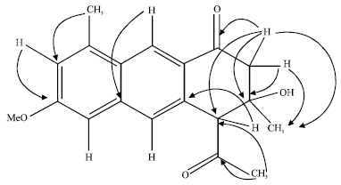 | |
| Fig. 2: | Some remarquable HMBC correlations of compound 1 |
On proton COSY, H-5, δ 6.22 showed a weak 4J correlation with H-10, δ 6.00. (v) As H-2b was a doublet of doublet, this suggest a weak coupling with H-4, confirmed from the 1H-1H COSY spectra, where a W type coupling was observed between the protons at δ 2.50 (H-2b) and 42.0 (H-4). Their chemical shift, downfield compared to a normal proton in their position, could be due to the effect of the neighbouring carbonyl. (vi) The stereochemistry was not determined. Taking into account the above comprehensive NMR interpretation and the comparison of these data to those of related compounds (Ingkaninan et al., 2000; Lee et al., 2001; Dagne et al., 1996), structure 1 was attributed to the compound which is a new dihydroanthracenone qualified as 4-acetyl-3,4-dihydro-3,8-dimethyl-3-hydroxy-6-methoxyanthracen-1(2H)-one, trivially named petersone A.
The 1H, 13C, IR and mass spectral data of 2 and 3 were similar to those previously reported from the phytochemical investigation of C. siamea (Ingkaninan et al., 2000). These compounds were then identified as 5-acetonyl-7-hydroxy-2-hydroxymethylenechromone (2) and 5-acetonyl-7-hydroxy-2-methylchromone (3).
Compound 4 was isolated as a white powder hardly soluble in most of the organic solvents. 1H and 13C NMR are characteristic of stigmasterol-3-O-β-D-glucoside (Neera and Wichtl, 1987), common in the plant kingdom.
The crude CH2Cl2/MeOH (1:1) leaf extract of Cassia petersiana showed antibacterial activity against all the three bacteria species used and the diameter of inhibition were 14-16 mm against S. typhi, 15-18 mm against S. paratyphi A and 17-18 mm against S. paratyphi B. Four compounds namely 4-acetyl-3,4-dihydro-3,8-dimethyl-3-hydroxy-6-methoxyanthracen-1(2H)-one, 5-acetonyl-7-hydroxy-2-hydroxymethylene-chromone, 5-acetonyl-7-hydroxy-2-methylchromone and stigmasterol-3-O-β-D-glucoside were isolated from the above crude extract and were tested for their antisalmonellal activities. The results obtained showed that compound 4 (stigmasterol-3-O-β-D-glucoside) was the only active compound with the following diameters of inhibition: 15-18 mm against S. typhi, 17-19 mm against S. paratyphi A and 19-21 mm against S. paratyphi B. Chloramphenicol, used as the standard, showed the diameters of 27-28, 25-27 and 26-28 mm against S. typhi, S. paratyphi A and S. paratyphi B, respectively (Table 2).
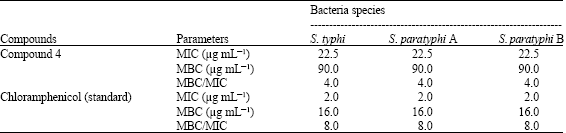
Compound 4, which showed antibacterial activity against all the three bacteria species used, was further studied using broth dilution technique and the following results were obtained: the MIC and MBC values were 22.5 and 90 μg mL-1, respectively, against all the three bacteria tested. For chloramphenicol the MIC and MBC values were 2 and 16 μg mL-1, respectively, against the same bacteria species (Table 3).
| Table 2: | Diameters of inhibition of S. typhi, S. paratyphi A and S. paratyphi B by the extract and compounds isolated from the leaves of Cassia petersiana |
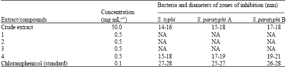 | |
| NA: Not Active; 1: 4-Acetyl-3-hydroxy-6-methoxy-3,8-dimethyl-dihydroanthracenone; 2: 5-Acetonyl-7-hydroxy-2-hydroxymethylene-chromone; 3: 5-Acetonyl-7-hydroxy-2-methylchromone and 4: Stigmasterol-3-O-β-D-glucoside | |
| Table 3: | Inhibition parameters (MIC, MBC) of compound 4, isolated from the leaves of C. petersiana |
 | |
| Compound 4: Stigmasterol-3β-D-glucoside; MIC: Minimum Inhibitory Concentration; MBC: Minimum Bactericidal Concentration | |
Antimicrobial substances are considered as bactericidal agents when the ratio MBC/MIC ≤4 and bacteriostatic agents when the ratio MBC/MIC>4 (Carbonnelle et al., 1987). For compound 4, the ratio MBC/MIC = 4, suggesting that it may be classified as bactericidal agent. Based on the MIC values, compound 4 was about eleven times less active than chloramphenicol.
CONCLUSIONS
Chromones were previously isolated from various species of the genus Cassia (Lee et al., 2001; Lu et al., 2001). The appearance of chromones derivatives in our results is of great interest for the investigation of members of this genus where they constitute a possible chemotaxonomic marker. In addition, the antisalmonellal activities of the CH2Cl2/MeOH (1:1) extract of the leaves of Cassia petersiana and the isolated compounds are here described for the first time and attest the traditional used of this plant against typhoid fever.
ACKNOWLEDGMENT
Authors are grateful to the Third World Academy of Science (TWAS) for the fellowship given to one of us (D.P.C.). Agence Universitaire de la Francophonie (AUF) is acknowledged for financial support of the work. We will like to thank Dr. Onana for plant identification.
REFERENCES
- Coetze, J., L. Mcieteka, E. Malan and D. Ferreira, 1999. Oligomeric flavonoid part 30, structure and synthesis of butiniflavan-epicatechin and epigallocatechin probutinidin. Phytochemistry, 52: 737-743.
Direct Link - Dagne, E., B.E. Van Wyk, M. Mueller and W. Steglich, 1996. Three dihydroanthracenones from gasteria bicolor. Phytochemistry, 41: 795-799.
Direct Link - Gatsing, D., R. Aliyu and W.B. Meli, 2003. Phytochemical profile and antisalmonellal properties of Allium sativum bulb extract. West Afr. J. Biol. Sci., 14: 29-36.
Direct Link - Gatsing, D., J.A. Mbah, I.H. Garba, P. Tane, P. Djemgou and B.F. Nji-Nkah, 2006. An antisalmonellal agent from the leaves of Glossocalyx brevipes Benth (Monimiaceae). Pak. J. Biol. Sci., 9: 84-87.
CrossRef - Ingkaninan, K., A.P. Ijzerman and R. Verpoorte, 2000. Luteolin, a compound with adenosine A(1) receptor-binding activity and chromone and dihydronaphthalenone constituents from Senna siamea. J. Nat. Prod., 63: 315-317.
Direct Link - Lee, C.K., P.H. Lee and Y.H. Kuo, 2001. The chemical constituents from the aril of Cassia fistula L. J. Chin. Chem. Soc., 48: 1053-1058.
Direct Link - Lu, T.S., Y.H. Yi, S.L. Mao, D.Z. Zhou, Q.Z. Xu, H.F. Tang and S.Y. Zhang, 2001. A new chromone glycoside from Cassia siamea Lam. Chinese Chem. Lett., 12: 703-704.
Direct Link - Moriyama, H., T. Iizuka, M. Nagai, H. Miyataka and T. Satoh, 2003. Antiinflamatory activity of heat-treated Cassia alata leaf extract and its flavanoid glycoside. Yakugaku Zasshi, 123: 607-611.
Direct Link - Rao, K.V., A.G. Damu, B. Jayaprakasam and D. Gunasekar, 1999. Flavonoid glycoside from Cassia hirsuta. J. Nat. Prod., 62: 305-306.
CrossRef - Viegas, Jr., C., S.V. Bolzani da, M. Furlan, E.J. Barreiro, M.C.M. Young, D. Tomazela and M.N. Eberlin, 2004. Further bioactive piperidine alkaloid from the flower and green fruits of Cassia spectabilis. J. Nat. Prod., 67: 908-910.
Direct Link