
A. Soni
Pre-Clinical Division, Venus Medicine Research Centre, Hill Top Industrial Estate, Bhatoli Kalan, Baddi, H.P.-173205, India
V.K. Dwivedi
Pre-Clinical Division, Venus Medicine Research Centre, Hill Top Industrial Estate, Bhatoli Kalan, Baddi, H.P.-173205, India
M. Chaudhary
Pre-Clinical Division, Venus Medicine Research Centre, Hill Top Industrial Estate, Bhatoli Kalan, Baddi, H.P.-173205, India
K. Malik
Pre-Clinical Division, Venus Medicine Research Centre, Hill Top Industrial Estate, Bhatoli Kalan, Baddi, H.P.-173205, India
V. Naithani
Pre-Clinical Division, Venus Medicine Research Centre, Hill Top Industrial Estate, Bhatoli Kalan, Baddi, H.P.-173205, India
S.M. Shrivastava
Pre-Clinical Division, Venus Medicine Research Centre, Hill Top Industrial Estate, Bhatoli Kalan, Baddi, H.P.-173205, India
Research Journal of Immunology
Year: 2010 | Volume: 3 | Issue: 1 | Page No.: 22-30


ABSTRACT
The present study was designed to determine the status of trace elements (Zn and Cu) and cytokines (IL-6, IL-1β and TNF-α) levels in severe burn rat model. Under brief anesthesia of ketamine, the shaved dorsum of the rats was exposed to 80°C (burn group) by hot wax for 5 min. The rats were fed standard pelleted diet and water ad libitum. The test room was air conditioned with temperature 23±2°C, humidity 65±5% and with artificial fluorescent light (10-14 h of light and dark), respectively. Rats were decapitated 24 h after burn injury and blood were taken for the determination of Zn, Cu and cytokines level. Present finding showed that level of Zn and Cu concentrations showed a significant trend of decrease in burned group and its levels were statistically increased (p<0.001) on 1, 7 and 14 days in ampucare treated group. Alternately, there was significant increased (p<0.01) in TNF-α, IL-6 and IL-1β in burned group on 1, 7 and 14 consecutive days as compared to control group. These levels become significantly lowered (p<0.001) on first, 7 and 14 days of treatment and come back to normal level when compared burned group. It was concluded that ampucare maintained levels of Zn and Cu as well as cytokines (IL-6, IL-1β and TNF-α) and increases its wound healing potential that would be a beneficial for the human who suffer from severe burn injury.
PDF Abstract XML References Citation
How to cite this article
A. Soni, V.K. Dwivedi, M. Chaudhary, K. Malik, V. Naithani and S.M. Shrivastava, 2010. Plasma Cytokines and Trace Element Level in Severe Burn Rat Model-With Special Reference to Wound Healing Potential of Ampucare. Research Journal of Immunology, 3: 22-30.
DOI: 10.3923/rji.2010.22.30
URL: https://scialert.net/abstract/?doi=rji.2010.22.30
DOI: 10.3923/rji.2010.22.30
URL: https://scialert.net/abstract/?doi=rji.2010.22.30
INTRODUCTION
Burn injury changes metabolic and immune responses that can be related to alterations in trace elements metabolism and cytokines. Trace elements, especially zinc (Zn) and copper (Cu), have important roles in human growth, development and immune function (Walker and Black, 2004). Guo et al. (2000) reported that these elements are essential in wound healing. These trace elements act as major antioxidant enzymes cofactors. Variation in Zn and Cu is important (Bang et al., 2000). There is evidence that infection affected the plasma’s Zn concentration (Brown, 1998). Some reports showed the alteration of trace elements metabolism in patients with burn injuries (Agay et al., 2005; Berger et al., 1998; Bang et al., 2000). The deficiencies of Zn and Cu have been reported in burn injuries (Guo et al., 1997; Cunnigham et al., 1991). Similarly, Clark and Kamen (1987) reported that during healing of the burn wound, the production and stimulation of cells belonging to the immune system are regulated by a network of cytokines. Interleukin-6 (IL-6), a vital cytokine, has multiple functions, including modification of the regulatory effect of other cytokines (Schindler et al., 1990; Schluter et al., 1991) and it reflects the severity of the morbid condition of the burn injury (Yamada et al., 1996). It is well known that the interleukin modulated the inflammatory response following trauma (Drost et al., 1993; Guo et al., 1990). Modulation of these cytokine levels effectively regulates the interlinked pathways responsible for healing. Heparin has been investigated and proved to modulate several phases of wound healing, including angiogenesis (Flint et al., 1994; Kratz et al., 1998; Folkman, 1985). It has a chemotactic effect on endothelial cells, with resultant stimulation of neovascularization (Terranova et al., 1985) and improvement of blood circulation subjective to the burn, thus collectively inducing repair mechanisms.
Ampucare is a oil based formulation product which applied externally for the treatment of wounds. It is an antimicrobial and ant-inflammatory polyherbal formulation with proven activity against E. coli, Pseudomonas aeruginosa, Proteus mirabilis, Staphylococcus aureus, Enterococcus aerogenes and Candida albicans (Saurab et al., 2008). It is herbal combination with Azadirachta indica and Curcuma longa as active ingredients. These two components have an anti-microbial, antioxidant, fungicidal and free radical scavenging properties. Dwivedi et al. (2008) reported an anti-inflammatory, immuno-modulatory and tissue regeneration properties of ampucare. Ampucare is also indicated for the treatment of dermal infections (Chaudhary et al., 2008a, b) and enhance the healing of different wound (Chaudhary et al., 2008c). We investigated the potential benefits of ampucare on burn wound healing in burned rats by assessing its modulatory effect on different cytokines (IL-6, IL-1β and TNF-α) and trace elements.
MATERIALS AND METHODS
Study Conduct
The study was carried out from 10th January 2009 to 20th April 2009 in pre-clinical division of Venus Medicine Research Centre, Venus Remedies Ltd., Baddi (India).
Plant Materials
Azadirachta indica, Curcuma longa and other herbs were used in the formulation of ampucare. These raw material were purchased from local vendor which was identified and approved by the botanist.
Chemicals
All the biochemicals used in the present study were procured from Sigma, St. Louis, MO, USA. Other chemicals purchased locally were of analytical grade.
Experimental Animals
Male albino rats of the same age, weighing between 250-300 g, were housed in separate cages under standard conditions, with a 14/12 h light-dark regimen. The rats were given standard rat chow and water ad libitum. All rats were housed in a filtered-air environment maintained at 23±2°C. The protocol was approved by institutional animal ethics committee.
Study Design
All of the rats were divided into 3 groups of 8 rats each as given below:
| • | Group-I (n = 8) : Control |
| • | Group-II (n = 8) : Burned group |
| • | Group-II (n = 8) : Burned + Ampucare treated group |
Ampucare was applied topically continued up to 14 consecutive days. Blood samples were collected on; 7 and 14th day. Blood samples were taken from the antecubital veins using stainless steel needles and trace element-free vacutainers. Serum was separated by centrifugation at 3,000 rpm for 10 min from remaining blood samples for the analysis of cytokines (IL-6, IL-1β and TNF-α) and trace elements (Zn and Cu).
Experimental Burn Model
The burn model was prepared according to the method of Holla et al. (1998) and Rao et al. (2000). Partial thickness of burn wounds were inflicted on overnight-starved animals under ketamine (10 mg kg-1, i.m.) anesthesia by pouring hot molten wax at 80°C. The wax was poured on the shaven back of the animal through a cylinder of 350 mm2 circular. The wax was allowed to stay on the skin till it gets solidified. Instantly after the injury and on subsequent days, the drugs were applied topically.
Estimation of Trace Elements
The Zn and Cu level were estimate by reference of Gümüs et al. (1999) using flame atomic absorption spectrophotometer (BRIAC FX-130, Beijing, China) with Zn and Cu hollow cathode lamp at wavelengths 213.9 and 327.9 nm, respectively. The 0.5 mL of serum was mixed with 4.5 mL of acidic glycerol HNO3 (1% and glycerol 5%). The absorption of solution was directly measured by atomic absorption spectrophotometer. The standard curves were prepared using 50, 100, 150, 200, 250 μg dL-1 solution of Cu and 10, 30, 40, 50 μg dL-1 solution of Zn in acidic glycerol. All chemical reagents and solutions were from Himedia Company (Mumbai, India).
Cytokines Assays
For IL-6, IL-1β and TNF-α, the assay uses the quantitative sandwich enzyme immunoassay technique (Invitrogen). A monoclonal antibody specific for rat IL-6, IL-1β and TNF-α were pre-coated onto a micro plate. Standards, controls and samples are pipetted into the wells and rat IL-6, IL-1β and TNF-α that present in the sample is bound to the immobilized antibody. After washing unbound substances were washed out, an enzyme-linked polyclonal antibody specific for rat IL-6, IL-1β and TNF-α was then added to the wells. Following washing was done to remove any unbound antibody-enzyme reagent, after that substrate solution was added to the wells. The enzyme reaction yields a blue product that turns yellow when the stop solution was added. The intensity of the color measured is in proportion to the amount of rat IL-6, IL-1β and TNF-α bound in the initial step. The sample values are then read off the standard curve.
Statistical Analysis
The results are expressed in Mean±SD. Statistical evaluation of the data was performed by one way-ANOVA followed by student Newman-Keuls using INSTAT 3.0 software. The statistical difference was analyzed between control, burned and ampucare treated group. The p<0.05 was considered statistically significant.
RESULTS
Trace Element Level
The Zn level in serum of the burned group were lowered (p<0.001) significantly when compared to control group on first, 7 and 14th consecutive days of treatment. This level increased significantly (p<0.001) in ampucare treated group when compared to burned group on the first, 7 and 14th days of interval and come near to control group. Similarly, the serum Cu concentration in burned group as significantly lowered (p<0.01) as compared to control group on different days of treatment. Its levels was statistically increased (p<0.001) in ampucare treated group when compared to burned group and come back to control level (Table 1).
Cytokines Parameters
There was significant increase (p<0.01) in level of TNF-α in serum of burned group as compared to control group in different days of interval. The level of TNF-α was decreased significantly (p<0.01) in ampucare treated group when compared to burned group and come near to control group (Fig. 1).
Similarly, the level of IL-6 in serum of burned group were increase significantly (p<0.001) as compared to control group on different days of interval. Its level become lowered (p<0.01) in ampucare treated group when compared to burned group and come back to normal level (Fig. 2).
| Table 1: | Comparative effects of Zn and Cu concentration in serum of control, burned and ampucare treated group |
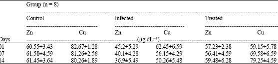 | |
| Values are expressed as Mean±SD | |
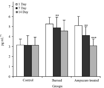 | |
| Fig. 1: | Comparative level of tumour necrosis factor (TNF-α) in serum of control, infected and ampucare treatment group. Values are expressed as Mean±SD. ***:Highly significant at p<0.001, **:Significant at p<0.01 |
Similarly, the level of IL-1β in burned group increase significantly (p<0.01) on different days of interval as compared to control group. Its levels become significantly lowered (p<0.001) on first, 7 and 14 days of ampucare treatment group and near to come back to normal level when compared burned group (Fig. 3).
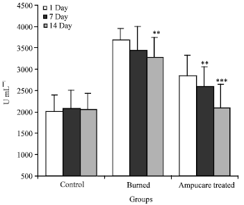 | |
| Fig. 2: | Comparative level of interleukin-6 (IL-6) in serum of control, infected and ampucare treatment group. Values are expressed as Mean±SD. ***:Highly significant at p<0.001, **:Significant at p<0.01 |
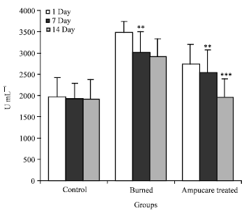 | |
| Fig. 3: | Comparative level of interleukin-β (IL-β) in serum of control, infected and ampucare treatment group. Values are expressed as Mean±SD. ***:Highly significant at p<0.001, **:Significant at p<0.01 |
DISCUSSION
Healing of a wound is a complex and protracted process of tissue repair and remodeling in response to injury. In order to balance degradative and regenerative processes, cell activation, cell division, chemotaxis, migration cell differentiation responsible control for various biochemical, cellular and immunological reaction cascades. They are mediated by locally released growth factors and cytokines, which may act in an autocrine or paracrine manner. All phases of wound healing are either directly or indirectly controlled by cytokines. It appears that it is the balance of these cytokines and other mediators rather than the mere presence or absence of one or more cytokines that plays a decisive role in regulating the initiation, progression and resolution of wounds. In addition, cell-cell and cell-matrix interactions, mediated, for example by various cell surface adhesion molecules, play an important role in wound healing.
The activation of the host immune system and the release of inflammatory mediators have been linked to physiological derangements observed in burn injury and other inflammatory conditions, increasing according to illness severity and the progression of systemic inflammatory response syndrome to multiple organ failure (Pavlos and Baltopoulos, 2008). Thus, it has been assumed that increased physiological responses parallel the intensity of cytokine production and the development of multiple organ failure and death. Since, the production or depression of several cytokines is related to physiological derangements commonly used in scoring systems, it seems reasonable to measure these circulating cytokines and use them as an additional tool to predict outcome. Yamada (1996) reported that tumour necrosis factor alpha, interleukin 6 and interleukin 8, as cytokines, showed high levels in patients with burn injury associated with sepsis and those who died. Cytokines are immunoregulatory proteins and glycoproteins that go through specific receptors on target cells in a paracrine, autocrine, or endocrine manner (Ohzato et al., 1993). These molecules are produced by a variety of cells, including those of the epidermis and include interleukins, TNFs and others. Cytokines are involved in the differentiation, activation and proliferation of both immune and non-immune cells involved in immuno-inflammatory reactions.
Ampucare is an antimicrobial and ant-inflammatory herbal formulation. The active components of ampucare are Azadirachta indica and Curcuma longa involved in wound healing (Fig. 4a, b). Azdirachta indica and Curcuma longa are reported to have significant anti-bacterial, immuno-modulatory and anti-inflammatory activities (Pai et al., 2004; Ali et al., 2001; Babu et al., 2003; Surh, 2002), which are complementary to wound healing process. Gupta et al. (1982) reported the moderate activity of Curcuma longa against TNE-[alpha] and IL-8. Kunchandy and Rao (1990) suggested that the anti-inflammatory activity of Curcuma longa might be due to its ability to scavenge oxygen radicals, which has been implicated in the inflammation process.
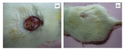 | |
| Fig. 4: | Wound healing effects of (a) burned and (b) ampucare treated group after 14 days of treatment |
Collectively these observations suggest that anti-inflammatory activity of Curcuma longa may be due to its effect on pro-inflammatory cytokines as well as its capacity to scavenge oxygen radicals. Thus, due to pro-inflammatory activity of Azadirachta indica and Curcuma longa in ampucare controls the level of TNF-α (Fig. 1), IL-6 (Fig. 2) and IL-1β (Fig. 3) levels continuously decreased on different days in serum of burned group and come back to normal level.
Trace elements plays an important role in antioxidant defense, inflammatory process and wound healing in burn injuries. The body distribution profile of these trace elements is changed following the burn injury and it is possibly due to contribution of Zn and Cu in the inflammatory process (Yamada et al., 1996).
Sometimes the low levels of plasma Zn may be related to hypoalbuminaemia; this protein is known to transport about 70% of Zn to the cells (Rowe and Bobilya, 2000). In this study, the loss of Zn continued on day 14 after the burn injury and did not return to near control level. Bang et al. (2000) reported that serum Cu concentration increased on days one and two and remained elevated on day ten following the burn injury. Similarly, present results indicated that after the treatment of ampucare in burn model, the Zn and Cu concentration in the plasma become increased and come near to the normal level (Table 1).
It was concluded that ampucare maintained levels of Zn and Cu as well as cytokines (IL-6, IL-1β and TNF-α) and increases its wound healing potential that would be a beneficial for the human who suffer from severe burn injury.
ACKNOWLEDGMENTS
Authors are thankful to Mr. Parveen Kumar (Lab Assistant) to support in experiment handling and financial Department of Venus Medicine Research Centre for financial support.
REFERENCES
- Agay, D., R.A. Anderson and C. Sandre, 2005. Alteration of antioxidant trace elements (Zn, Se, Cu) and related metallo-enzymes in plasma and tissues following burn injury in rats. Burns, 31: 366-371.
CrossRef - Ali, M., E. Ravinder and R. Ramachandram, 2001. A new flavonoid from the aerial parts of Tridax procumbens. Fitoterapia, 72: 313-315.
CrossRef - Babu, G., K. Sanjeeva and L. Bairy, 2003. Effect of tridax procumbens on burn wound healing. Indian Drugs, 40: 488-491.
Direct Link - Bang, R.L., A.B. Mattappallil, H.M. Dashti and A. Albade, 2000. Thermal injury and changes in the trace elements. J. Trace Elem. Exp Med., 13: 255-264.
Direct Link - Berger, M.M., F. Spertini, A. Shenkin, C. Wardle, L. Wiesner, C. Schindler and R.L. Chiolero, 1998. Trace elements supplementation modulates pulmonary infection rates after major burns: A double-blind, placebo-controlled trial. Am. J. Clin. Nutr., 68: 365-371.
Direct Link - Brown, K., 1998. Effects of infections on plasma zinc concentration and implications for zinc status assessment in low-income countries. Am. J. Clin. Nutr., 68: 425S-429S.
Direct Link - Chaudhary, M., S.M. Shrivastava and V. Naithani, 2008. A study to evaluate dermal sensitization potential of ampucare. Life Sci. Bull., 5: 51-54.
Direct Link - Chaudhary, M., V.K. Dwivedi and V. Niathani, 2008. Clinical trail survey report of Ampucare done on patients with different wound. J. Ecophysiol. Occup. Hlth., 8: 89-97.
Direct Link - Clark, S.C. and R. Kamen, 1987. The human haematopoietic colony-stimulating factors. Science, 236: 1229-1237.
PubMed - Cunnigham, J.J., M.K. Lyndon, S.E. Briggs and M. DeCheke, 1991. Zinc and copper status of severely burned children during TPN. J. Am. Coll. Nutr., 10: 57-62.
Direct Link - Drost, A.C., D.G. Burleson, W.G. Cioffi, B.S. Jordan, A.D. Mason and B.A. Pruitt, 1993. Plasma cytokines following thermal injury and their relationship with patient mortality, burn size and time post-burn. J. Trauma., 35: 335-339.
Direct Link - Flint, N., F.L. Cove and G.S. Evans, 1994. Heparin stimulates the proliferation of intestinal epithelial cells in primary culture. J. Cell Sci., 107: 401-411.
Direct Link - Folkman, J., 1985. Regulation of angiogenesis: A new function of heparin. Biochem. Pharmacol., 34: 905-909.
PubMed - Gumus, N., Z.B. Safran, S. Acarturk and A. Abdulrezzak, 1999. Examination of serum zinc, copper, magnesium, and iron levels in patients with electric and flame/scald burns. Annl. Burns Fire Disasters, 12: 142-145.
Direct Link - Guo, Y., C. Dickerson, F.J. Chrest, W.H. Adker and A.M. Munster, 1990. Increased levels of circulating interleukin-6 in burn patients. Clin. Immun. Immunopathol., 54: 361-371.
PubMed - Kratz, G., M. Back, C. Arnander and O. Larm, 1998. Immobilized heparin accelerates the healing of human wounds in vivo. Scand. J. Plast. Reconstr. Hand Surg., 32: 381-385.
PubMed - Kunchandy, E. and M.N.A. Rao, 1990. Oxygen radical scavenging activity of curcumin. Int. J. Pharm., 58: 237-240.
CrossRefDirect Link - Ohzato, H., M. Monden, K. Yoshizaki, A. Ogata and N. Nishimoto et al., 1993. Systemic production of IL-6 following acute inflammation. Biochem. Biophys. Res. Commun., 197: 1556-1562.
Direct Link - Pavlos, M.M. and G.J. Baltopoulos, 2008. Circulating cytokines and outcome prediction of burned children with concomitant inhalation injury. Crit. Care, 12: 155-157.
CrossRef - Rao, C.M., K.M. George, K.L. Bairy and S.N. Somayaji, 2000. An apprantial of the healing profiles of oral and external (gel) Metronidazole on partial thickness Burn wounds. Indian J. Pharmacol., 32: 282-287.
Direct Link - Rowe, D.J. and D.J. Bobilya, 2000. Albumin facilitates zinc acquisition by endothelial cells. PSEBM, 224: 178-186.
Direct Link - Schindler, R., J. Mancilla, S. Endres, R. Ghorbani, S.C. Clark and C.A. Dinarello, 1990. Correlations and interactions in the production of interleukin-6 (IL-6), IL-1, and tumour necrosis factor (TNF) in human blood mononuclear cells: IL-6 suppresses IL-1 and TNF. Blood, 75: 40-47.
PubMed - Schluter, B., B. Konig, U. Bergmann, F.E. Muller and W. Konig, 1991. Interleukin 6-a potential mediator of lethal sepsis after major thermal trauma: Evidence for increased IL-6 production by peripheral blood mononuclear cells. J. Trauma., 31: 1663-1670.
PubMed - Surh, Y.J., 2002. Anti-tumor promoting potential of selected spice ingredients with antioxidative and anti-inflammatory activities: A short review. Food Chem. Toxicol., 40: 1091-1097.
Direct Link - Terranova, V.P., R. Difloria, R.M. Lyall, S. Hic, R. Friesel and T. Maciag, 1985. Human endothelial cells are chemotactic to endothelial cell growth factor and heparin. J. Cell Biol., 101: 2330-2334.
PubMed - Walker, C.F. and R.E. Black, 2004. Zinc and the risk for infectious disease. Ann. Rev. Nutr., 24: 255-275.
CrossRef - Yamada, Y., S. Endo and K. Inada, 1996. Plasma cytokine levels in patients with severe burn injury with reference to the relationship between infection and prognosis. Burns, 22: 587-593.
CrossRef