
N. Balambigai
Department of Zoology, P.G. and Research, Government Arts College (Autonomous), Coimbatore- 641018, Tamil Nadu, India
D. Aruna
Department of Zoology, P.G. and Research, Government Arts College (Autonomous), Coimbatore- 641018, Tamil Nadu, India
Research Journal of Environmental Toxicology
Year: 2011 | Volume: 5 | Issue: 2 | Page No.: 141-146


ABSTRACT
The main aim of the present study was to investigate the effects of copper sulphate on the acetylcholine content, acetylcholinesterase and Na+K+ ATPase activity in various tissues of the fish Cyprinus carpio. These parameters were analysed in fishes exposed to the lethal concentration of copper sulphate for a period of 15, 30 and 45 min and subsequently for every one hour up to a period of 24, 48 and 72 h. Results indicate that the ACh levels, AChE and Na+K+ATPase activity showed variable pattern of increase and decrease, depending on the duration of exposure and the type of tissue involved. These tools are essential for improving the risk assessment process by understanding the sites and mechanisms of neurotoxicity and the effects of neurotoxicants.
PDF Abstract XML References Citation
Received: February 25, 2011;
Accepted: April 25, 2011;
Published: June 03, 2011
How to cite this article
N. Balambigai and D. Aruna, 2011. Impact of Copper Sulphate, an Essential Micronutrient on ACh, AChE and Na+K+ATPase in Various Tissues of the Fish Cyprinus carpio (L.). Research Journal of Environmental Toxicology, 5: 141-146.
DOI: 10.3923/rjet.2011.141.146
URL: https://scialert.net/abstract/?doi=rjet.2011.141.146
DOI: 10.3923/rjet.2011.141.146
URL: https://scialert.net/abstract/?doi=rjet.2011.141.146
INTRODUCTION
Copper enters the aquatic environment through several pathways, including runoff from mineral deposits, mining operations and industrial activities. Besides that, loading and off loading of fishes, cleaning of boats and ships, ballasting, painting and repairing boats as well as large ships and cargo also would proportionally increase the Cu levels in the aquatic environment (Kamaruzzaman et al., 2008). Copper is a trace metal essential for living organisms but at high concentrations it becomes one of the most toxic heavy metals to fish and are bio-accumulate. Fish are the final trophic link of hydro ecosystems which most easily accumulates pollutants Cepanko et al. (2006). Ahmad and Afzal (2001) showed that higher concentration of metals was accumulated in the muscles of fish. Ronagh et al. (2009) showed that the highest concentration of copper found in liver of the fish. So, heavy metals are taken up through different organs of the fish and are concentrated at various levels in the fish body (Papagiannis et al., 2004). These trace elements are not metabolized (Kan and Meijer, 2007) and carry-over of toxic substances from feed to food of animal origin (meat, organs, milk and eggs) (Kaplan et al., 2011).
Recent evident indicated that trace amount of copper in water, along with cholesterol might be important factor in the etiology of Alzheimers Disease (AD) (Sparks and Schreurs, 2003). Acetylcholine, is one of the principal neurotransmitter of the cholinergic neurons, involved in neurodegenerative diseases (Wacker et al., 2005) and is related to cognitive functions involved in the learning and memory process. Fish acetylcholinesterase (AChE) activity is often referred as one of the most successful example of clinical test in aquatic toxicology where it is responsible for the degradation of the neurotransmitter acetylcholine in the synaptic cleft. AChE is used as a biomarker of the cholinergic function, since its activity is inhibited by different toxic agents such as heavy metals (Frasco et al., 2007). Biological membranes are the first fence that has to be overcome by toxic compounds targeting the cell. Copper accumulates in fish tissues and predominantly leads to ionoregulation disturbances (De Boeck et al., 2004). The Na-K pump is also present in the membranes of nerve cells. Adenosine tri-phosphatase is essential for maintenance of membrane potential in excitable cells and it is involved in diverse physiological functions viz neurotransmitter release and their uptake (Anbarasi et al., 2005). Any insight into the mechanism of action of heavy metals on neural mechanism will serve to find out ways to finding out methods of correcting irregular neural transmission, a burning problem of the present day. Hence, the present work was undertaken to study the effect of Copper sulphate on ACh content, AChE and Na+K+ ATPase activity in various tissues of the fish Cyprinus carpio.
MATERIALS AND METHODS
Fingerlings of Cyprinus carpio were collected from the TamilNadu Fisheries Development Corporation Ltd., Azhiyar for the experiment during 2009. After acclimatization for two weeks, the lethal concentration for Copper sulphate at 72 h was found out using the Probit analysis method of Finney (1947). The experimental design adopted in the present study was the sacrifice method of Maynard and Loosli (1962). Fish exposed to lethal concentration of copper sulphate for 15 min, 30 min, 45 min and every one hour upto 24, 48 and 72 h. Acetylcholine content, AChE and Na+K+ ATPase activity were analysed in tissues samples of liver brain and muscle of control and copper sulphate treated fish. Acetylcholine was estimated following the method of Hestrin (1949). The intensity was measured at 530 nm and the values were expressed as μg/g. AChE, Acetylcholinesterase (EC.3.1.1.7) was estimated by the method of Metcalf (1951). The intensity was measured at 545 nm and the values were represented in μmoles ACh hydrolysed/mg tissue/hour. The activity of Na+K+ ATPase enzyme activity was determined following the procedure of LeBel et al. (1978) and the intensity was measured at 870 nm. The results were expressed as μg Pi liberated/mg protein/min. The data obtained were analysed by following Duncan’s Multiple Range Test (DMRT) (Duncan, 1955).
RESULTS AND DISCUSSION
The lethal concentration (LC50) at 72 h was found to be 8 ppm for the fish C.carpio exposed to copper sulphate.
On exposure to the lethal concentration of Copper sulphate the liver, brain and muscle tissues at 15, 30 and 45 min and everyone hour observation continuously thereafter revealed a intermittent decrease and increase in up to 24, 48 and 72 h. The results of the present study indicate a steady decline in the Acetylcholine content after an initial period of accumulation (Fig. 1a-c). The reason for declined activity may be due to the AChE inhibition or due to the function of any other mechanisms. In the schematic events of neural transmission, the level of ACh is regulated by the balance between the catabolic AChE and anabolic ChAT pathways . The major portion of the choline which acts as substrate for ACh synthesis in vivo is produced from ACh hydrolysis (enzymatic) and recaptured by the presynaptic nerve terminal (Browning, 1976) through cationic channels. So, choline availability becomes a rate limiting factor for the synthesis of acetylcholine. Therefore, if AChE is inhibited by copper homeostasis alteration, ACh will not be synthesized and thus will impair the functioning of the nerve transmission.
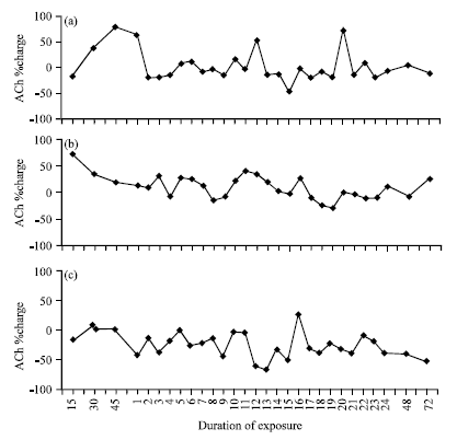 | |
| Fig. 1: | Percent change in ACh contant of various tissues of fish treated with copper sulphate; (a) Liver, (b) Brain and (c) Muscle |
The results differ from those reported by Saluja and Kumar (2005) found a increase in the ACh content in the stomach of Rattus, treated with copper sulphate for 15 days and 30 days. This difference is possibly by the duration of exposure.
In the present study, the AChE activity was declined in all the tissues at all exposure periods following the exposure of copper sulphate after which the animal succumbs (Fig. 2a-c). Frasco et al. (2005) suggest that the heavy metal copper ions induced inhibition of the AChE activity. The interference of metal ions with thiol groups of thiocholine, a product of the hydrolysis of the substrate acetylcholine and with certain buffers such as phosphate. Another valid explanation could be the metal ions cause conformational changes that result in loss of catalytic activity (Del-Ramo et al., 1993). Franciscato et al. (2009) observed that invitro high concentration of copper decreased the cerebrum and cerebellum AChE activity of the suckling rats. These results differ from Romani et al. (2003), observed an increased of AChE activity of brain and muscle in the fish Sparus auratus exposed for 20 days of sublethal concentrations of CuSo4. Differences among the results may be related to the species of the animals, concentrations of toxicant and duration of exposure, respectively.
In the present study, following copper toxicity, Na+K+ ATPase shows the declined activity in liver, brain and muscle tissues at all exposure periods except for few h (Fig. 3a-c). The results of this study also in agreement with the findings of Prasanthi et al. (2006) and Maiti et al. (2010) who discovered that decreased activity of Na+K+ATPase and energy depletion when the fish exposed to lead in a dose dependent manner. Eriyamremu et al. (2008) also reported in their study that the Na+K+ ATPase activity in liver tissues of rabbits were reduced when treated with Cadmium. Hashemi et al. (2008) also found decreased activity of Na+K+ ATPase in the gills of Cyprinus carpio within the first days of copper exposure with significant reduction in activity at the third day of exposure. Heavy metals exposure inhibits Na+K+ ATPase activity because of its high affinity to sulfhydryl groups found in transport enzymes and its interaction with Mg2+-binding sites.
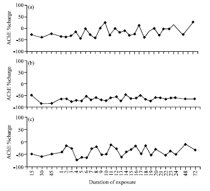 | |
| Fig. 2: | Percent change in AChE activity of various tissues of fish treated with copper sulphate; (a) Liver, (b) Brain and (c) Muscle |
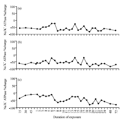 | |
| Fig. 3: | Percent change in Na+K+ ATPase activity of various tissues of fish treated with copper sulphate; (a) Liver, (b) Brain and (c) Muscle |
CONCLUSION
Present experiments indicate that essential metal copper sulphate have caused toxic effects at LC50 concentration. If there is an abundance of essential heavy metals, the homeostatic mechanisms cease to function and the essential heavy metals act in an either acutely or chronically toxic manner. Inhibition of ACh, AChE and Na, K-ATPase may be severe for the animal health point of view. Thus, research of this type is of practical importance, the results of which could be used in the fields of environmental protection and health care.
ACKNOWLEDGMENT
My sincere gratitude to our Principal, Government Arts College (Autonomous) Coimbatore, TamilNadu, India for providing necessary support to the present study.
REFERENCES
- Anbarasi, K., G. Vani, K. Balakrishna an C.S. Devi, 2005. Effect of Bacoside A on membrane-bound ATPases in the brain of rats exposed to cigarette smoke. J. Biochem. Mol. Toxicol., 19: 59-65.
CrossRefPubMedDirect Link - Cepanko, V., R.L. Idzelis, V. Kesminas and R. Ladigiene, 2006. Radiological investigation of roach and perch from some lakes in Lithuania. J. Environ. Eng. Landscape Manage., 14: 199-205.
Direct Link - De Boeck, G., W. Meeus, W. De Coen and R. Blust, 2004. Tissue-specific Cu bioaccumulation patterns and differences in sensitivity to waterborne Cu in three freshwater fish: Rainbow trout (Oncorhynchus mykiss), common carp (Cyprinus carpio) and gibel carp (Carassius auratus gibelio). Aquat. Toxicol., 70: 179-188.
CrossRefPubMed - Eriyamremu, G.E., S.E. Ojimogho, S.O. Asagba and O. Lolodi, 2008. Changes in Brain, liver and kidney lipid peroxidation, antioxidant enzymes and ATPases of rabbits exposed to cadmium ocularly. J. Biol. Sci., 8: 67-73.
CrossRefDirect Link - Franciscato, C., F.R. Goulart, N.M. Lovatto, F.A. Duarte and E.M.M. Flores et al., 2009. Zncl2 exposure protects against behavioural and acetylcholinesterase changes induced by HgClM2. Int. J. Dev. Neurosci., 27: 459-468.
CrossRef - Frasco, M.F., D. Fournier, F. Carvalho and L. Guilhermino, 2005. Do metals inhibit acetylcholinesterase (AChE)? Implementation of assay conditions for the use of AChE activity as a biomarker of metal toxicity. Biomarker, 10: 360-375.
PubMed - Hashemi, S., R. Blust and G. De Boeck, 2008. The effect of starving and feeding on copper toxicity and uptake in Cu acclimated and non-acclimated carp. Aquat. Toxicol., 86: 142-147.
CrossRef - Hestrin, S., 1949. The reaction of acetylcholine and other carboxylic acid derivatives with hydroxylamin and its analytical application. J. Biol. Chem., 180: 249-261.
Direct Link - Kamaruzzaman, B.Y., M.C. Ong and K.C.A. Jalal, 2008. Levels of copper, zinc and lead in fishes of mengabang Telipot River, terengganu, Malaysia. J. Boil. Sci., 8: 1181-1186.
CrossRefDirect Link - Kan, C.A. and G.A.L. Meijer, 2007. The risk of contamination of food with toxic substances present in animal feed. Anim. Feed Sci. Technol., 133: 84-108.
CrossRefDirect Link - Kaplan, O., N.C. Yildirim, N. Yildirim and M. Cimen, 2011. Toxic elements in animal products and environmental health. Asian J. Anim. Vet. Adv., 6: 228-232.
CrossRefDirect Link - Ahmad, M.G.T. and H. Afzal, 2001. Concentration levels of heavy and trace metals in the fish and relevant water from Rawal and Mangla lakes. J. Biol. Sci., 1: 414-416.
CrossRefDirect Link - Papagiannis, I., I. Kagalou, J. Leonardos, D. Petridis and V. Kalfakakou, 2004. Copper and zinc in four freshwater fish species from lake pamvotis (Greece). Environ. Int., 30: 357-362.
CrossRefDirect Link - Prasanthi, R.P.J., G.H. Reddy, C.S. Chetty and G.R. Reddy, 2006. Influence of calcium and zinc on lead-induced alterations in ATPases in the developing mouse brain. J. Pharmacol. Toxicol., 1: 270-277.
CrossRefDirect Link - Romani, R., C. Antognelli, F. Baldracchini, A. Santis, G. Isani, E. Giovannini and G. Rosi, 2003. Increased acetylcholinesterse activites in specimens of Sparus auratus exposed to sublethal concentrations. Chem. Biol. Interact., 145: 321-329.
PubMed - Sparks, D.L. and B.G. Schreurs, 2003. Trace amount of copper in water induce β- amyloid plaques and learning deficits in a rabbit model of Alzheimer`s disease. Proc. Natl. Acad. Sci. USA., 100: 11065-11069.
CrossRef - Ronagh, M.T., A. Savari, F. Papahn and M.A. Hesni, 2009. Bioaccumulation of heavy metlas in Euryglossa orientalis from the Hendijan Seaport (Coastal of Persian Gulf), Iran. J. Biol. Sci., 9: 272-275.
CrossRef