
Wannee Jiraungkoorskul
Department of Pathobiology, Faculty of Science, Mahidol University, Bangkok 10400, Thailand
Somphong Sahaphong
Mahidol University International College, Mahidol University,
Salaya Campus, Nakhonpathom 73170, Thailand
Piya Kosai
Department of Pathobiology, Faculty of Science, Mahidol University, Bangkok 10400, Thailand
Myung-Huk Kim
Mahidol University International College, Mahidol University,
Salaya Campus, Nakhonpathom 73170, Thailand
Research Journal of Environmental Toxicology
Year: 2007 | Volume: 1 | Issue: 1 | Page No.: 27-36


ABSTRACT
Micronucleus formation in fish cells, as an indicator of chromosomal damage, has been increasingly used to detect the genotoxic potential of heavy metal exposure. This study was investigated the effects of cadmium (Cd) and ascorbic acid (AA) on the red-tailed tinfoil barb (Puntius altus) using the micronucleus (MN) and nuclear abnormality (NA) tests for the period of 24, 48, 72 and 96 h. The MN frequencies in the erythrocytes, gill, liver and fin cells were analyzed comparatively to evaluate the sensitivity and suitability of these different cell types. NA shapes in erythrocytes were scored into blebbed nuclei (BL), lobed nuclei (LB), notched nuclei (NT) and binuclei (BN). It was observed that, fish showed significant sensitivity to the different treatments. In general, the highest value of both MN and NA cells were significantly increased in the Cd treated group followed by the combination of Cd and AA treated group. On the other hand, the MN and NA frequencies in erythrocytes were the most sensitive to the treatment and could provide valuable information than those in gill, liver and fin cells. The frequencies of each NA shape in erythrocytes of all treatments were observed in the following NT > LB > BN > BL. Results of MN and NA revealed the highest number after 48 h treatment in all cases and decreased within the longer time exposure. Our results demonstrated the efficacy of AA in reducing genotoxicity in fish induced by Cd. Otherwise; they showed the sensitivity and suitability of MN and NA frequencies in erythrocytes for pollution biomarkers.
PDF Abstract XML References
How to cite this article
Wannee Jiraungkoorskul, Somphong Sahaphong, Piya Kosai and Myung-Huk Kim, 2007. Micronucleus Test: The Effect of Ascorbic Acid on Cadmium Exposure in Fish (Puntius altus). Research Journal of Environmental Toxicology, 1: 27-36.
DOI: 10.3923/rjet.2007.27.36
URL: https://scialert.net/abstract/?doi=rjet.2007.27.36
DOI: 10.3923/rjet.2007.27.36
URL: https://scialert.net/abstract/?doi=rjet.2007.27.36
INTRODUCTION
The increasing levels of pollution caused by a wide variety of toxic substances in various water bodies. The important problems are those that tend to accumulate in organisms, those that are persistent because of their stability or slow biodegradability. Heavy metals possess all of these characteristics and are one of the major contributors to the pollution of Thailand’s natural aquatic ecosystem. Cadmium (Cd) is an extremely toxic element of continuing concern because its environmental levels have risen steadily (WHO, 1992; Goering et al., 1995). They are being used in a wide variety of industrial processes in Thailand, for example, the use of Cd as a coloring agent, a stabilizer and in alloy mixtures.
Ascorbic acid (AA), commonly known as vitamin C, is essential for many aquatic animal species, as they cannot synthesize this micronutrient and depend on an exogenous supply. The function of AA is a strong reducing agent in many tissues and is therefore involved in several physiological processes including growth, reproduction, immunity and the response to stress and infectious agents (Verlhac and Gabaudan, 1994). It has also been reported to have anticarcinogenic (Pauling et al., 1985), anticlastogenic (Gebhart et al., 1985) and even antimutagenic (Shamberger, 1984) roles in a variety of test systems, but its role in modulating cytogenetic damage in any fish has few reported (Guha and Khuda-Bukhsh, 2002).
It has been suggested that a variety of biomarkers and bioassays in the laboratory and field studies be used in determining the effects of genotoxic pollution. These include the presence of DNA adducts, chromosomal aberrations, DNA strand breaks and measurement of micronuclei frequencies. Among the currently available test systems, the micronucleus assay is the most widely applied method due to its simplicity, reliability, sensitivity and proven suitability for fish species. Although originally developed for its application in mouse, it was subsequently modified by Hoofman and de Raat for the application in the laboratory to fish (Hoofman and Raat, 1982). The micronucleus test is a measure of subcellular process such as induced chromosomal breaks (clastogenesis) or cell spindle malfunction (aneugenesis) (Heddle et al., 1991). Otherwise, in most cases the method of exposure employed has been intraperitoneal injection of the product assayed. Some experiments with exposure to pollutants by immersion have been described for different species: for example, Nile tilapia, Oreochromis niloticus, to glyphosate (Jiraungkorskul et al., 2003, 2002). Butterfish, Poronotus triacanthus, to copper (Jiraungkorskul et al., 2006).
Most of the piscine micronuclei surveys have been carried out in erythrocytes, but an alternative possibility could be to try micronucleus test in other tissues, like gill and liver cells (Cavas et al., 2005) and fin cells (Arkhipchuk and Garanko, 2005). Since the liver is the center of xenobiotic metabolism, it can be considered as a suitable tissue for micronucleus tests. Use of cells from liver tissue, however, has some limitations mainly due to its low mitotic index. On the other hand, gill cells also have some advantages over erythrocytes, because they are continuously dividing and are also directly exposed to contaminants (Al-Sabti and Metcalfe, 1995). Fin cells could be used for in vivo cytological investigations without causing any damage to the fish organism and its functioning (Arkhipchuk and Garanko, 2005).
The application of environmental toxicology studies among non-mammalian vertebrates is rapidly expanding and, for aquatic systems, fish have become indicators for the evaluation of the effects of these compounds. Several species of freshwater fish have been reported to be good targets for biomonitoring of rivers and lakes using the micronucleus test as a genotoxicity indicator: rainbow trout, Oncorhynchus mykiss (De Flora et al., 1993), brown trout, Salmo trutta (Sanchez-Galan et al., 1999). None of these species is appropriate to biomonitor Southeast Asia freshwater ecosystems because they are not native to them. Red-tailed tinfoil barb (Puntius altus) is one of the commercialized freshwater fish in Thailand. At the present time, this species is the first Southeast Asia fish investigated in relation to heavy metal exposure. It can provide a good model to study responses and possible adaptations of local fish populations to aquatic pollutants.
The present study, aimed to evaluate the effect of ascorbic acid associated with acute exposed to cadmium in red-tailed tinfoil barb (P. altus). We investigated of the sensitivity and suitability of erythrocytes, gill, liver and fin cells for micronucleus test. We also investigated the new parameter in erythrocytes as the incidence of abnormal shape nuclei.
MATERIALS AND METHODS
Animals
Red-tailed tinfoil barb (P. altus) 27.61±5.17 g in body weight and 13.50±0.77 cm in total length, were purchased from a commercial hatchery in Bangkok, Thailand. Tap water was filtered with activated charcoal (Aquapur, thysen, FRG) to eliminate chemical contamination. The physicochemical characteristics of water were measured daily, according to the experimental procedures described in Standard Methods for the Examination of Water and Wastewater (APHA, 2005). Conductivity was measured with Hanna instruments Model 3 DiST WP (Hanna Instruments Inc., USA). The pH was measured with a Cyberscan 510 (Eutech Instruments Inc., USA) and the temperature was measured with a glass mercury thermometer. A 16:8 h light-dark cycle was maintained throughout.
Acclimatization to laboratory conditions for 7 days was done using dechlorinated tap water that had the following physicochemical characteristics: temperature = 29.0±1.0°C, pH = 6.6-7.0, total hardness = 68-80 mg L-1 (as CaCO3), alkalinity = 75-80 mg L-1 and conductivity = 190-220 μmhos cm-1. Chlorine residual and ammonia were below detection limits. Fish were fed twice a day with 37%-protein commercial fish food (Charoen Pokphand Group, Bangkok, Thailand). The quantity of food was 2% of the initial body weight per day.
Experimental Design
Fish (n = 40) were randomly assigned to four equally sized groups. Each fish was injected intraperitoneal 1 mL/100 g BW with dose selected through range-finding trials that produced fairly quantifiable changes in cytogenetic protocol used as follows: (1) distilled water; (2) group II, 0.05% CdCl2.H2O (Sigma, Germany, CAS No. 10108-64-2); (3) 0.05% CdCl2.H2O plus 0.05% AA (Sigma, Germany, CAS No.50-81-7) and (4) 0.05% AA. The injected fish were kept separately in the glass flow-through aquaria (50 x 50 x 120 cm) with continuous aeration were filled with 200 L of dechlorinated tap water whose physicochemical characteristics were the same as those described previously.
At different times (24, 48, 72 and 96 h), 2 fish of each group were anesthetized with 0.2 g L-1 MS-222 (tricaine methan sulphonate, Sigma, Germany, CAS No.886-86-2), weighed and measured. The organs (blood, gill, liver and fin cells) were removed and prepared for micronuclei and nuclear abnormality tests.
Micronuclei (MN) and Nuclear Abnormality (NA) Tests: Giemsa staining
Peripheral blood samples were obtained by caudal vein puncture using a heparinized syringe. Blood was smeared immediately on clean grease free microscope slides, air dried for 12 h and then fixed in absolute ethanol for 20 min. Each slide was stained with 5% Giemsa solution for 30 min.
Gill arches were removed and placed into vials containing Carnoy’s fixative (3:1 methanol:acetic acid). Gills were transferred into 20% acetic acid solution for 15 min for tissue maceration. After this chemical maceration, epithelial cells were then scraped off the gills and placed on clean slides. After tissue clumps on slides were removed, slides were air-dried and stained with 5% Giemsa solution for 30 min.
A portion of liver tissues were removed and placed into vials containing Carnoy’s fixative. Small pieces of tissues were transferred into vials containing 45% acetic acid solution for 30 min for tissue maceration. After maceration, tissues were gently minced and filtered to obtain a cell suspension. The obtained cells were smeared on a clean slide, air-dried and stained with 5% Giemsa solution for 30 min.
The edge of caudal fins was cut at a depth of 2-3 mm and placed into vials containing Carnoy’s fixative. Small pieces of tissue were transferred into vials containing 45% acetic acid solution for 30 min for tissue maceration. Then tissues were gently minced and filtered to obtain a cell suspension. The obtained cells were smeared on a clean slide, air-dried and stained with 5% Giemsa solution for 30 min.
Three slides were prepared for each organ. From each slide 1000 cells were scored under 1000x magnification using a Nikon E600 light microscope and photographed using a Nikon DXM 1200 digital camera (Tokyo, Japan). Slides were scored by a single observer using blind review. Frequencies of micronucleated (MN) and nuclear abnormality (NA) cells were expressed per 1000 cells (%).
Micronuclei and Nuclear Abnormality Cells Scoring
Only the cells clearly isolated from the surrounding cells were scored. The criteria for the identification of MN were earlier described: (a) MN must be smaller than one-third of the main nuclei, (b) MN must be clearly separated from the main nuclei, (c) MN must be on the same plane of focus and have the same color. Cells having two nuclei with approximately equal sizes were considered as binucleates (Fenech et al., 2003). Nuclear abnormality shapes were scored into one of the following categories: blebbed nuclei (BL), lobed nuclei (LB), notched nuclei (NT) and binuclei (BN) (Carrasco et al., 1990). The result was expressed as the mean value (%) of the sum for all the individual abnormality observed.
Statistical Analysis
All data were expressed as means±SD. A two-way analysis of variance was used to determine the significance of micronuclei and nuclear abnormalities test. The least-significant difference (LSD) was used for determination of significant differences at p<0.05.
RESULTS
Normal erythrocyte, approximately diameter 7 μm, was contained mainly elliptical nuclei (Fig. 1A). The small non-refractile circular or ovoid particle lying in the cytoplasm and resembling a nucleus with respect to staining properties was considered as micronuclei. The size of the micronuclei varied to some extent (between 1/25th and 1/5th that of nuclear size) but the number was always one. The position of the micronuclei in the cytoplasm also varied, some located very near to the nucleus or some located vary far even at the periphery of the cell (Fig. 1B). Some of the nuclei clearly deviated from their normal shape and were either blebbed (BL, Fig. 1C), lobed (LB, Fig. 1D), notched (NT, Fig. 1E) and binucleated cells (BN, Fig. 1F). All abnormalities of nuclei were scored. In briefly, cells with two nuclei were considered as binucleates. The two nuclei should be approximately equal size, staining pattern and staining intensity, within the same cytoplasmic boundary. Blebbed nuclei presented a relatively small evagination of the nuclear membrane, which contained euchromatin. Evaginations larger than the blebbed nuclei, which could have several lobes, were classified as lobed nuclei. Nuclei with depth into a nucleus were recorded as notched nuclei.
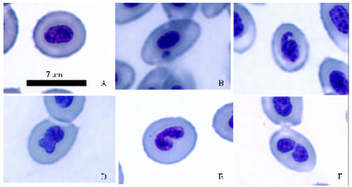 | |
| Fig. 1: | Photomicrographs of erythrocytes with normal nucleus (A); with micronuclei (B) and with nuclear abnormalities: blebbed nuclei (C); lobed nuclei (D); notched nuclei (E); binuclei (F) of P. altus |
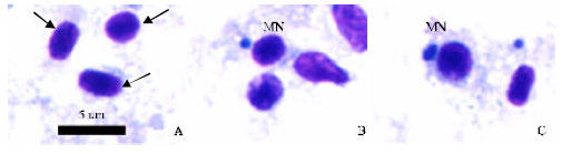 | |
| Fig. 2: | Photomicrographs of gill cells with normal nucleus marked by arrows (A) and with micronuclei indicated by MN (B-C) of P. altus |
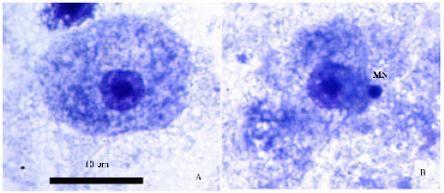 | |
| Fig. 3: | Photomicrographs of liver cells with normal nucleus (A) and with micronuclei indicated by MN (B) of P. altus |
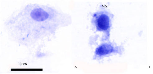 | |
| Fig. 4: | Photomicrographs of fin cells with normal nucleus (A) and with micronuclei indicated by MN (B) of P. altus |
Normal gill cell approximately diameter 4-5 μm was contained mainly elliptical nuclei (Fig. 2A). Similarly to MN in periphery blood, micronuclei of gill cells were small non-refractile circular or ovoid particle lying in the cytoplasm and resembling a nucleus with respect to staining properties (Fig. 2B-C). Liver cells with normal nucleus and with micronuclei were shown in Fig. 3A-B, respectively. Fin cells with normal nucleus and with micronuclei were shown in Fig. 4A-B, respectively.
| Table 1: | Frequencies (%) of micronuclei in erythrocytes, gill, liver and fin cells of P. altus exposed to different time and treatments (mean±SD) |
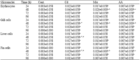 | |
| Cont. = Control group; Cd = Cadmium group; Mix = Cadmium plus ascorbic acid; AA = Ascorbic acid, a = The mean difference was significant in row when compared the control group (p≤0.05), b = The mean difference was significant in row when compared the cadmium group (p≤0.05), c = The mean difference was significant in row when compared the mix group (p≤0.05), * = The mean difference was significant in column when compared the erythrocytes (p≤0.05), # = The mean difference was significant in column when compared the gill cells (p≤0.05) | |
| Table 2: | Frequencies (%) of nuclear abnormalities cells in erythrocytes of P. altus exposed to different time and treatments (mean±SD) |
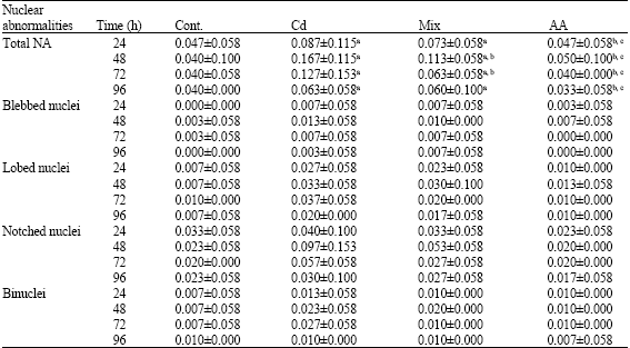 | |
| Cont. = Control group; Cd = Cadmium group; Mix = Cadmium plus ascorbic acid; AA = Ascorbic acid, a = The mean difference was significant in row when compared the control group (p≤0.05), b = The mean difference was significant in row when compared the cadmium group (p≤0.05), c = The mean difference was significant in row when compared the mix group (p≤0.05) | |
The frequencies of MN observed in various treated and control fish have been summarized in Table 1. It is evident that MN was not significantly induced in the control group at 24, 48, 72 and 96 h. Otherwise; the differences were statistically significant in treated group when compared with those of the control group at all the time intervals. The frequencies of MN were significant highest number in fish treated with Cd alone. However, when both Cd and AA were conjointly administered, the MN appeared to be reduced to some extent, the difference being statistically significant at 48 h. The frequencies of NA observed in various treated and control fish have been summarized in Table 2. Similarly to MN, the frequencies of NA were also significant greater number in fish treated with Cd. On the other hand, the MN frequencies in erythrocytes were the most sensitive to the treatments and could provide valuable information than those in gill, liver and fin cells. When NA in erythrocytes were analyzed separately, it was observed that the frequency of each abnormality shapes were found in the following order: NT > LB >BN > BL. Results of MN and NA revealed the gradually increased with the time up to 48 h in all cases and decreased to some extent at 72 and 96 h.
DISCUSSION
MN is cytoplasmic chromatin masses with the appearance of small nuclei that arise from chromosome fragments or intact whole chromosomes lagging behind in the anaphase stage of cell division. Their presence in cells is a refection of structural and/or numerical chromosomal aberrations arising during mitosis (Heddle et al., 1991). MN and NA tests in fish are generally performed in enucleated peripheral blood erythrocytes mainly due to its technical feasibility. In most studies short-term exposure period ranging between 24 and 96 h were reported to be sufficient to induce micronuclei and erythrocytes have been reported to be a sensitive biomarker of genotoxicity (Heddle et al., 1991; Ayllon and Garcia-Vazquez, 2000).
There are some assays in fish aimed to evaluate the differential sensitivity of diverse species to different chemicals (Allon and Gardia-Vazquez, 2000). But in most cases the method of exposure has been intraperitoneal injection as described in this study and the previous reports, for the example, cadmium significantly induced micronuclear expression in brown trout, S. trutta (Sanchez-Galan et al., 1999) and rainbow trout, O. mykiss (Castano et al., 1998). In the present study, the peak values for MN and NA in erythrocytes have been obtained after 48 h. Similar time-dependent effects were also previously reported in fish erythrocytes exposed to textile mill effluent (Cavas and Ergene-Gozukara, 2003) and cadmium chloride (Ayllon and Garcia-Vazquez, 2000). Pollution by heavy metals is an important problem due to their stable and persistent existence in the environment. It is well known that heavy metals interferes the regular chromosome segregation during cell division mainly by inhibition of polymerization of actin tubules, an essential structure of the mitotic spindle (Miura and Imura, 1987). It was suggested that the mechanism of Cd genotoxicity is mainly conditioned by single strand breaks in DNA through the direct cadmium-DNA interactions as well as by the action of incision nucleases and/or DNA-glycosylase during DNA repair (Privezentsev et al., 1996). Correspondingly, most of the toxic chemicals that produce genotoxic effects have been known to form reactive oxygen species as well as electrophilic free-radical metabolites that interact with DNA to cause disruptive changes. It has been suggested that during the heavy metal exposure, electrophilic ions and radicals are produced, interacting with nucleophilic sites in DNA and leading to breaks and other related damage in the latter.
Further, it would be revealed from the results of this study that Cd produced genotoxic effects. Interestingly the injection of ascorbic acid appeared to modulate the genotoxic effects of Cd at all time intervals. The exact mechanism which ascorbic acid minimizes the genetoxic of Cd is not known. However, it is known that ascorbic acid has marked nucleophilic properties it might intercept reactive electrophilic metabolites produced by Cd, thereby preventing their attack on nucleophilic sites on DNA and hence blocking adduct formation (Liehr et al., 1989). Otherwise ascorbic acid is an anti-oxidant which might inhibit the oxidative metabolism of Cd and thus could prevent the production of mutagenic electrophilic metabolites (Goncharova and Kuzhir, 1989). Also as part of a redox buffer system ascorbic acid can scavenge harmful free radical metabolites or reactive oxygen species (Sato et al., 1990). Thus, the general protective effect of ascorbic acid observed against Cd induced genotoxicity could actually be accomplished through one or many of these inhibition mechanisms.
In the present study, the MN and NA tests were carried out on the different types of cells. On the erytrocytes, those are the most commonly used for these tests in the fish. On the gill cells, those are more directly exposed to environmental contaminants. Since genotoxic activation of xenobiotics usually occurs in the liver, the hepatic MN and NA assays are also thought to be a sensitive tool for detecting genotoxic responses. The hepatic micronuclei technique is rarely used in fish and there are only several studies available in the literature (Cavas et al., 2005; Al-Sabti, 1995). In the present study, the MN frequencies in erythrocytes were the most sensitive to the treatments and could provide valuable information than those in gill, liver and fin cells. There are contradictory results about the sensitivity of these cells in the MN and NA tests. Cavas et al. (2005) reported that gill and liver cells were found to be more sensitive tissues than peripheral blood cells. The reason for this is their higher mitotic index values. The same situation might also be considered for fin cells. Arkhipchuk and Garanko (2005) reported the highest increases of cells having micronuclei were observed in blood after fish exposure to cadmium ions and in the fin tissue after the copper impact. In the present study, there was a general tendency of occurrence of notched and lobed types in higher frequencies. An analysis revealed spontaneous frequencies of nuclear abnormalities in erythrocytes were found in the following order: NT > LB > BN > BL. Thus, our results seem to be in agreement with previous studies (Cavas and Ergene-Gozukara, 2003; Mallick and Khuda-Bukhsa, 2003).
In conclusion, the results presented in this study show that the efficacy of ascorbic acid in reducing genotoxicity in fish associated with acute exposed to cadmium. Otherwise, they showed the sensitivity and suitability of micronuclei and nuclear abnormality frequencies in erythrocytes for pollution biomarkers. Our data indicated that the fish micronucleus test and the new parameter in erythrocytes as the incidence of abnormal shape nuclei are the valid technique for monitoring genotoxic effects induced by heavy metals.
ACKNOWLEDGMENTS
This study was funded by the Thailand Research Fund (TRF) and the Commission on Higher Education: the New Researchers Grant 2006 and in part by Mahidol University International College and Faculty of Science, Mahidol University.
REFERENCES
- Al-sabti, K., 1995. An in vitro binucleated blocked hepatic cell technique for genotoxicity testing in fish. Mutat. Res., 335: 109-120.
Direct Link - Al-Sabti, K. and C.D. Metcalfe, 1995. Fish micronuclei for assessing genotoxicity in water. Mutat. Res./Genet. Toxicol., 343: 121-135.
CrossRefPubMedDirect Link - APHA, 2005. Standard Methods for the Examination of Water and Wastewater. 18th Edn. American Public Health Association/American Water Works Association/Water Pollution Control Federation, Washington, DC., USA.
Direct Link - Arkhipchuk, V.V. and N.N. Garanko, 2005. Using the nucleolar biomarker and the micronucleus test on in vivo fish fin cells. Ecotox. Environ. Safety, 62: 42-52.
CrossRef - Ayllon, F. and E. Garcia-Vazquez, 2000. Induction of micronuclei and other nuclear abnormalities in European minnow Phoxinus phoxinus and mollie Poecilia latipinna: An assessment of the fish micronucleus test. Mutat. Res., 467: 177-186.
Direct Link - Carrasco, K.R., K.L. Tilbury and M.S. Myers, 1990. Assessment of the piscine micronucleus test as an in situ biological indicator of chemical contaminant effects. Can. J. Fisher. Aquat. Sci., 47: 2123-2136.
CrossRefDirect Link - Castano, A., G. Carbonell, M. Carballo, C. Fernandez, S. Boleas and J.V. Tarazoma, 1998. Sublethal effects of repeated intraperitoneal cadmium injection on rainbow trout. Ecotoxicol. Environ. Sef., 41: 29-35.
CrossRefDirect Link - Cavas, T. and S. Ergene-Gozukara, 2003. Micronuclei nuclear lesions and interphase silver-stained nucleolar organizer Agnors as cyto-genotoxicity indicators in Oreochromis niloticus exposed to textile mill effluent. Mutat. Res., 538: 81-91.
Direct Link - Cavas, T., N.N. Garanko and V.V. Arkhipchuk, 2005. Induction of micronuclei and binuclei in blood, gill and liver cells of fishes subchronically exposed to cadmium chloride and copper sulphate. Food Chem. Toxicol., 43: 569-574.
CrossRefDirect Link - De Flora, S., L. Vigano, F. D`Agostini, A. Camoirano and M. Bagnasco et al., 1993. Multiple genotoxicity biomarkers in fish exposed in situ to polluted river water. Mutat. Res., 319: 167-177.
CrossRefDirect Link - Fenech, M., W.P. Chang, M. Kirsch-Volders, N. Holland, S. Bonassi and E. Zeiger, 2003. Humn project detailed description of the scoring criteria for the cytokinesis-block micronucleus assay using isolated human lymphocyte cultures. Mutat. Res., 534: 65-75.
CrossRef - Gebhart, E., H. Wagner, K. Grziwok and H. Behnsen, 1985. The action of anticlastogens in human lymphocyte cultures and their modification by rat liver S9 II. Studies with vitamin C and E. Mutat. Res., 149: 83-94.
Direct Link - Guha, B. and A.R. Khuda-Bukhsh, 2002. Efficacy of vitamin-C (L-ascorbic acid) in reducing genotoxicity in fish (Oreochromis mossambicus) induced by ethyl methane sulphonate. Chemosphere, 47: 49-56.
CrossRefDirect Link - Heddle, J.A., M.C. Cimino, M. Hayashi, F. Romagna and M.D. Shelby et al., 1991. Micronuclei as an index of cytogenetic damage: Past, present and future. Environ. Mol. Mutag., 18: 277-291.
CrossRefDirect Link - Hoofman, R.N. and W.K. de Raat, 1982. Induction of nuclear anomalies (micronuclei) in the peripheral blood erythrocytes of the eastern mudminnow Umbra pygmaea by ethyl methanesulphonate. Mutat. Res. Lett., 104: 147-152.
CrossRefDirect Link - Jiraungkoorskul, W., E.S. Upatham, M. Kruatrachue, S. Sahaphong, S. Vichasri-Grams and P. Pokethitiyook, 2002. Histopathological effects of Roundup, a glyphosate herbicide, on Nile tilapia (Oreochromis niloticus). ScienceAsia, 28: 121-127.
Direct Link - Jiraungkoorskul, W., E.S. Upatham, M. Kruatrachue, S. Sahaphong, S. Vichasri-Grams and P. Pokethitiyook, 2003. Biochemical and histopathological effects of glyphosate herbicide on Nile tilapia (Oreochromis niloticus). Environ. Toxicol., 18: 260-267.
CrossRefDirect Link - Mallick, P. and A.R. Khuda-Bukhsa, 2003. Nuclear anomalies and blood protein variations in fish of the Hooghly-Matlah river system, India, as an indicator of genotoxicity in water. Bull. Environ. Contam. Toxicol., 70: 1071-1082.
CrossRefDirect Link - Pauling, L., J.C. Nixon, F. Stiff, R. Marcuson and W.B. Dunham et al., 1985. Effect of dietary ascorbic acid on the incidence of spontaneous mammary tumors in RIII mice. Proc. Natl. Acad. Sci. USA., 82: 5185-5189.
Direct Link - Privezentsev, K.V., N.P. Sirota and A.I. Gaziev, 1996. The genotoxic effects of cadmium studied in vivo. Tsitol. Genet., 30: 45-51.
PubMedDirect Link - Sanchez-Galan, S., A.R. Linde, J.I. Izquierdo and E. Garcia-Vazquez, 1999. Brown trout and European minnow as target species for genotoxicity test: Differential sensitivity to heavy metals. Ecotoxicol. Environ. Sef., 43: 301-304.
Direct Link - Sato, K., E. Niki and H. Shimasaki, 1990. Free radical-mediated chain oxidation of low density lipoprotein and its synergistic inhibition by vitamin E and vitamin C. Arch. Biochem. Biophys., 279: 402-405.
CrossRefDirect Link - Shamberger, R.J., 1984. Genetic toxicology of ascorbic acid. Mutat. Res./Rev. Genet. Toxicol., 133: 135-159.
CrossRefDirect Link - Verlhac, V. and J. Gabaudan, 1994. Influence of vitamin C on the immune system of salmonids. Aquacult. Fish Manage., 25: 21-36.
CrossRefDirect Link