
A. Al-Soqeer
Department of Plant Production and Protection, Faculty of Agriculture and Veterinary Medicine, Qassim University, P.O. Box 6622, Buraidah, 51452, Qassim, Saudi Arabia
Research Journal of Botany
Year: 2011 | Volume: 6 | Issue: 1 | Page No.: 11-20


ABSTRACT
Hot-water extracts of Capparis spinosa and Artemisia monosperma were studied against lipid peroxidation induced by lead acetate in rats. Ten rats were assigned to each of four groups. Group 1 was negative control, group 2 received only lead acetate (0.6%) in drinking water (positive control), group 3 received hot-water extract from Artemisia (5%) with lead acetate in drinking water (0.6%) and group 4 received hot-water extract from Capparis (5%) with lead acetate in drinking water (0.6%) experiment continued for 6 weeks before scarifying the animals and blood samples were collected. Chemical composition and antioxidant activities of hot-water extract from Artemisia and Capparis sp. were estimated using chemical methods. Total Antioxidants in Artemisia and Capparis extracts were 675.33, 115.66 μmol Trolox per 100 mL, respectively. Activity of the antioxidant enzyme glutathione-S-transferase activity decreased after lead administration but extracts from Artemisia and Capparis maintained the level to normal. Lead administration resulted in significant increases in the concentration of serum triglycerides, urea and aspartate transaminase, alanine transaminase (AST and ALT). Extracts from Artemisia and Capparis reduced the elevated concentrations to normal values. The obtained results showed that the biochemical alterations produced by lipid peroxidation after lead administration returned to normal values or even improved due to high levels of total antioxidants in extracts of Artemisia and Capparis sp. Data concluded that the protective effects of hot-water extracts from Artemisia and Capparis sp. may play a role in protection against lead acetate in rats. This protective effect may be due to high level of total antioxidant contents in these plants.
PDF Abstract XML References Citation
Received: June 15, 2010;
Accepted: October 17, 2010;
Published: April 09, 2011
How to cite this article
A. Al-Soqeer, 2011. Antioxidant Activity and Biological Evaluation of Hot-water Extract of Artemisia monosperma and Capparis spinosa Against Lead Contamination. Research Journal of Botany, 6: 11-20.
URL: https://scialert.net/abstract/?doi=rjb.2011.11.20
URL: https://scialert.net/abstract/?doi=rjb.2011.11.20
INTRODUCTION
In recent years the therapeutic importance of herbal drugs has gained considerable attention. It has been found that utilization of cheap plant sources that are without side effects is a trend in all countries.
In Saudi Arabia herbal medicine is widely spread and many local plants are used for treatment of various ailments. One of these plants is Capparis spinosa (CS) (Capparidaceae), locally known as Shafalah. Scientific classification and uses of Capparis spinosa are shown in Fig. 1. They are native shrubs widely distributed in Al-Qassim region. This plant was found to be common to the Mediterranean basin and has been used by the traditional medicine for its diuretic and antihypertensive effects and also in certain pathological conditions related to uncontrolled lipid peroxidation. The extract contains many constituents, in particular some flavonoids (kaempferol and quercetin derivatives) and hydrocinammic acids with several known biological effects such as the anti-inflammatory and the antioxidant ones (Panico et al., 2005).
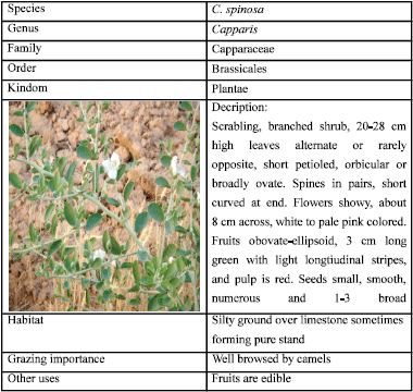 | |
| Fig. 1: | Scientific classification and uses of Capparis spinosa |
The phytotherapy knowledge of this plant has been of interest to many investigators. The main constituents of CS have been demonstrated by other workers to be flavonoids, alkaloids, lipids and glucosinolates and these constituents may vary from one locality to another (Calis et al., 2002; Sharaf et al., 2000; Brevard et al., 1992).
Shafalah has been used since ancient times in Morocco in the treatment of digestive disorders and was commonly used in phytomedicine as an antibacterial (Singh et al., 2002), antiulcerogenic (Khayyal et al., 2001) antiproliferative (Nakano et al., 1998).
Recently, Yang et al. (2010) found that fruits of Capparis spinosa contain a significant amount of compounds with many health benefits. Three new alkaloids, (1) Capparisine A, (2) Capparisine B, (3) Capparisine C and (4) two known alkaloids, (4) 2-(5-hydroxymethyl-2-formylpyrrol-1-yl) propionic acid lactone and (5) N-(3'-maleimidy1)-5-hydroxymethyl-2-pyrrole formaldehyde were isolated from the fruits of C. spinosa.
Another important plant is Artemisia species. Scientific classification and uses of Artemisia monosperma (Athir) are presented in Fig. 2.
The herb, Artemisia monosperma, is a perennial fragrant plant which grows widely and wildly in the Arabian deserts. The leaves of the plant are taken in folk medicine by certain women of Jordan for abortion induction (Hijazi and Salhab, 2010). In a phytochemical study of Artemisia monosperma three compounds were characterised by spectroscopic means. Compounds were evaluated for their ability to inhibit 12-lipoxygenase, as antibacterials and in a cytotoxicity assay (Stavri et al., 2005).
The antioxidant activities of various fractions of Artemisia species were investigated, especially, the n-butanol extract that was found to causes significant increase in the rat liver cytosolic superoxide dismutase (SOD) and catalase activities (Juteau et al., 2002; Kim et al., 2003). The Artemisia oil has a characteristic flavor, due to the presence of many components with strong sensory properties at a low threshold, such as trans-ethyl cinnamate (20.8%) and thus could be suitable for using as antioxidant and flavoring agent in the food industry (El-Massry et al., 2002).
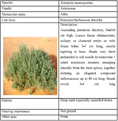 | |
| Fig. 2: | Scientific classification and uses of Artemisia monosperma (Athir) |
It was claimed by many researchers that the risk from contamination by heavy metals is mainly due to the oxidative stress produced by these metals and consequently depletion of body antioxidants (Ercal et al., 2001).
Because many synthetic antioxidants (butylated hydroxyanisole and butylated hydroxytoluene) have been proved to have undesirable side effects (Jayaprakasha et al., 2007). There has been an increasing interest in the use of medicinal plants rich in antioxidants to reduce free radicals-induced tissue injury (Zhishen et al., 1999). Oxidative stress is the excess formation of and/or incomplete removal of highly reactive molecules such as Reactive Oxygen Species (ROS). In vivo some of ROS play positive role such as energy production, phagocytosis and regulation of cell growth (Halliwell and Gutteridge, 1999). On the other hand, ROS are capable of damaging a wide range of essential biomolecules such as proteins, DNA and lipids (Farber, 1994). Cells have several antioxidant defense mechanisms that help to prevent the destructive effects of ROS. These defense mechanisms include anti oxidative enzymes, such as superoxide dismutase, catalase and glutathione peroxidase and of small molecules such as glutathione and vitamin C and E (Maser et al., 2002). The efficiency of the antioxidant defense system is altered under pathological conditions and therefore the ineffective scavenging and/or over production of free radicals may play a crucial role in determining tissue damage (Aruoma, 1994; Halliwell, 1994).
From the foregoing considerations, it can be concluded that Phyto chemicals content in plants are promising alternatives to synthetic drugs provided that certain precautions were taken in consideration. The present investigation aims at analyzing the chemical composition and nutritive value of Capparis spinosa and Artemisia monosperma and test their efficacy against lead contamination in rats.
MATERIALS AND METHODS
The study to be described was carried out at the College of Agriculture and Veterinary Medicine, Qassim university, Saudi Arabia during the period 2009/2010.
Plants
Capparis spinosa (CS): Scientific classification and uses of Capparis spinosa is shown in Fig. 1. Scientific classification and uses of Artemisia monosperma (Athir) is shown in Fig. 2.
Preparation of pressurized hot water extraction: Hot water extraction of Artemisia and Capparis were prepared according the methods described by Abdel-Salam et al. (2009): Approximately 50 g of each of Artemisia and Capparis materials were cut into small pieces, melded and placed in a flask (2 L) with 1000 mL of distilled water and boiled for 15 min, then the mixture was filtered twice, first through cheese-cloth (50% cotton/50% polyester) and then through filter paper (Whatman No. 2). The final concentrations of the prepared of Artemisia or Capparis were 5% as a total solids. The amounts of obtained Artemisia and Capparis extracts were preserved in sterile dark bottles (500 mL) in a cool environment (4°C) until further use.
Antioxidant activity determination in Artemisia monosperma and Capparis spinosa: Antioxidant activity was determined according to the method of Sanchez-Moreno et al. (1998). Antioxidant capacity was quantified as μg Trolox equivalents using a standard curve.
Proximate analysis and nutritive value: The vitamin C content was determined by the titrimetric method according to the method of AOAC (1990). Lipid content of these plants was determined by the direct extraction method. Three grams of dried sample was extracted by 50 mL of petroleum ether at 60°C for 60 min in a soxtec system HT (series 1043 Extractor unit, Tecator). The lipid value was obtained by mean of weighing. Protein content was determined by the Kjeldahl method. Other determinations were carried out according to Benjapak et al. (2008).
Animals: Forty male Swiss albino rats weighting about 120-150 g were randomly divided into four-test groups (each containing 10 rats). Animals were placed in cages and were assigned to specific diets.
Diets: The composition of the basal diet used in this study is as follows: milk protein (12%), sucrose (5%), fat (10%), vitamin mixtures (1%), salt mixtures (4%), fiber (4%) and starch (64%). Rats were divided into the following groups.
| • | The first group (G1) was fed on the basal diet. without lead acetate (control negative) |
| • | The second group (G 2) was fed on the basal diet with lead acetate in drinking water (0.6%) (control positive) |
| • | The third group (G3) was fed on basal diet + hot-water extract from Artemisia (5%) with lead acetate in drinking water (0.6%) |
| • | The fourth fed (G 4) on basal diet + hot-water extract from capparis (5%) with lead acetate in drinking water (0.6%) |
The feeding experiment continued for 6 weeks and at the end of this period, rats were anesthetized by diethyl ether, bled and sacrificed. Blood samples were collected and frozen at (4-6°C) for 2 h before centrifugation at 3000X g for 30 min. Sera were harvested, labeled and stored deep frozen (-20°C) until used for assays.
Biochemical analysis: Urea was determined in serum according to the methods of Tietz (1970) and Bonsnes. Triglyceride were determined according to the methods of Stein and Myers (1995).
Alanine aminotransaminase (ALT) and Aspartate aminotransaminase (AST) activities were determined according to the method of Reitman and Frankel (1957). Glutathione-S-Transferase (GST) activity was determined according to Habig et al. (1974). GST activity was defined as the amount of enzyme catalyzing the formation of 1 mol of products per min under condition of assay.
Activity = Å 340 nm/(9.6)x1000 = M/min |
Statistical analysis: Mean and standard deviation of the obtained data from each different experimental group were calculated and conducted according the method described by Miller and Miller (1992).
RESULTS
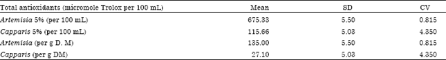
Total antioxidant activity in Artemisia and Capparis extracts: Total antioxidants in Artemisia, Capparis, are shown in Table 1. The mean values were 675.33, 115.66 μmol Trolox per 100 mL for Artemisia, Capparis, respectively.
When total antioxidants are expressed as micromole Trolox per gram D.M, it was found that Artemisia contained higher antioxidants (135.66 ) when compared with Capparis (27.10) micromole Trolox per gram D.M.
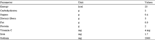
Nutritional value of Capparis are presented in Table 2. As can be noticed from the Table 2 Capparis is a rich source of vitamin C. Each 100 g contained 4 mg of Ascorbic acid. It is also a rich source of iron and sodium.
| Table 1: | Total antioxidants in Artemisia and Capparis extracts |
 | |
| Table 2: | Nutritional value per 100 g of Capparis spinosa |
 | |
| Table 3: | Serum urea concentrations in control and in rats treated with Artemisia monosperma and Capparis spinosa water extracts |
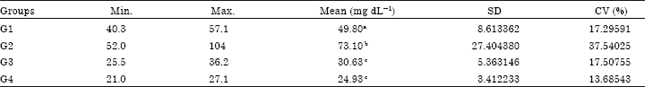 | |
| Means in the same column with different superscript are significantly different (p<0.05) | |
| Table 4: | Serum triglyceride concentrations in control rats and in rats treated with Artemisia monosperma and Capparis spinosa water extracts |
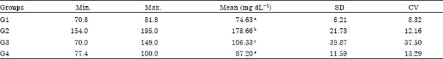 | |
| Means in the same column with different superscript are significantly different (p<0.05) | |
| Table 5: | Activities of AST and ALT in serum of control rats and in rats treated Artemisia monosperma and Capparis spinosa water extracts |
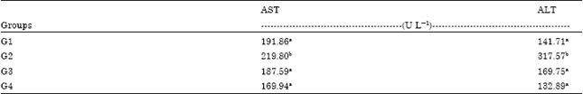 | |
| Means in the same column with different superscript are significantly different (p<0.05) | |
Serum urea: As shown in Table 3 there was a significant (p<0.05) increase in the concentration of urea in the positive control group. Administration of water extracts of Artemisia monosperma and Capparis spinosa resulted in a significant (p<0.05) decrease in levels of urea indicating usefulness of these medicinal plant extracts. Concentration of serum urea was 49.8 mg dL-1 in the control group (negative control) that increased to 73.10 dL in the positive control that received no plant extracts. Concentrations of serum urea decreased significantly (p<0.05) to 30.63 and 24.93 mg dL-1 after administration of Artemisia monosperma and Capparis spinosa, respectively.
Serum triglycerides: Serum triglycerides concentration increased significantly following lead acetate administration (Table 4). Triglycerides concentration in the negative control group was 74.63 mg dL-1 that increased to 178.66 mg dL-1 in the positive control. Treatment with water extracts from Artemisia monosperma and Capparis spinosa decreased the concentration of serum triglycerides to 106.33 and 87.20 mg dL-1, respectively.
As presented in Table 5. Lead acetate administration increased significantly (p<0.05) the activity of AST. The activity of this enzyme in the control group was 191.86 U L-1 that increased after lead administration to 219.80 U L-1 in the positive control group. This activity was significantly reduced to 187.59 and 169.94 U L-1 following administration of Artemisia monosperma and Capparis spinosa water extracts.
Activity of Glutathione-S-transferase in serum is shown in Table 6, the activity in serum of control rats, when expressed as Activity = A/9.6*1000 M min-1, was found to be 16.94 that decreased significantly (p<0.05) to 11.25 in the positive control. When water extract of Artemisia monosperma and Capparis spinosa was administered the activity of this enzyme increased significantly (p<0.05) to 18.61and 21.35 A/9.6 1000 M min-1, respectively.
| Table 6: | Glutathione-S-transferase activity in serum of control rats and in rats treated with Artemisia monosperma and Capparis spinosa water extracts |
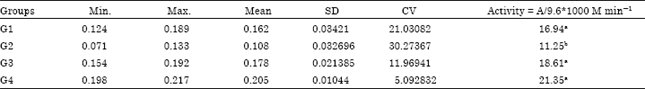 | |
| Means in the same column with different superscript are significantly different (p<0.05) | |
DISCUSSION
In various cultures especially in the developing world, people are dependant on plants as medicine against various ailments. In Greek popular medicine a herbal tea made of caper root and young shoots is considered to be beneficial against rheumatism.
In the present study, it was found that lead ingestion disturbs many biological functions in rats mostly through free radicals generation, The antioxidant capacity in the body of rats as in other animals-is limited and usually exhausted by free radicals generated.
Lead administration increased the level of triglycerides and urea in blood. Ingestion in drinking water of Artemisia monosperma and Capparis spinosa water extracts lowered the concentrations of these metabolites. This may be due to the presence of high levels of antioxidants in Artemisia monosperma and Capparis spinosa (Table 1) and also due to high level of vitamin C. Nutrient antioxidants may act together to reduce reactive oxygen species levels more effectively than single dietary antioxidants, because they can function as synergists (Eberhardt et al., 2000; Rossetto et al., 2002; Podsedek, 2007).
The antioxidant activities of Artemisia species were investigated by Juteau et al. (2002) and Kim et al. (2003) and they found that extracts from Artemisia caused significant increases in the rat liver cytosolic superoxide dismutase (SOD) and catalase (CAT) that are highly involved in the antioxidant system of the body. The increase in the activities of these enzymes will explain the improvement in the pathological changes achieved in kidneys of lead intoxicated rats. It was found that Artemisia extract significantly increased gastrointestinal transit time and the reaction time to thermal stimuli, but had no effect on the activity of alkaline phosphatase or concentrations of creatinine and urea in plasma (Marrif et al., 1995). The plant extract showed weak antimicrobial activity. On the other hand, Qureshi et al. (1990) reported that ethanolic extracts of the aerial parts of Artemisia abyssinica and A. inculta when subjected to acute toxicity observations in mice for 24 h and chronic toxicity evaluation for 3 months. It was found that external morphological changes, visceral toxicity, haematological changes, spermatogenic dysfunction and effect on body weight and vital organ weight were recorded. damage was observed in A. abyssinica treated mice while A. inculta failed to produce any significant spermatotoxic effects.
Administration of lead acetate resulted in elevated activities of AST and ALT enzymes that may be due to liver damage caused by the free radicals, however, ingestion of Artemisia monosperma and Capparis spinosa water extracts in drinking water decreased the activities of these enzymes indicating a protective effect against liver damage. This is in agreement with the finding of Gadgoli and Mishra (1999). They reported that p-Methoxy benzoic acid isolated from the methanolic soluble fraction of the aqueous extract of Capparis spinosa L. (Capparidaceae) was found to possess significant antihepatotoxic activity against carbon tetrachloride and paracetamol induced hepatotoxicity in vivo and thioacetamide and galactosamine induced hepatotoxicity in isolated rat hepatocytes, using in vitro technique. through physicochemical and spectral studies. Isolation, identification and antihepatotoxic activity of the compound is reported for the first time in the plant.
On the other hand lead acetate intoxication reduced the activity of the enzyme glutathione-S-transferase. Glutathione-S-transferase (GST) provides glutathione (GSH) and helps to neutralize the toxic neutrophiles (Halliwell, 1994). Treatment with hot water extract of either Artemisia monosperma and Capparis spinosa improved the activity of this enzyme leading to improved formation of the very potent antioxidant, glutathione.
Previous phytochemical studies on Artemisa have yielded polyyne, sesquiterpene (Stavri et al., 2005) and acetophenone natural products (Bohlmann and Ehlers, 1977). Bohlmann and Ehlers (1977) and known metabolites including the sesquiterpene spathulenol, an hydroxylated polyyne and the flavonoid eriodyctiol-7-methyl ether. These compounds were evaluated by those authors for their activity in 12-lipoxygenase, antibacterial and found to protect against cytotoxicity. The mechanism(s) of protection offered by Artemisia monosperma and Capparis spinosa water extract are not clear at present and further work is needed.
It can be concluded that Artemisia and Capparis extracts contain high antioxidant compounds that protected rats against lipid peroxidation generated by lead. Isolation of individual compounds in these two plants is necessary for further studies.
ACKNOWLEDGMENTS
This study was supported by a research grant from the Deanship of Scientific Research, Qassim University, Saudi Arabia. The author would like to thank Dr. Hassan M. Mousa and Dr. Ahmed Abdel-Salam from the Food Science and Human Nutrition Department, College of Agriculture and Veterinary Medicine for their valuable help with the Proximate and biochemical analysis and nutritive value.
REFERENCES
- Hijazi, A.M. and A.S. Salhab, 2010. Effects of Artemisia monosperma ethanolic leaves extract on implantation, mid-term abortion and parturition of pregnant rats. J. Ethnopharmacol., 128: 446-451.
CrossRef - Abdel-Salam, A.M., A.S. Ammar and W.K. Galal, 2009. Evaluation and properties of formulated low calories functional yoghurt cake. Int. J. Food Agric. Environ., 7: 218-221.
Direct Link - Panico, A.M., V. Cardile, F. Garufi, C. Puglia, F. Bonina and G. Ronsisvalle, 2005. Protective effect of Capparis spinosa on chondrocytes. Life Sci., 77: 2479-2488.
CrossRefDirect Link - Brevard, H., M. Brambilla, A. Chaintreau, J.P. Marison and H. Diserens, 1992. Occurrence of elemental sulphur in capers (Capparis spinosa L.) and first investigation of the flavour profile. Flavour Fragrance J., 7: 313-321.
CrossRefDirect Link - Calis, I., A. Kuruuzum-Uz, P.A. Lorenzetto and P. Ruedi, 2002. (6S)- Hydroxy-3-oxo-alpha-ionol glucosides from Capparis spinosa fruits. Phytochemistry, 59: 451-457.
PubMed - Eberhardt, M.V., C.Y. Lee and R.H. Liu, 2000. Nutrition: Antioxidant activity of fresh apples. Nature, 405: 903-904.
CrossRefDirect Link - Ercal, N., H. Gurer-Orhan and N. Aykin-Burns, 2001. Toxic metals and oxidative stress part I: Mechanisms involved in metal-induced oxidative damage. Curr. Top. Med. Chem., 1: 529-539.
CrossRefPubMedDirect Link - Farber, J.L., 1994. Mechanisms of cell injury by activated oxygen species. Environ. Health Perspect., 102: 17-24.
PubMed - Habig, W.H., M.J. Pabst and W.B. Jakoby, 1974. Glutathione S-transferases: The first enzymatic step in mercapturic acid formation. J. Biol. Chem., 249: 7130-7139.
CrossRefPubMedDirect Link - Halliwell, B., 1994. Free radicals and antioxidants: A personal view. Nutr. Res., 52: 253-265.
CrossRefPubMedDirect Link - Jayaprakasha, G.K., K.K. Mandadi, S.M. Poulose, Y. Jadegoud, G.A. Nagana-Gowda and B.S. Patil, 2007. Inhibition of colon cancer cell growth and antioxidant activity of bioactive compounds from Poncirus trifoliata (L.) Raf. Bioorg. Med. Chem. 15: 4923-4932.
PubMed - Juteau, F., V. Masotti, J.M. Bessiere, M. Dherbomez and J. Viano, 2002. Antibacterial and antioxidant activities of Artemisia annua essential oil. Fitoterapia, 73: 532-535.
Direct Link - El-Massry, K.F., A.H. El-Ghorab and A. Farouk, 2002. Antioxidant activity and volatile components of Egyptian Artemisia judaica L. Food Chem., 79: 331-336.
CrossRef - Khayyal, M.T., M.A. El-Ghazaly, S.A. Kenawy, M. Seif-el-Nasr, L.G. Mahran, Y.A. Kafafi and S.N. Okpanyi, 2001. Antiulcerogenic effect of some gastrointestinally acting plant extracts and their combination. Arzneimittelforschung, 51: 545-553.
PubMedDirect Link - Kim, K.S., S. Lee, Y.S. Lee, S.H. Yung, Y. Park, K.H. Shin and B.K. Kim, 2003. Anti-oxidant activities of the extracts from the herbs of Artemisia apiacea. J. Ethnopharmacol., 85: 69-72.
CrossRefDirect Link - Maser, R.L., D. Vassmer, B.S. Magenheimer and J.P. Calvet, 2002. Oxidant stress and reduced antioxidant enzyme protection in polycystic kidney disease. J. Am. Soc. Nephrol., 13: 991-999.
Direct Link - Marrif, H.I., B.H. Ali and K.M. Hassan, 1995. Some pharmacological studies on Artemisia herba-alba (Asso.) in rabbits and mice. J. Ethnopharmacol., 49: 51-55.
CrossRefDirect Link - Nakano, Y., H. Matsunaga, T. Saita, M. Mori, M. Katano and H. Okabe, 1998. Antiproliferative constituents in Umbelliferae plants II. Screening for polyacetylenes in some Umbelliferae plants and isolation of panaxynol and falcarindiol from the root of Heracleum moellendorffii. Biol. Pharm Bull., 21: 257-261.
PubMedDirect Link - Podsedek, A., 2007. Natural antioxidants and antioxidant capacity of brassica vegetables: A review. LWT-Food Sci. Technol., 40: 1-11.
CrossRefDirect Link - Qureshi, S., A.M. Ageel, M.A. Al-Yahya, M. Tariq, J.S. Mossa and A.H. Shah, 1990. Preliminary toxicity studies on ethanol extracts of the aerial parts of Artemisia abyssinica and A. inculta in mice. J. Ethnopharmacol., 28: 157-162.
CrossRef - Reitman, S. and S. Frankel, 1957. A colorimetric method for the determination of serum glutamic oxalacetic and glutamic pyruvic transaminases. Am. J. Clin. Pathol., 28: 56-63.
CrossRefPubMedDirect Link - Sanchez-Moreno, C., J.A. Larrauri and F. Saura-Calixto, 1998. A procedure to measure the antiradical efficiency of polyphenols. J. Sci. Food Agric., 76: 270-276.
CrossRefDirect Link - Sharaf, M., M.A. El-Ansari and N.A.M. Saleh, 2000. Quercetin triglycoside from Capparis spinosa. Fitoterapia, 71: 46-49.
CrossRefDirect Link - Singh, G., I.P.S. Kapoor, S.K. Pandey, U.K. Singh and R.K. Singh, 2002. Studies on essential oils: Part 10; antibacterial activity of volatile oils of some spices. Phytother. Res., 16: 680-682.
CrossRefDirect Link - Stein, E.A. and G.L. Myers, 1995. National cholesterol education program recommendations for triglycerides measurements: Executive summary. The National Cholesterol Education Program Working Group on Lipoprotein Measurement. Clin. Chem., 41: 1421-1426.
PubMed - Yang, T., C.H. Wang, G.X. Chou, T. Wu, X.M. Cheng and Z.T. Wang, 2010. New alkaloids from Capparis spinosa: Structure and X-ray crystallographic analysis. Food Chem., 123: 705-710.
CrossRefDirect Link - Zhishen, J., T. Mengcheng and W. Jianming, 1999. The determination of flavonoid contents in mulberry and their scavenging effects on superoxide radicals. Food Chem., 64: 555-559.
CrossRefDirect Link