
Sahar Y. Al-Okbi
Department of Nutrition and Food Sciences, National Research Centre, Dokki, Cairo, Egypt
LiveDNA: 20.16948
Doha A. Mohamed
Department of Nutrition and Food Sciences, National Research Centre, Dokki, Cairo, Egypt
LiveDNA: 20.18316
Eman E. Abd-Elhady
Department of Nutrition and Food Sciences, Faculty of Home Economics, Al-Azhar University, Egypt
Ahmed M.S. Hussein
Department of Food Technology, National Research Centre, Dokki, Cairo, Egypt
Enas S.K. Al-Siedy
Department of Nutrition and Food Sciences, National Research Centre, Dokki, Cairo, Egypt
Pakistan Journal of Biological Sciences
Year: 2018 | Volume: 21 | Issue: 7 | Page No.: 359-368


ABSTRACT
Background and Objectives: Non-alcoholic fatty liver disease (NAFLD) is accused as inducer of both cardiovascular and chronic liver diseases. The aim of the present study was to evaluate the therapeutic effect of combined freeze dried orange juice with its dried pulp supplemented with methionine, as functional food, in comparison to orange bioactive constituents, as parallel formula, in NAFLD rat model. Materials and Methods: Proximate composition, dietary fibers, minerals, total phenolics, fatty acids and phytosterols were determined in the orange functional food. The NAFLD was induced in rats through feeding high fructose diet. The prepared functional food and its parallel formula were evaluated in NAFLD rats through determination of liver fat and plasma lipid profile, malondialdehyde, tumor necrosis factor-α, leptin, insulin and glucose as well as liver and kidney function with histopathological examination of the liver. Insulin resistance (IR) and total cholesterol/high density lipoprotein cholesterol were calculated. Results: Orange functional food was shown to contain 9.17% dietary fibers, 1.4% potassium, 1.4 phenolic content as mg gallic acid/g, oleic acid as the major fatty acid (29.75% of total fatty acids) and 11.97% phytosterols from unsaponifiable matter. The studied formulas produced reduction of liver and plasma lipids, inflammatory and oxidative stress biomarkers, IR and leptin with improving liver function and histopathology pointing to potential management of NAFLD. Conclusion: Orange functional food and its parallel formula were promising in management of NAFLD; with superiority to orange functional food. Phenolic compounds, dietary fibers, phytosterols and mono and poly-unsaturated fatty acids could be responsible to the bioactivity of orange formula.
PDF Abstract XML References Citation
Copyright: © 2018. This is an open access article distributed under the terms of the creative commons attribution License, which permits unrestricted use, distribution and reproduction in any medium, provided the original author and source are credited.
How to cite this article
Sahar Y. Al-Okbi, Doha A. Mohamed, Eman E. Abd-Elhady, Ahmed M.S. Hussein and Enas S.K. Al-Siedy, 2018. Comparative Study of Orange and its Main Bioactive Constituents as Remedy for Non-alcoholic Fatty Liver in Rats. Pakistan Journal of Biological Sciences, 21: 359-368.
DOI: 10.3923/pjbs.2018.359.368
URL: https://scialert.net/abstract/?doi=pjbs.2018.359.368
DOI: 10.3923/pjbs.2018.359.368
URL: https://scialert.net/abstract/?doi=pjbs.2018.359.368
INTRODUCTION
Nonalcoholic fatty liver disease (NAFLD) is one of the most common liver diseases all over the world with an estimated prevalence of 1 billion and refers to hepatic steatosis through which hepatic deposition of fats is above 5% of the overall weight of the liver, which is not caused by excessive intake of alcohol other known causes of chronic liver disease1-3. The NAFLD is considered nowadays as the most common causative factor for chronic liver diseases in developed nations as well as in developing countries. It is growing rapidly at an alarming rate4. The NAFLD encompasses a wide spectrum of clinical disease from benign and non-progressive fatty infiltration of the liver to hepatic steatosis, accompanied by distinctive balloon degeneration, inflammation and fibrosis (nonalcoholic steatohepatitis or NASH)5. NASH is associated with progressive liver damage leading to cirrhosis. The NAFLD is associated with diabetes mellitus, metabolic syndrome as well as obesity. There is mounting evidence that NAFLD is a risk factor for cardiovascular disease (CVD). The NAFLD contributes to the progression of early atherosclerosis and endothelial dysfunction independently of traditional CVD risk factors6. The ideal treatment for NAFLD is weight loss, dietary treatment and exercise7. Recent evidence suggested that sedentary lifestyle and diets high in sugar (from sucrose and/or high fructose corn syrup) not only increase the risk of NAFLD, but also NASH8. Nutraceuticals and functional foods research is an emerging era in the prevention and treatment of chronic diseases. Equally nutraceuticals and functional foods have physiological benefits and reduce the risk of chronic disease beyond basic nutritional functions9,10. The effects of nutraceuticals on NAFLD have received increasing attention and many types of these agents have been suggested for the treatment of NAFLD/NASH. A number of clinical trials outlined an improvement in liver function and a possible positive influence on liver histology11. It is well known that fruits and vegetables are rich in non-digestible components and phytochemicals (carotenoids, polyphenols, flavonoids, vitamin C and others) that individually or in combination, may act synergistically to contribute to nutritional and health welfare12. It was reported previously that phenolic compounds in fruits and vegetables promote health12. Consumption of fruits that are high in antioxidants could represent a useful means to improve the antioxidant status of NAFLD patients13. The aim of the present study was to evaluate the therapeutic effect of combined freeze dried orange juice with its dried pulp supplemented with methionine, as functional food formula, in comparison to orange bioactive constituents, as parallel formula, in NAFLD rat model. The aim included studying the bioactive constituents in orange.
MATERIALS AND METHODS
Materials
Plants: Egyptian orange (Abu sorra) Citrus sinensis L. Osbek was used in the present study and it was purchased from local market, Egypt.
Chemicals: Quercetin, methionine and pectin, all were purchased from Sigma (USA). Diethyl ether and petroleum ether 40-60°C were obtained from BDH Chemical Co., England. All other chemicals were of high analytical grades.
Animals: White male albino rats of Sprague-Dawley strain of body weight ranged between 135-150 g were used in the study. The rats were kept individually in stainless steel cages at room temperature of about 25 ±26°C, food and water were supplied ad libitum with 12 h light/dark cycle. The animals were obtained from the Central Animal House of the National Research Centre, Egypt. The experimental procedure was carried out according to the ethical committee of National Research Centre, Cairo, Egypt for care and use of laboratory animals (publication No. 85-23 revised 1985).
METHODS
Preparation of plant materials: Orange was washed by tap water. The thin outer coloured layer of orange was peeled off and discarded. The prepared orange was squeezed using fruit juicer. Orange juice was freeze dried. The pulp of orange together with the white orange skin was dried in an air-circulated oven at 40°C. The dried orange pulp and white orange skin were reduced into powder form as far as possible.
Preparation of functional foods: The contents in Table 1 showed the composition of the functional food (FF) that was prepared utilizing concentrated form of orange of functional food ingredient proposed to possess health benefits towards fatty liver.
| Table 1: | Composition of orange functional food and its parallel formula |
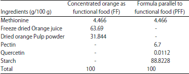 | |
The FF contains freeze dried orange juice and dried orange pulp powder with the same ratio of their occurrence in the parent orange; in addition of methionine. A parallel formula (PFF) made from similar quantities of bioactive constituents that present in orange according to previous literature. Also, the quantity of bioactive constituents given to rats in the present study was based on previous studies14-16. Ingredients of functional food were homogeneously mixed with the least quantity of water and refreeze dried. The parallel formula was just mixed separately to be made into powder form.
Sensory evaluation of functional food: Organoleptic characteristics of the prepared functional food were sensory evaluated by 20 panelists. Each panelist was asked to assign scores 0-20 for color, taste, flavor, homogeneity and overall acceptability. Before sensory evaluation of functional food 10 g of functional food was added to fresh water to make 84 cm solution.
Assessment of the chemical composition of the prepared functional food (FF): The prepared functional food was analyzed for moisture, protein, fat, crude fiber, ash and dietary fibers contents using standard AOAC17 procedure. The content of P, K, Ca, Mg, Na, Mn, Zn and Cu were analyzed in the ash of the functional food17 using atomic absorption Perkin-Elmer 1100B spectrophotometer apparatus (Model Perkin Elmer P Lambda 2). Different chemical analysis were carried out in triplicate and averaged.
Determination of total phenolic content (TPC): The TPC were extracted from the dry sample of functional food according to the method of Velioglu et al.18. Total phenolics were determined colorimetrically in the extracted samples using Folin-Ciocalteu reagent19. The reaction was conducted in triplicate and results were averaged. The TPC was expressed as gallic acid equivalents (GAE) in milligrams/g dry sample.
Assessment of fatty acids, hydrocarbons and phytosterols contents in functional foods: The unsaponifiable fraction and fatty acid methyl esters of the petroleum ether extracts of functional foods were prepared according to AOAC17 to be subjected to gas liquid chromatography (GLC) analysis of fatty acids, hydrocarbons and phytosterols.
Preparation of diets:. A balanced diet, high fructose diet (for induction of fatty liver according to Kawasaki et al.20, functional food diet (FFD) and parallel formula diet (PFFD) were shown in Table 2. For preparing FFD and PFFD; the functional food and the parallel formula were added as 20% from the balanced diet, respectively on the expense of starch.
Experimental design: Thirty rats were divided into two groups. Group one included 6 rats and fed on balanced diet for 9 weeks representing normal control (NC). The second group consisted of 24 rats fed on high fructose diet for 5 weeks to induce fatty liver. At the end of the 5 weeks; rats of the second group were divided into four sub-groups; each sub-group included 6 rats. Rats of the first and second sub-groups were fed on high fructose (Fatty liver control group; FLC) and balanced diets (fatty liver shifted to balanced diet; FLSB), respectively for 4 weeks. Rats of the third and fourth sub-groups were fed on FFD and PFFD diets, respectively for 4 weeks. Rats were fasted 12-14 h and blood samples were taken for determination of the different biochemical parameters. Livers were separated and weighed. A part from each liver was preserved in 10% formalin for histopathology examination and the other part was kept in deep freeze until analyzed.
| Table 2: | Composition of different experimental diets (g/100 g) |
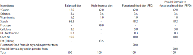 | |
| *Quantity of casein that contains 10 g protein as determined by AOAC17 | |
Blood sampling: Blood samples were collected by draining from the orbital vein of anaesthetized rats by using heparinized capillary tubes. Blood samples were received in centrifuge in heparinized tubes. Plasma of different blood samples were separated by centrifugation at 3000 rpm for 15 min. Plasma lipids profile including total cholesterol (T-Ch)21, low density lipoprotein cholesterol (LDL-Ch)22, high density lipoprotein cholesterol (HDL-Ch)23 and triglycerides (TG)24 were determined. The ratio T-Ch/HDL-Ch was calculated as indicative of cardiovascular risk. Also plasma activity of alkaline phosphatase25 and transaminases (ALT and AST)26 were assessed as liver function biomarkers. Plasma leptin27 and the inflammatory biomarker tumor necrosis factor-α (TNF- α)28 were determined adopting ELISA. Plasma malondialdyhyde29 was estimated as indicator of lipid peroxidation. Plasma insulin30 and blood glucose31 were determined. Insulin resistance was calculated based on homeostasis model assessment of insulin resistance (HOMA-IR), according to Cacho et al.32. The equation was:
Kidney function was assessed through estimation of plasma urea33 and creatinine34.
Statistical analysis: Data of animal experiments were expressed as Mean±SE and were statistically analyzed by one way analysis of variance ANOVA followed by Duncan test. In all cases, p<0.05 was used as the criterion of statistical significance. Results of proximate analysis, dietary fibers and sensory evaluation were expressed as Mean±SE.
RESULTS
Sensory evaluation of functional food: When the prepared functional food was sensory evaluated, it could be noticed that it was accepted by panelists (Table 3).
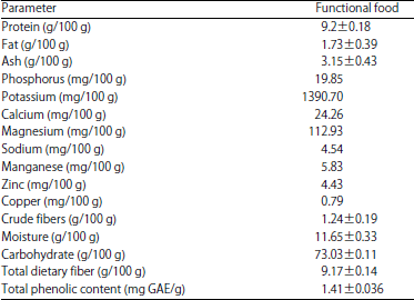
Proximate composition, minerals, dietary fibers and total phenolic in the prepared functional food: The data in Table 4 showed the proximate analysis, minerals, dietary fibers and total phenolic of the prepared functional food. It can be noticed that protein was higher than fat content. Potassium was of the highest level while cupper was of the lowest concentration among minerals. Total dietary fibers were present in moderate level. Total phenolic contents were 1.41 mg GAE/g sample.
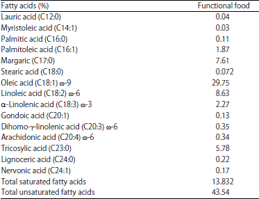
Fatty acids and unsaponifiable constituents of functional food: The data in Table 5 showed the fatty acid content of the prepared functional food (as percentage of total fatty acids). It can be noticed that the total saturated fatty acid was extremely lower than the total unsaturated fatty acid. Monounsaturated fatty acids were present as 31.95% represented by myristoleic, palmitoleic, oleic, gondoic and nervonic acid. Oleic acid was the highest unsaturated fatty acid, while linolenic acid (ω-3 fatty acid) was the lowest one. Linoleic acid which is an ω-6 fatty acid was 8.63%. The polyunsaturated fatty acids (Dihomo-γ-linolenic acid and arachidonic) were only present in traces in the prepared functional food.
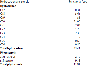
It can see in Table 6 the GLC analysis of unsaponifiable matter of the prepared functional food. It can be noticed that the total hydrocarbon was 43.41% while total phytosterols was 11.97%, represented by stigmasterol and β-sitosterol.
Biological experiment
Biochemical parameters of the biological experiment: The data in Table 7 showed biochemical parameters of the different experimental groups. It can be noticed that T-Ch, TG, LDL-Ch and T-Ch/HDL-Ch of FLC group were of the highest level among all the studied groups and were significantly higher than that of NC. Plasma level of HDL-Ch of FLC group was of the lowest level among all the studied groups. When rats of FLS-B group compared to FLC group there was significant reduction in plasma T-Ch/HDL-Ch with insignificant reduction in T-Ch, TG and LDL-Ch and insignificant increase in HDL-Ch. Feeding the functional food diet and its parallel diet produce significant reduction in total cholesterol, triglycerides, LDL-Ch and TCh/HDL-Ch with significant increase in HDL-Ch compared to FLC group. The groups of rats fed on FFD and PFFD showed significant decrease in LDL-Ch and T-Ch /HDL-Ch with significant increase in HDL-Ch compared to fatty liver shifted to balanced diet group. While as regard to TG; there was a significant decrease in FFD group along with insignificant decrease in PFFD group compared to FLS-B group. Also there was insignificant decrease in T-Ch in FFD and PFFD groups compared to FLS-B.
| Table 3: | Sensory evaluation of orange functional food (FF) |
| Table 4: | Proximate composition, dietary fibers, phenolic content (Mean±SE) and minerals of orange functional food |
 | |
| Table 5: | Fatty acids contents of orange functional food (as percentage of total fatty acids) |
 | |
There were no significant changes between different lipid parameters when comparing the group fed on functional food diet with its parallel functional food. Despite the improvement in plasma lipids on feeding the functional food diet or its parallel functional food diet the level of plasma T-Ch, TG, LDL-Ch, HDL-Ch and T-Ch/HDL-Ch still not matching that of NC.
Liver total fat, liver T.Ch and liver TG of FLC group were significantly higher than NC group. When rats with fatty liver shifted to balanced diet were compared to FLC group there were no significant changes in liver total fat, liver T-Ch and liver TG. Feeding the functional food diet and its parallel diet produced significant reduction in liver total fat, liver T-Ch and liver TG compared to FLC group and FLS-B group. Comparing the group fed on functional food diet with its parallel group showed non-significant changes in hepatic total fat, total cholesterol and triglycerides.
| Table 6: | GLC analysis of unsaponifiable matter of orange functional food (as percentage of total unsaponifiable matter) |
 | |
It can be noticed that MDA, TNF-α and leptin of FLC group were of the highest levels among all the studied groups and were significantly higher than that of all the studied groups except for MDA and leptin in case of FLS-B that showed non-significant change from FLC group. Groups fed on the functional food and its parallel diet, showed significant reduction in MDA, TNF-α and leptin compared to FLS-B group. The MDA levels of rats fed on the functional food and its parallel diet match that of the rats fed on balanced diet. Feeding the two functional foods could not reduce the levels of leptin and TNF-α to the normal levels of rats fed on the balanced diet. Comparing plasma levels of MDA, TNF-α and leptin of functional food group with its parallel group showed non-significant changes.
Plasma insulin, glucose and IR of FLC group were of the highest level among all the studied groups and were significantly higher than control rats fed on balanced diet. When rats of FLB-S group were compared to FLC group there was significant reduction in IR and insignificant reduction in insulin and glucose. Feeding the functional food diet and its parallel diet produced significant reduction in plasma insulin, glucose and IR compared to FLC and FLS-B groups; however the levels of such parameters still not matching that of NC. Also comparing plasma glucose, insulin and insulin resistance of functional food group with its parallel group showed non-significant changes.
Plasma ALT, AST, ALP and creatinine of FLC group were significantly higher than that of the NC. When rats of FLS-B compared to FLC group there was significant reduction in AST. Feeding FFD and its parallel diet produce significant reduction in ALT, AST, ALP and creatinine compared to control FLC group. Groups of rats fed on FFD and its parallel diet showed significant reduction in ALT, AST and ALP compared to FLS-B, except for AST in PFFD group that showed non-significant change compared to FLS-B.
| Table 7: | Biochemical parameters of different experimental groups |
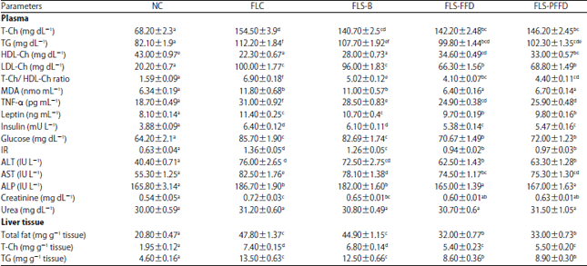 | |
Data are expressed as Mean±SE, Values with different superscript letters in the same column are significantly different at p<0.05 levels, NC: Normal control, FLC: Fatty liver control, FLS-B: Fatty liver rats shifted to balanced diet, FLS-FFD: Fatty liver rats shifted to functional food diet, FLS-PFFD: Fatty liver rats shifted to parallel functional food diet | |
There was insignificant change of urea among all experimental groups. Rats fed on FFD and its parallel diet showed significant higher level of ALT and AST compared to normal control while ALP and creatinine matched the normal level.
Histopathological examination of the liver: The histopathological examination of liver of rat of control group that fed on balanced diet showed normal appearance of hepatic plates, central veins and portal areas. The hepatocytes appeared oval in shape with homogenous eosinophilic cytoplasm and centrally located nucleus (Fig. 1a). The liver of rats of FLC group showed multiple focal areas of severe fatty changes in hepatocytes. The hepatocytes appeared swollen with presence of circumscribed vacuoles occupied the cytoplasm and pushing the nucleus to the peripheral giving the signet ring appearance. Generalized inflammation with coagulative necrosis and hyperplasia were noticed in FLC group (Fig. 1b). In Fig. 1c the liver of rat of FLS-B group showed areas of coagulative necrosis in which the outlines of hepatocytes appeared while cellular details are lost. Small fat droplets appeared in some hepatocytes of FLS-B group. Congestion of blood vessels associated with areas of hemorrhages was recorded with focal aggregation of mononuclear cells in between the hepatocytes in FLS-B. These changes could show some very slight improvement on replacing the high fructose diet by balanced diet, but still there is fatty accumulation, inflammation and coagulative necrosis. The liver of FF group showed individual inflammatory cells aggregation in between the hepatocytes with slight fatty changes in few numbers of hepatocytes (Fig. 1d). The liver of rats of PFF group showed diffuse fatty changes in most of hepatocytes along with congestion of blood vessels (Fig. 1e). Histopathological examination showed variable degrees of improvement on feeding the functional food diets compared to liver of the group continued feeding high fructose diet and FLS-B group. The FF diet was promising in the improvement of fatty liver based on histopathological examination, however still liver tissues did not restore the exact normal appearance by feeding the test diets.
DISCUSSION
In the present study; preparation and evaluation of functional food prepared from pulp of orange together with the white orange skin supplemented by methionine was studied in NASH rat model in comparison to a formula containing the main bioactive constituents that present in orange. The NASH is considered as one of the risk factors for CVD where elevation of oxidative stress, inflammation and dyslipidemia result from the increased synthesis and accumulation of cholesterol and triglycerides in liver35.
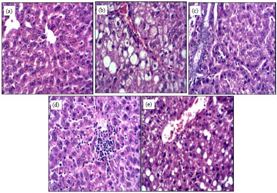 | |
| Fig. 1(a-e): | Section of rat liver of different groups (H and E X400), (a): Liver of rat of the NC group fed on balanced diet showing normal appearance of hepatocytes, (b): Liver of the FLC group shows the signet ring appearance of fat vacuoles, (c): Liver of FLS-B group showing fat vacuoles in some of hepatocytes, (d): Liver of FLS-FFD group showed focal aggregation of inflammatory cells and few numbers of hepatocytes showed fatty changes, (e): Liver of FLS- PFFD group showing diffuse fatty changes in most of hepatocytes, NC: Normal control, FLC: Fatty liver control, FLS-B: Fatty liver rats shifted to balanced diet, FLS-FFD: Fatty liver rats shifted to functional food diet, FLS-PFFD: Fatty liver rats shifted to parallel functional food diet |
The proximate composition of the functional food in the current study differs greatly than that of raw orange according to USDA data base which certainly might be due to the concentrated form of orange used in the present study and the difference in orange variety. Also, the added methionine could participate in protein levels in the FF. The prepared functional food contains phenolic compounds, linoleic and linolenic acids of reported health benefits which agreed with a previous study36. It was reported that orange fruits contain β-sitosterol, campesterol, stigmasterol, β-sitostanol and campestanol while the present study showed only the presence of β-sitosterol and stigmasterol.
Feeding high fructose diet with saturated fat in the present study produced dyslipidemia, elevated inflammatory marker (TNF-α), oxidative stress biomarker (MDA) and leptin. High fructose diet also led to liver dysfunction and increased level of creatinine, glucose, insulin and IR with significant increase of hepatic deposition of fat, triglycerides and cholesterol. Histopathological changes that noticed in the liver tissue in the present study proved the presence of fatty liver with inflammation and starting of necrosis pointed to the induction of steatohepatitis with fibrosis. The histopathological results coincided with the biochemical results. These changes verify the induction of steatohepatitis by such diet. Previous studies showed similar results concerning high fructose diet due to that fructose is a highly lipogenic of profound metabolic effects in the liver and as dyslipidemic 37,38 because it stimulates de novo lipogenesis. Saturated fat-rich diet was also reported to induce insulin resistance, dyslipidemia, steatosis, promotes endoplasmic reticulum stress and hepatocyte injury39. So the presence of both saturated fat and fructose in the diet in the present study could have a synergistic effect in induction of fatty liver. Progression of fatty liver to steatohepatitis as the case in the present study could involve the presence of inflammation, mitochondrial dysfunction, enhanced oxidative stress induced by ROS, lipid peroxidation and production of adipokines which cause hepatocyte damage and fibrosis. Deposition of liver fat lead to increased secretion of VLDL, hypertriglyceridemia, decreased HDL and high LDL40 as noticed in the control fatty liver group in the present study showing the cardiovascular diseases risk due to fatty liver that further emphasized by the elevated T-Ch/HDL-Ch. High IR was reported to impair lipid metabolism in the liver by affecting the master regulators of hepatic de novo lipogenesis41 that lead to further increase the deposition of hepatic lipids. On the other hand; TG accumulation activates serine kinase cascade that inhibits insulin signaling, leading to IR in the liver42. This increase in IR goes in line with the elevated IR in the present study.
Patients with NAFLD had high persistent levels of alanine aminotransferase elevation. AST has a milder elevation than ALT in NAFLD43. This finding is in agreement with the results of NAFLD control group in the present study. Also the increased ALP in fatty liver control group agreed with a previous study44. Leptin resistance was reported in steatohepatitis45, which could be deduced from the present study since though leptin is an insulin sensitizer, there was elevated IR indicating the occurrence of leptin resistance which could contribute in the induction of hepatocyte inflammation and injuries.
It could be noticed that the prepared functional food and its parallel formula used in the present study have beneficial effect towards fatty liver through amelioration of the different biochemical and histopathological changes. It was reported that orange juice showed beneficial effects towards NAFLD patients13 and mice feeding high fat diet46 through enhancement of IR and reduction of the levels of liver enzymes, TG, T-Ch and body mass index. Drinking orange juice greatly modified liver steatosis through stimulating the expression of peroxisome proliferator-activated receptor-α and the target gene acylCoA-oxidase. Also inhibit the expression of liver X receptor-α and the target gene fatty acid synthase, with restored liver glycerol-3-phosphate acyltransferase 1 activity46.
Orange is an excellent source of vitamin C and diverse phytochemicals, including carotenoids (b-carotene, lutein and b-cryptoxanthin), flavonoids (e.g., naringenin) and choline47. These constituents of orange might be responsible of the remedial effect of orange in fatty liver rats of the present study. Methionine is an essential amino acid that plays a vital role in protein synthesis and is an intermediate in synthesis of S-adenosylmethionine and glutathione, two important antioxidants48. Therefore methionine deficiency predisposes to oxidative stress, inflammation and plays a causal role in the pathogenesis of NASH and liver fibrosis49. So supplementation of methionine in FF could participate in fatty liver treatment in the present study.
Quercetin which is one of the bioactive constituents in orange in FF reduces inflammatory cytokine levels and improves lipid peroxidation and insulin resistance in animal models of NAFLD50. Quercetin showed hepatoprotective effect in rats treated with CCl4 by improving liver enzymes (AST, ALT), reducing the expression of profibrogenic genes (TGF-β, CTGF, and collagen-1α), inhibition of activated hepatic stellate cells and slowing down of NF-κB51. Phenolic contents, phytosterols, dietary fibers and unsaturated fatty acids especially polyunsaturated and monounsaturated fatty acids might render the functional food and its parallel formula the therapeutic effect towards fatty liver and its risk factors to CVD. Phenolic compounds were reported to reduce NAFLD, cardiovascular disease and diabetes52. These therapeutic effects of phenolic compounds might be attributed to their antioxidant and anti-inflammatory activity. Dietary fibers that present in the functional food including pectin may also participate in improving plasma lipid profile, plasma glucose, IR and steatosis as reported previously53. So the presence of dietary fibers in the prepared functional food studied in the present work might participate in their therapeutic effect towards fatty liver. Monounsaturated fatty acids are extremely beneficial for subjects with NAFLD. Monounsaturated fatty acids lower LDL, oxidized LDL, triglycerides levels, serum glucose and VLDL and elevate HDL levels. Monounsaturated fatty acids enhance oxidation of other FA via stimulation of peroxisome proliferator-activated receptors (PPARs) alpha and gamma thereby enhance degradation of fat and decrease of lipogenesis through diminished activation of sterol regulatory element binding protein39.
CONCLUSION
Rats fed high fructose diet supplemented with saturated fat are a good model for studying NAFLD. The prepared functional food and its parallel formula afford cardio and hepatoprotection against NAFLD. The bioactivity of functional food could be attributed to the presence of phenolic compounds, dietary fibers, phytosterols, poly and mono-unsaturated fatty acids in the prepared functional food.
SIGNIFICANCE STATEMENT
This research pointed to the possible use of orange functional food (Juice and pulp mixed with methionine) and its parallel formula (mixture of methionine, pectin and quercetin) for management of NAFLD and reducing the risk of cardiovascular diseases; with superiority to orange functional food. Phenolic compounds, dietary fibers, phytosterols and mono and poly-unsaturated fatty acids could be responsible to the bioactivity of orange functional food.
ACKNOWLEDGMENT
The present research is financed by National Research Centre.
REFERENCES
- Farrell, G.C., V.W.S. Wong and S. Chitturi, 2013. NAFLD in Asia-as common and important as in the West. Nat. Rev. Gastroenterol. Hepatol., 10: 307-318.
CrossRefDirect Link - Sattar, N., E. Forrest and D. Preiss, 2014. Non-alcoholic fatty liver disease. Br. Med. J., 349: 24-28.
CrossRefDirect Link - Younossi, Z.M., A.B. Koenig, D. Abdelatif, Y. Fazel, L. Henry and M. Wymer, 2016. Global epidemiology of nonalcoholic fatty liver disease-meta-analytic assessment of prevalence, incidence, and outcomes. Hepatology, 64: 73-84.
CrossRefDirect Link - Perumpail, B.J., R. Cholankeril, E.R. Yoo, D. Kim and A. Ahmed, 2017. An overview of dietary interventions and strategies to optimize the management of non-alcoholic fatty liver disease. Diseases, Vol. 5, No. 4.
CrossRefDirect Link - Rinella, M.E., 2015. Nonalcoholic fatty liver disease: A systematic review. J. Am. Med. Assoc., 313: 2263-2273.
CrossRefDirect Link - Sodhi, K., N. Puri, G. Favero, S. Stevens and C. Meadows et al., 2015. Fructose mediated non-alcoholic fatty liver is attenuated by HO-1-SIRT1 module in murine hepatocytes and mice fed a high fructose diet. PLoS One, Vol. 10, No. 6.
CrossRefDirect Link - Oliveira, C.P., P. de Lima Sanches, E.O. de Abreu-Silva and A. Marcadenti, 2015. Nutrition and physical activity in nonalcoholic fatty liver disease. J. Diabetes Res., Vol. 2016.
CrossRef - Jensen, T., M.F. Abdelmalek, S. Sullivan, K.J. Nadeau and M. Green et al., 2018. Fructose and sugar: A major mediator of nonalcoholic fatty liver disease. J. Hepatol., 68: 1063-1075.
CrossRefDirect Link - Hardy, G., 2000. Nutraceuticals and functional foods: Introduction and meaning. Nutrition, 16: 688-689.
CrossRefPubMedDirect Link - Das, L., E. Bhaumik, U. Raychaudhuri and R. Chakraborty, 2012. Role of nutraceuticals in human health. J. Food Sci. Technol., 49: 173-183.
CrossRefDirect Link - Del Ben, M., M. Fabiani, L. Loffredo, L. Polimeni and R. Carnevale et al., 2012. Oxidative stress mediated arterial dysfunction in patients with obstructive sleep apnoea and the effect of continuous positive airway pressure treatment. BMC Pulmonary Med., Vol. 12, No. 1.
CrossRefDirect Link - Spinelli, S., L. Lecce, D. Likyova, M.A. Del Nobile and A. Conte, 2018. Bioactive compounds from orange epicarp to enrich fish burgers. J. Sci. Food Agric., 98: 2582-2586.
CrossRefDirect Link - Ekhlasi, G., F. Shidfar, S. Agah, S. Merat and A.F. Hosseini, 2016. Effects of pomegranate and orange juice on antioxidant status in non-alcoholic fatty liver disease patients: A randomized clinical trial. Int. J. Vitam. Nutr. Res., 85: 292-298.
CrossRefDirect Link - Crome, P., J.A. Vale, G.N. Volans, B. Widdop and R. Goulding, 1976. Oral methionine in the treatment of severe paracetamol (Acetaminophen) overdose. Lancet, 308: 829-830.
CrossRefPubMedDirect Link - National Toxicology Program, 1992. Toxicology and carcinogenesis studies of quercetin (CAS No. 117-39-5) in F344 rats (Feed studies). Natl. Toxicol. Program. Tech. Rep. Ser., 409: 1-171.
PubMedDirect Link - Tiwary, C.M., J.A. Ward and B.A. Jackson, 1997. Effect of pectin on satiety in healthy US Army adults. J. Am. Coll. Nutr., 16: 423-428.
CrossRefDirect Link - Velioglu, Y.S., G. Mazza, L. Gao and B.D. Oomah, 1998. Antioxidant activity and total phenolics in selected fruits, vegetables and grain products. J. Agric. Food Chem., 46: 4113-4117.
CrossRefDirect Link - Singleton, V.L. and J.A. Rossi, 1965. Colorimetry of total phenolics with phosphomolybdic-phosphotungstic acid reagents. Am. J. Enol. Vitic., 16: 144-158.
CrossRefDirect Link - Kawasaki, T., K. Igarashi, T. Koeda, K. Sugimoto and K. Nakagawa et al., 2009. Rats fed fructose-enriched diets have characteristics of nonalcoholic hepatic steatosis. J. Nutr., 139: 2067-2071.
CrossRefDirect Link - Watson, D., 1960. A simple method for the determination of serum cholesterol. Clin. Chim. Acta, 5: 637-643.
CrossRefDirect Link - Schriewer, H., U. Kohnert and G. Assmann, 1984. Determination of LDL cholesterol and LDL apolipoprotein B following precipitation of VLDL in blood serum with phosphotungstic acid/MgCl2. J. Clin. Chem. Clin. Biochem., 22: 35-40.
CrossRefDirect Link - Burstein, M., H.R. Scholnick and R. Morfin, 1970. Rapid method for the isolation of lipoproteins from human serum by precipitation with polyanions. J. Lipid Res., 11: 583-595.
PubMedDirect Link - Megraw, R.E., D.E. Dunn and H.G. Biggs, 1979. Manual and continuous-flow colorimetry of triacylglycerols by a fully enzymatic method. Clin. Chem., 25: 273-278.
Direct Link - Gout, J., D. Sarafian, J. Tirard, A. Blondet and M. Vigier et al., 2008. Leptin infusion and obesity in mouse cause alterations in the hypothalamic melanocortin system. Obesity, 16: 1763-1769.
CrossRefDirect Link - Stepaniak, J.A., K.E. Gould, D. Sun and R.H. Swanborg, 1995. A comparative study of experimental autoimmune encephalomyelitis in Lewis and DA rats. J. Immunol., 155: 2762-2769.
PubMedDirect Link - Satoh, K., 1978. Serum lipid peroxide in cerebrovascular disorders determined by a new colorimetric method. Clin. Chim. Acta, 90: 37-43.
CrossRefPubMedDirect Link - Turkington, R.W., A. Estkowski and M. Link, 1982. Secretion of insulin or connecting peptide a predictor of insulin dependence of obese diabetics. Arch. Intern. Med., 142: 1102-1105.
CrossRefDirect Link - Trinder, P., 1969. Determination of glucose in blood using glucose oxidase with an alternative oxygen acceptor. Ann. Clin. Biochem., 6: 24-27.
CrossRefDirect Link - Cacho, J., J. Sevillano, J. de Castro, E. Herrera and M.P. Ramos, 2008. Validation of simple indexes to assess insulin sensitivity during pregnancy in wistar and sprague-dawley rats. Am. J. Physiol. Endocrinol. Metab., 295: 1269-1276.
CrossRefDirect Link - Fawcett, J.K. and J.E. Scott, 1960. A rapid and precise method for the determination of urea. J. Clin. Pathol., 13: 156-159.
CrossRefPubMedDirect Link - Al-Okbi, S.Y., D.A. Mohamed, T.E. Hamed and A.E. Edris, 2013. Potential protective effect of Nigella sativa crude oils towards fatty liver in rats. Eur. J. Lipid Sci. Technol., 115: 774-782.
CrossRefDirect Link - Letaief, H., H. Zemni, A. Mliki and S. Chebil, 2016. Composition of Citrus sinensis (L.) Osbeck cv «Maltaise demi-sanguine» juice. A comparison between organic and conventional farming. Food Chem., 194: 290-295.
CrossRefDirect Link - Angulo, P. and K.D. Lindor, 2002. Non‐alcoholic fatty liver disease. J. Gastroenterol. Hepatol., 17: S186-S190.
CrossRefDirect Link - Dekker, M.J., Q. Su, C. Baker, A.C. Rutledge and K. Adeli, 2010. Fructose: A highly lipogenic nutrient implicated in insulin resistance, hepatic steatosis and the metabolic syndrome. Am. J. Physiol. Endocrinol. Metab., 299: 685-694.
CrossRefDirect Link - Zivkovic, A.M., J.B. German and A.J. Sanyal, 2007. Comparative review of diets for the metabolic syndrome: Implications for nonalcoholic fatty liver disease. Am. J. Clin. Nutr., 86: 285-300.
CrossRefDirect Link - Cusi, K., 2012. Role of obesity and lipotoxicity in the development of nonalcoholic steatohepatitis: Pathophysiology and clinical implications. Gastroenterology, 142: 711-725.
CrossRefDirect Link - Fotbolcu, H. and E. Zorlu, 2016. Nonalcoholic fatty liver disease as a multi-systemic disease. World J. Gastroenterol., 22: 4079-4090.
CrossRefPubMedDirect Link - Popov, V.B. and J.K. Lim, 2015. Treatment of nonalcoholic fatty liver disease: The role of medical, surgical and endoscopic weight loss. J. Clin. Transl. Hepatol., 3: 230-238.
CrossRefPubMedDirect Link - AlShaalan, R., M. Aljiffry, S. Al-Busafi, P. Metrakos and M. Hassanain, 2015. Nonalcoholic fatty liver disease: Noninvasive methods of diagnosing hepatic steatosis. Saudi J. Gastroenterol., 21: 64-70.
CrossRefPubMedDirect Link - Al-Okbi, S.Y., D.A. Mohamed and T.E. Hamed, 2015. Protection from steatohepatitis and its risk factors by plants food mixtures. Res. J. Pharm. Biol. Chem. Sci., 6: 1602-1613.
Direct Link - Lee, J.H., J.J. Lee, W.K. Cho, N.H. Yim, H.K. Kim, B. Yun and J.Y. Ma, 2016. KBH-1, an herbal composition, improves hepatic steatosis and leptin resistance in high-fat diet-induced obese rats. BMC complement. Altern. Med., Vol. 16, No. 1.
CrossRefDirect Link - Salamone, F., G.L. Volti, L. Titta, L. Puzzo and I. Barbagallo et al., 2012. Moro orange juice prevents fatty liver in mice. World J. Gastroenterol., 18: 3862-3868.
CrossRefPubMedDirect Link - Aschoff, J.K., S. Kaufmann, O. Kalkan, S. Neidhart, R. Carle and R.M. Schweiggert, 2015. In vitro bioaccessibility of carotenoids, flavonoids and vitamin C from differently processed oranges and orange juices [Citrus sinensis (L.) Osbeck]. J. Agric. Food Chem., 63: 578-587.
CrossRefDirect Link - Mato, J.M., M.L. Martinez-Chantar and S.C. Lu, 2008. Methionine metabolism and liver disease. Annu. Rev. Nutr., 28: 273-293.
CrossRefDirect Link - Henao-Mejia, J., E. Elinav, C. Jin, L. Hao and W.Z. Mehal et al., 2012. Inflammasome-mediated dysbiosis regulates progression of NAFLD and obesity. Nature, 482: 179-185.
CrossRefDirect Link - Zhang, M.H., Z.Q. Liang, Q. Qin, S.L. Li, D.S. Zhou and L. Tang, 2013. Effects of quercetin on serum levels of resistin and IL-18 and on insulin resistance in nonalcoholic fatty liver disease rats. Chin. J. Hepatol., 21: 66-70.
CrossRefDirect Link - Hernandez-Ortega, L.D., B.E. Alcantar-Diaz, L.A. Ruiz-Corro, A. Sandoval-Rodriguez, M. Bueno-Topete, J. Armendariz-Borunda and A.M. Salazar-Montes, 2012. Quercetin improves hepatic fibrosis reducing hepatic stellate cells and regulating pro‐fibrogenic/anti‐fibrogenic molecules balance. J. Gastroenterol. Hepatol., 27: 1865-1872.
CrossRefDirect Link - Tripoli, E., M.L. Guardia, S. Giammanco, D. Di Majo and M. Giammanco, 2007. Citrus flavonoids: Molecular structure, biological activity and nutritional properties: A review. Food Chem., 104: 466-479.
CrossRefDirect Link - Kellow, N.J., M.T. Coughlan, G.S. Savige and C.M. Reid, 2014. Effect of dietary prebiotic supplementation on advanced glycation, insulin resistance and inflammatory biomarkers in adults with pre-diabetes: A study protocol for a double-blind placebo-controlled randomised crossover clinical trial. BMC Endocrine Disord., Vol. 14, No. 1.
CrossRefDirect Link