
Ahmad Ghorbani
Pharmacological Research Center of Medicinal Plants, School of Medicine, Mashhad University of Medical Sciences, Mashhad, Iran
Mousa-Al-Reza Hadjzadeh
Neurocognitive Research Center, Department of Physiology, School of Medicine, Mashhad University of Medical Sciences, Mashhad, Iran
Ziba Rajaei
Department of Physiology, School of Medicine, Isfahan University of Medical Sciences, Isfahan, Iran
Seyed Bamdad Zendehbad
Neurocognitive Research Center, Department of Physiology, School of Medicine, Mashhad University of Medical Sciences, Mashhad, Iran
Pakistan Journal of Biological Sciences
Year: 2014 | Volume: 17 | Issue: 4 | Page No.: 523-528


ABSTRACT
Several studies support hypolipidemic effect of fenugreek in normal and diabetic subjects. However, very little is known about the possible direct action of fenugreek on adipose tissue. The present study was designed to investigate the effects of fenugreek seeds on adipogenesis and lipolysis. Preadipocytes were isolated from adipose tissue of normal rats and differentiated to adipocyte in the presence of ethanolic extract of fenugreek seeds. The effect of this extract on lipolysis was also evaluated in fat tissue isolated from diabetic rats. Fenugreek led to a significant reduction in lipid droplet accumulation as evaluated with Oil Red O staining. Incubation of preadipocytes with the extract for 24 h resulted in significant decrease in cell viability. The extract, even at high concentrations (up to 1000 μg mL-1), had virtually no significant effect on lipolysis. The present data demonstrated that fenugreek seed inhibits formation of new differentiated adipocytes from precursor cells through an anti-proliferative effect on preadipocytes.
PDF Abstract XML References Citation
Received: March 16, 2013;
Accepted: April 04, 2013;
Published: November 26, 2013
How to cite this article
Ahmad Ghorbani, Mousa-Al-Reza Hadjzadeh, Ziba Rajaei and Seyed Bamdad Zendehbad, 2014. Effects of Fenugreek Seeds on Adipogenesis and Lipolysis in Normal and Diabetic Rats. Pakistan Journal of Biological Sciences, 17: 523-528.
DOI: 10.3923/pjbs.2014.523.528
URL: https://scialert.net/abstract/?doi=pjbs.2014.523.528
DOI: 10.3923/pjbs.2014.523.528
URL: https://scialert.net/abstract/?doi=pjbs.2014.523.528
INTRODUCTION
Adipose tissue is a highly dynamic organ that has an important role in regulation of energy metabolism. The adipose mass depends on the number and the size of adipocytes and is determined by the rate of preadipocytes proliferation; differentiation of the preadipocytes into mature adipocytes; the balance between lipogenesis and lipolysis within individual mature adipocyte; and the rate of adipocyte apoptosis (Smith and Ravussin, 2006). Expansion of fat mass can be occurring by an increase in both fat cell size and number of adipocytes. Obesity is a result of body fat mass expansion and is still one of the main health problems all over the world. Based on World Health Organization reports, in 2008, about 1.4 billion people in the world suffer from overweight. Also, it is estimated that 2.8 million men and women die annually from obesity related diseases (WHO, 2013). Numerous studies have shown that obesity increases risk of cardiovascular diseases, stroke, metabolic syndrome and type-2 diabetes (Chiang et al., 2011; Lavie et al., 2009). The mass of adipose tissue can be reduced by increasing lipolysis, inhibiting adipogenesis and inducing apoptosis of fat cells (Rayalam et al., 2008).
Several plants have been shown to have direct effects on lipolysis, preadipocytes differentiation and adipocyte life cycle (Rayalam et al., 2008; Andersen et al., 2010). Trigonella foenum-graecum (fenugreek), an annual herb in the family Fabaceae, has a long history for amelioration of abnormalities in glucose and lipid homeostasis (Fatima et al., 2004; Ghorbani and Rakhshandeh, 2012). Many studies support hypolipidemic effect of fenugreek seed in diabetic subjects (Kassaian et al., 2009). It has also reported that fenugreek reduces the body weight gain and hyperlipidaemia induced by high-fat diet (Handa et al., 2005; Al-Matubsi et al., 2011). However, very little is known about the possible direct action of fenugreek on adipose tissue. Up to now, decrease of fat accumulation in differentiating 3T3-L1 cell line is the only report on action of fenugreek on adipocytes (Vijayakumar et al., 2010). Therefore, the present study was designed to investigate the effects of fenugreek seeds on adipogenesis and lipolysis in normal and diabetic animals, respectively.
MATERIALS AND METHODS
Chemicals and reagents: Dulbecco’s Modified Eagles Medium (DMEM) and fetal calf serum was purchased from Gibco (Carlsbad, CA). Streptozotocin (STZ) was obtained from Enzo Life (USA). Penicillin-streptomycin, type-II collagenase, 3-(4,5-Dimethyl-2-thiazolyl)-2,5- Diphenyl-2H-tetrazolium bromide (MTT), fatty acid-free bovine serum albumin fraction V, 4-(2-hydroxyethyl) piperazine-1-ethanesulfonic acid sodium salt (HEPES), glycerol assay reagent, and isoproterenol were provided from Sigma (USA). Dimethyl sulfoxide (DMSO) and 3-isobutyl-1-methylxanthine (IBMX) were purchased from Fluka Chemical Co. Indomethacin and human insulin were kindly provided by EXIR Company (Iran).
Preparation of extracts: The fenugreek seeds were purchased from a local market. The seeds were cleaned and grounded to fine powder with a blender. The macerated extract was prepared by suspension of 100 g of the powder in 70% ethanol and incubation for 72 h at 37°C. The extract was then dried on a water bath and the yield dissolved in DMSO.
Animals: Male albino Wistar rats (280-330 g) were used for each experiment. They were housed in a room with controlled lighting (12 h light/12 h darkness) and temperature (22 ±2°C). The animals were given standard pellets diet and water ad libitum. All animal experiments were done according to the ethical guidelines of the animal care of the Mashhad University of Medical Sciences, Iran. Effect of fenugreek extract on adipogenesis and lipolysis was studied on adipose tissue isolated from normal and diabetic rats, respectively. For induction of diabetes, the animals were given a single dose of STZ (55 mg kg-1, i.p.). Two days after STZ injection, induction of diabetes was confirmed by measuring fasting blood glucose. Rats were considered to be diabetic if they had blood glucose concentration of 250 mg dL-1 or higher (Shafiee-Nick et al., 2012).
Preadipocyte preparation and culture: Subcutaneous adipose tissue sample was excised from normal rats under ether anesthesia. The tissue was sliced into small pieces and washed with phosphate-buffered saline. The tissue pieces were then digested in phosphate-buffered saline containing collagenase (2 mg mL-1) under shaking (60 cycles min-1) at 37°C. After centrifugation, the floated adipocytes were discarded and the stromal cells were suspended in DMEM medium supplemented with 10% fetal calf serum, 100 units mL-1 penicillin and 100 μg mL-1 streptomycin and seeded (104 cells/well) in 12-well plates. After 24 h of incubation, the medium was changed into differentiation medium consisting of DMEM supplemented with 3% fetal calf serum, 250 μM IBMX, 66 μM biotin, 34 μM d-pantothenate, 1 μM dexamethasone, 0.2 μM insulin and 5 μM indomethacin. The cells were maintained in the differentiation medium for 3 days and then exposed to the adipocyte maintenance medium consisting of DMEM supplemented with 3% fetal calf serum, 66 μM biotin, 34 μM d-pantothenate, 1 μM dexamethasone and 0.2 μM insulin. The cells were cultured in adipocyte maintenance medium for 9 days and the medium was changed every 3 days (Yu et al., 2011). To study effect of fenugreek extract on adipogenesis, the differentiation and adipocyte maintenance medium were supplemented with varying concentrations of the extract or vehicle (1% DMSO).
Oil red O staining: Oil Red O was used to stain intracellular triglyceride droplets in differentiated adipocytes. After 12 days of differentiation, the cells were fixed using 10% formalin and then stained by Oil Red O solution. After several washing with distilled water, the stain was eluted from cells using isopropanol and its optical density was read at 540 nm (Yu et al., 2011).
Cell viability assay: The effect of fenugreek extract on viability of the stromal cells was determined using MTT colorimetric assay as previously described (Mortazavian and Ghorbani, 2012; Mortazavian et al., 2012). The cells were seeded (5000/well) in 96-well culture plates containing DMEM medium supplemented with 10% fetal calf serum, 100 units/mL penicillin and 100 μg mL-1 streptomycin. After 24 h, the medium was changed by fresh one containing various concentrations of fenugreek extract and the cells were further incubated for 24 h. At the end of treatment, the MTT was added to the cell media at final concentration of 0.5 mg mL-1 and the cells incubated for 2 h. Then, the absorbance of formazan dye was measured at 545 nm. The assay was done in triplicate and repeated twice times.
Lipolysis studies: The effect of fenugreek extract on lipolysis was evaluated using an ex-vivo organ culture method (Ghorbani et al., 2011, 2013). After seven days of STZ injection, the retroperitoneal adipose tissues were excised from diabetic rats. The tissues were minced into uniform small pieces of about 5 mg. The tissue pieces were washed, dried on the gauze, and weighted precisely. The tissues were then distributed into 24-well culture plate (100 mg/well) and bathed with 1 mL Krebs-Ringer bicarbonate buffer containing 5.5 mM glucose, 25 mM HEPES and 2% (w/v) bovine serum albumin. The wells were left untreated (basal lipolysis) or treated with isoproterenol (stimulated lipolysis) and incubated in the absence or presence of fenugreek extract at 37°C in a humidified chamber under constant shaking for 90 min. At the end of the incubation, concentration of glycerol in the media was measured by an enzymatic method.
Statistical analysis: The results are presented as the mean±standard error. The values were compared using the one-way analysis of variance followed by Tukey’s post hoc test. Results were considered to be statistically significant, if the p-values were under 0.05.
RESULTS
Effect of fenugreek on adipogenesis: Exposure of differentiating cells to the fenugreek extract led to a significant reduction in lipid droplet accumulation as evaluated with Oil Red O staining (Fig. 1). The presence of 50, 200 and 400 μg mL-1 of the extract in the culture medium decreased the lipid droplet content from 100±11% (untreated cells) to 57±12% (p < 0.05), 64±6.6% (p<0.05) and 60±5% (p<0.05), respectively (Fig. 2).
Effect of fenugreek on viability of preadipocytes: Incubation of preadipocytes in the presence of fenugreek extract resulted in significant decrease in cell viability (Fig. 3).
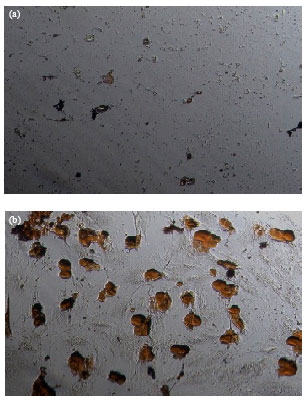 | |
| Fig. 1(a-b): | Oil Red O staining of differentiated adipocytes after treatment with fenugreek extract. Preadipocytes were isolated from normal rats and cultured for 12 days in the presence (a) or absence (b) of fenugreek extract (50 μg mL-1) in adipogenic media. Lipid-containing Oil Red O-positive cells appear in non-treated cells (original magnification,x200) |
The extract at 10, 50, 100, 200 and 400 μg mL-1 led to 12%, 39% (p <0.001), 54% (p<0.001), 51% (p<0.001) and 50% (p<0.001) decrease in cell viability, respectively.
Effect of fenugreek on lipolysis: Figure 4a demonstrates the effect of fenugreek extract on basal lipolysis.
 | |
| Fig. 2: | Effect of fenugreek extract on lipid droplet accumulation in differentiating preadipocyte isolated from normal rats. The lipid accumulation was estimated by measuring the optical density of Oil Red O stain eluted from cells. Data are mean±SEM of two independent experiments performed in triplicate. *p<0.05 vs vehicle |
 | |
| Fig. 3: | Effect of fenugreek extract on viability of preadipocytes isolated from normal rats. The cells cultured for 24 h in the presence of the extract. The bars show percent of cell viability as compared with untreated cells (vehicle). Data are mean±SEM of two independent experiments performed in triplicate. *p<0.001 vs vehicle |
 | |
| Fig. 4(a-b): | Effects of fenugreek extract on basal and stimulated lipolysis in diabetic rats, (a): Lipolysis in retroperitoneal adipose tissue was assessed in the presence of 100 μg mL-1 fenugreek extract or 1 μM isoproterenol, (b) and (c), Isoproterenol-induced lipolysis in the adipose tissue was assessed in the presence or absence of fenugreek extract. Results are expressed relative to basal and isoproterenol-induced lipolysis. The data are presented as means±SEM of 6 independent experiments. *p<0.001 vs vehicle |
The presence of this extract (100 μg mL-1) in the tissue medium did not change basal glycerol release (95±13% and 100±3.4% for fenugreek and vehicle, respectively). To examine the effect of fenugreek on stimulated lipolysis, the lipolytic activity was also evaluated in the presence of isoproterenol, a nonselective beta adrenergic receptor agonist which induces lipolysis. As expected, isoproterenol led to a significant elevation (2.87 fold, p<0.001) in lipolysis.
As shown in Fig. 4b, the extract at concentrations of 10, 100 and even 1000 μg mL-1 had virtually no significant effect on the stimulated lipolysis. Therefore the level of glycerol release was still higher than that of basal level.
DISCUSSION
The mass of adipose tissue is determined with the balance between accumulation of lipid droplet (through lipogenesis and formation of new adipocytes from precursor cells) and reduction of fat content (through lipolysis and adipocyte apoptosis). The present study showed that macerated extract of fenugreek inhibits formation of new differentiated fat cells from preadipocytes. This finding is in agreement with earlier reports that fenugreek inhibits lipid accumulation in differentiating 3T3-L1 cells (Vijayakumar et al., 2010) and reduces the body weight gain induced by high-fat diet (Handa et al., 2005). The results from MTT assay demonstrated that anti-proliferative action of the extract is most probably responsible for inhibition of new adipocytes formation. In line with this finding, Al-Daghri and coworkers reported that fenugreek extract induces cellular death at concentrations of ≥30 μg mL-1 (Al-Daghri et al., 2012).
Breakdown of triglycerides (lipolysis) in adipose tissue is a highly regulated process. The regulation of this process is essential for maintaining body homeostasis and prevention of various diseases development. Although, a variety of factors affect on lipolysis, insulin and catecholamines are the main anti-lipolytic and pro-lipolytic hormones, respectively (Large et al., 2004). Previously it has been shown that fenugreek stimulates insulin signaling pathways in some tissue (Mohammad et al., 2006; Vijayakumar et al., 2005). To test whether this plant can induce anti-lipolytic effect in fat tissue, hydrolysis of triglyceride was investigated in the tissue incubated with the seed extract. This investigation was done using an ex vivo organ culture model which has certain advantages over isolated adipocyte because it retains autocrine, paracrine, cell-cell and cell-matrix interactions (Ghorbani et al., 2013). Yet, because the limited amount of adipose tissue obtained from diabetic rats, it was impossible to test varying concentration of the extract on basal lipolysis. Beside, the results showed that fenugreek (with the tested concentrations) has no effect on basal or catecholamine-stimulated lipolysis in adipose tissue isolated from diabetic rats. Therefore, its beneficial effect on diabetic dyslipidemia is mediated by other mechanism suggested above.
In conclusion, the present study demonstrated that macerated extract of fenugreek seed inhibits formation of new differentiated adipocytes from precursor cells. This effect of fenugreek is mediated through its anti-proliferative action on preadipocytes.
ACKNOWLEDGMENTS
This work was supported by a grant from Research Council of Mashhad University of Medical Sciences, Mashhad, IRAN. The authors declare that they have no conflict of interest.
REFERENCES
- Al-Daghri, N.M., M.S. Alokail, K.M. Alkharfy, A.K. Mohammed and S.H. Abd-Alrahman et al., 2012. Fenugreek extract as an inducer of cellular death via autophagy in human T lymphoma Jurkat cells. BMC Complement. Altern. Med., Vol. 12.
CrossRef - Al-Matubsi, H.Y., N.A. Nasrat, G.A. Oriquat, M. Abu-Samak, K.A. Al-Mzain and M. Salim, 2011. The hypocholesterolemic and antioxidative effect of dietary diosgenin and chromium chloride supplementation on high-cholesterol fed Japanese quails. Pak. J. Biol. Sci., 14: 425-432.
CrossRefDirect Link - Fatima, N., I.U. Siddiqui, F. Perveen and Z.T. Maqsood, 2004. Among few commonly used anti-diabetic herbs: Fenugreek is the best. Pak. J. Biol. Sci., 7: 966-970.
CrossRefDirect Link - Handa, T., K. Yamaguchi, Y. Sono and K. Yazawa, 2005. Effects of fenugreek seed extract in obese mice fed a high-fat diet. Biosci. Biotechnol. Biochem., 69: 1186-1188.
CrossRef - Kassaian, N., L. Azadbakht, B. Forghani and M. Amini, 2009. Effect of fenugreek seeds on blood glucose and lipid profiles in type 2 diabetic patient. Int. J. Vitamin Nutr. Res., 79: 34-39.
CrossRefDirect Link - Large, V., O. Peroni, D. Letexier, H. Ray and M. Beylot, 2004. Metabolism of lipids in human white adipocyte. Diabetes Metab., 30: 294-309.
CrossRef - Mohammad, S., A. Taha, K. Akhtar, R.N.K. Bamezai and N.Z. Baquera, 2006. In vivo effect of Trigonella foenum graecum on the expression of pyruvate kinase, phosphoenolpyruvate carboxykinase and distribution of glucose transporter (GLUT4) in alloxan-diabetic rats. Can. J. Physiol. Pharmacol., 84: 647-654.
CrossRefPubMed - Mortazavian, S.M. and A. Ghorbani, 2012. Antiproliferative effect of viola tricolor on neuroblastoma cells in vitro. Aust. J. Med. Herbalism., 24: 93-96.
Direct Link - Mortazavian, S.M., A. Ghorbani and T.G. Hesari, 2012. Effect of hydro-alcoholic extracts of viola tricolor and its fractions on proliferation of cervix carcinoma cells. Iran. J. Obstetric Gyncol. Infertil., 15: 9-16.
Direct Link - Rayalam, S., M.A. Della-Fera and C.A. Baile, 2008. Phytochemicals and regulation of the adipocyte life cycle. J. Nutr. Biochem., 19: 717-726.
CrossRefPubMedDirect Link - Vijayakumar, M.V., S. Singh, R.R. Chhipa and M.K. Bhat, 2005. The hypoglycaemic activity of fenugreek seed extract is mediated through the stimulation of an insulin signalling pathway. Br. J. Pharmacol., 146: 41-48.
CrossRefPubMedDirect Link - Vijayakumar, M.V., V. Pandey, G.C. Mishra and M.K. Bhat, 2010. Hypolipidemic effect of fenugreek seeds is mediated through inhibition of fat accumulation and upregulation of LDL receptor. Obesity, 18: 667-674.
CrossRefPubMedDirect Link - Yu, G., Z.E. Floyd, X. Wu, T. Hebert, Y.D.C. Halvorsen, B.M. Buehrer and J.M. Gimble, 2011. Adipogenic Differentiation of Adipose-Derived Stem Cells. In: Adipose-Derived Stem Cells: Methods and Protocols, Methods in Molecular Biology, Gimble, J.M. and B.A. Bunnell (Eds.). Springer, New York, pp: 193-200.