
Tapabrata Saha
Department of Veterinary Epidemiology and Preventive Medicine, West Bengal University of Animal and Fishery Sciences, 68 K.B. Sarani, Kolkata-700 037, West Bengal, India
Chanchal Guha
Department of Veterinary Epidemiology and Preventive Medicine, West Bengal University of Animal and Fishery Sciences, 68 K.B. Sarani, Kolkata-700 037, West Bengal, India
Dhruba Chakraborty
Institute of Animal Health and Veterinary Biologicals, Government of West Bengal, 68 K.B. Sarani, Kolkata-700 037, West Bengal, India.
Biplab Pal
Institute of Animal Health and Veterinary Biologicals, Government of West Bengal, 68 K.B. Sarani, Kolkata-700 037, West Bengal, India.
Ujjwal Biswas
Department of Veterinary Epidemiology and Preventive Medicine, West Bengal University of Animal and Fishery Sciences, 68 K.B. Sarani, Kolkata-700 037, West Bengal, India
Amaresh Chatterjee
Department of Veterinary Epidemiology and Preventive Medicine, West Bengal University of Animal and Fishery Sciences, 68 K.B. Sarani, Kolkata-700 037, West Bengal, India
Patricia Koenig
Institute of Diagnostic Virology, Friedrich-Loeffler-Institut, S�dufer10, 17493 Greifswald-InselRiems, Germany.
Martin Beer
Institute of Diagnostic Virology, Friedrich-Loeffler-Institut, S�dufer10, 17493 Greifswald-InselRiems, Germany.
Pakistan Journal of Biological Sciences
Year: 2013 | Volume: 16 | Issue: 15 | Page No.: 720-725


ABSTRACT
Infectious Bovine Rhinotracheitis (BoHV-1) is the most important emerging disease of cattle in India. With an aim to reactivate BoHV-1 from latently infected sero-positive cattle for molecular characteristics of the isolates prevalent in tropical and sub-tropical countries like India and further epidemiological investigations on IBR infections this study had been conducted. Artificial stress with dexamethasone at the dose rate of 0.1 mg kg-1 body weight for 5 consecutive days was induced in BoHV-1 sero-positive cows. Then isolation from nasal swabs was attempted in Madin Darby Bovine Kidney (MDBK) cell line to find out the prevalent strain in India. The virus was isolated from all the three cows. All the three isolates were typed as BoHV-1.2 (Strain India 4, India 5 and India 6). The reactivation obtained in this study with dexamethasone suggests the usefulness of BoHV-1 cow latency model for epidemiological investigations on BoHV-1 infections in tropical and sub-tropical countries like India, Pakistan etc.
PDF Abstract XML References Citation
Received: October 04, 2012;
Accepted: January 16, 2013;
Published: April 04, 2013
How to cite this article
Tapabrata Saha, Chanchal Guha, Dhruba Chakraborty, Biplab Pal, Ujjwal Biswas, Amaresh Chatterjee, Patricia Koenig and Martin Beer, 2013. Isolation and Characterization of BoHV-1 from Seropositive Cows after Inducing Artificial Stress in West Bengal, India. Pakistan Journal of Biological Sciences, 16: 720-725.
DOI: 10.3923/pjbs.2013.720.725
URL: https://scialert.net/abstract/?doi=pjbs.2013.720.725
DOI: 10.3923/pjbs.2013.720.725
URL: https://scialert.net/abstract/?doi=pjbs.2013.720.725
INTRODUCTION
Extensive artificial insemination in cattle since few decades in India has not only facilitated the exchange of genetic characteristic both nationally and internationally but also increased their susceptibility to various diseases. The most important among them is Infectious Bovine Rhinotrachietis (IBR) caused by BoHV-1 (Gibbs and Rweyemamu, 1977; Kilari et al., 2000; Dhand et al., 2002). IBR seropositive cows in different states of India were regularly reported by different scientists like Mehrotra and Rajya (1981), Gill et al. (1987), Satyanarayana and SuriBabu, (1987), Vaid et al. (1991), Suresh et al. (1999), Chinchkar et al. (2002), Dhand et al., 2002), Rajesh et al. (2003) and others. SuriBabu et al. (1984) and Mohan et al.(1989) indicated the presence of IBR in the bovine population in most of the organized farms in India. Like most herpesviruses, IBR virus becomes latent (trigeminal ganglia in case of respiratory infection and sacral ganglion after genital infection) following a primary infection with field virus or vaccination with an attenuated strain as mentioned by Davies and Carmichael (1973) and Radostits et al. (2000). Stressful situation, such as transport, parturition, high and low ambient temperature (specially in pure and cross-breeds), high milk yield and artificial stress induced by steroid injection cause reactivation of the latent virus from the ganglia and consequently intermittent shedding of virus into the environment thereby act as a potent source of infection to other healthy cattle. In India, the first report of IBR was made long ago by Mehrotra et al. (1976) as an outbreak of kerato-conjunctivitis in cross breed calves. There were previous records regarding reactivation of BoHV-1 with stress, most of which were after experimental inoculation of the virus in animal and thereafter inducing stress with steroids and isolation of the reactivated virus (Pastoret et al., 1980; Ackermann et al., 1982; Rock et al., 1992; Hage et al., 1998; Six et al., 2001). But the present study was aimed in isolation of BoHV-1 by artificial stress only from seropositive cattle which were naturally infected with field strain virus. Dexamethasone being an immunosuppressant was injected in the sero-positive cattle to induce stress. Further characterization of the isolated virus was done in International Reference Laboratory. Although Saha et al. (2010) had isolated BoHV-I (Strain India 3) from nasal discharge of cattle, there was no record of isolation of BoHV-1 after inducing artificial stress in cattle in India. Thus this study has a great value, which can provide valuable clues to epidemiological investigations on BoHV-1 infections in tropical and sub-tropical countries like India, Pakistan and others.
MATERIALS AND METHODS
In India particularly in state like West Bengal till now no routine vaccination against Infectious Bovine Rhinotracheitis (Bovine Herpes virus type-1) was practiced in cattle thereby any BoHV-1 isolate from cattle would be the field strain virus. Thus in this study dexamethasone being an immunosuppressant was injected in BoHV-1 seropositive cattle for reactivation and further isolation and characterization of the field strain virus. This study was conducted during 2006-’09.
Seroprevalence analysis: Seroprevalence analysis were conducted by Virus Neutralization Test (VNT) as the method recommended by OIE (2009) to detect the BoHV-1 seropositive cattle in an organized cattle farm of Kolkata, West Bengal, India. Virus neutralization test was performed in Madin Darby Bovine Kidney (MDBK) cell line in Eagle’s Minimum Essential Medium (EMEM) with antibiotics (Penicillin at the rate of 1, 00, 000 I.U. and Streptomycin at the rate of 100 mg L-1 of medium) and fetal calf serum (10%).The Standard Strain of Bovine Herpesvirus type-1 (BoHV-1 strain 2204) virus procured from Central Animal Disease Research and Diagnosis (CADRAD) Centre, Indian Veterinary Research Institute, Uttar Pradesh at the Institute of Animal Health and Veterinary Biologicals, Kolkata, was used as standard virus where the entire work was conducted. The virus was propagated in MDBK cell line and infectivity titer in cell culture i.e., TCID50 per mL was recorded at regular interval. No vaccination against IBR (BoHV-1) was undertaken in that farm.
Animal selection and induction of artificial stress: Three dry, non-pregnant, healthy seropositive cows (Animal identification No. 301, 616 and 665) from a private cattle farm were selected randomly for this study. To induce artificial stress dexamethasone was injected intravenously at 0.1 mg kg-1 b.wt. for 5 consecutive days following the dose applied by Pastoret et al. (1986) and Six et al. (2001). Straub and Lorenz (1991) and Pastoret et al. (1980) had also applied different immunosuppressive drugs including dexamethasone to latently infected cattle to re-isolate the inoculated BoHV-1 from them. After injecting dexamethasone for five consecutive days, isolation of BoHV-1 from the animals was attempted. The cows were observed regularly for any unusual signs. Paired serum samples were also collected for a possible rise in antibody titer against BoHV-1 after 21 days.
Isolation and identification of BoHV-1: Collection, processing of the sample and isolation of the virus was attempted as the method described by OIE (2009).
Samples of nasal swab were taken in viral transport medium i.e., Eagle’s Minimum Essential Medium (EMEM) with antibiotics (Penicillin at the rate of 1,00,000 I.U. and Streptomycin at the rate of 100 mg L-1 of medium) and fetal calf serum (5%).
Nasal swabs were collected after dexamethasone injection when the cows’ shows nasal secretion indicating immunosuppression. Nasal swabs were collected with commercially available sterile swabs when the secretion was serous rather than mucopurulent in nature. Nasal swabs were collected by vigorously rubbing cotton swabs against mucosal surfaces.
After submerging into viral transport medium, the nasal swabs were transported to the laboratory in ice packs. In the laboratory, swabs were agitated in the transport medium to elute virus and left at room temperature for 30 min. The swabs were removed and the transport medium was clarified by centrifugation at 1500 g for 10 min. The supernatants were filtered through 0.45 μm filters and a 100 μL volume of supernatant from the processed swabs was inoculated into MDBK cell culture monolayers (tissue culture flask of 25 mL volume) and incubated at 37°C for 2 h. After 2 h adsorption, the cells were rinsed, maintenance medium was added and the cultures were subsequently incubated at 37°C in an incubator with 5% CO2.
The inoculated cell culture was observed daily for CPE under inverted microscope. CPE was characterized by grape like clusters of rounded cells gathered around a hole in the monolayer. In cases, where no CPE was observed within 7 days, the inoculated cell culture was frozen and thawed thrice, clarified by centrifugation and the supernatant was used for inoculation into fresh monolayers. In case of negative to CPE after 7 days with this 1st passage, the sample was regarded as negative to BoHV-1.
The isolated virus was then freeze dried in glass ampoule for transportation. The freeze-dried virus samples were sent to the OIE and National Reference Laboratory for BoHV-1 (Friedrich-Loeffler-Institut, Institute of Diagnostic Virology, Federal Research Institute for Animal Health, InselRiems, Germany) for confirmation and subsequent typing in dry ice. Cell culture, immunofluorescence study, real time PCR, Restriction Fragment Length Polymorphism (RFLP) analysis and sequencing of the virus sample was performed.
RESULTS
Clinical symptoms of the cattle after artificial stress: Serous nasal discharge was observed from 7th day in three cows (No. 301 and 616) except in one animal (No. 665) which showed nasal secretion from the end of 5th day (last day of dexamethasone injection). Nasal secretion gradually decreased from 10th day onward. The cows showed mild depression but their appetite was normal. The animals showed no rise of temperature, which was almost subnormal (below 101°F) in all the animals. The animals showed mild diarrhea for 3-4 days starting from 8th day and their nasal mucosa were prominently pink. The animal appeared almost normal but milk production suddenly dropped.
Virus isolation and identification: Herpesvirus was recovered from all the three seropositive cattle showing the nasal discharge and the duration of excretion of the virus varies with the individual cattle. The results of virus isolation are presented in Table 1.
It was observed that the shedding of BoHV-1 virus was prolonged in those animals whose antibody titers rise comparatively less in the convalescent serum (Table 2).
The isolated virus showed characteristic CPE and was neutralized by respective convalescent serum. The BoHV-1 reference virus (BoHV-1 strain 2204) was neutralized to a comparable extent by the same convalescent serum (Kumar et al., 1994) and there was four fold to seven fold increases in antibody titers in paired sera samples (Table 2). Electron microscopy study showed icosahedrons shaped virus (Fig. 1) of 100 nm in diameter.
In immunofluorescence analysis all the isolates stained clearly and homogenously positive for BoHV-1 gB, gD, gG, gC and gE. These tests were performed for infected monolayer as well as for single viral plaques grown under semisolid medium to exclude the possibility of mixtures of different strains within the samples.
Quantitative real time PCR tests showed distribution of equal amounts of gE and gB in each of the samples. These findings exclude the concomitance of BoHV-5 and verified the classification as Bovine herpesvirus type 1.
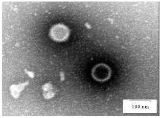 | |
| Fig. 1: | BoHV-1 under electron microscope |
| Table 1: | Isolation of IBR virus (BoHV-1) from nasal secretions by inducing artificial stress with dexamethasone in seropositive cattle |
 | |
To clarify whether the isolates exhibited different capabilities of cell to cell spread, serial dilutions were grown in cell culture under semisolid medium and stained for BoHV-1 gB. Diameters of least 30 plaques from each isolate were determined using a graduated ocular and correlated to the plaque size of BoHV-1 Schönböken which was set as 100%. Plaque sizes were analyzed and no satisfactory significant difference could be demonstrated among the isolates.
Restriction Fragment Length Polymorphism (RFLP) analysis identified all isolates as Bovine herpesvirus type 1. All the 3 isolates were assigned to genotype BoHV-1.2 (Strains India 4, India 5 and India 6).
Nucleotide sequencing: Parts of the regions encoding gH, gB and gC were amplified and sequenced on a 3130 Genetic Analyzer (ABI). In gH sequence analysis Strains India 4, 5 and 6 showed identical sequences. In gB sequence analysis all the 3 isolates from India showed an identical sequence in the analyzed 450 base pair of the gB gene. The sequence was named “India gBcons”. In gC sequence analysis all the 3 isolates from India showed an identical sequence in the analyzed 158 base pair spanning overlapping region of gC gene. The sequence was named “India gCcons”.
| Table 2: | Antibody titres against IBR virus (BoHV-1) in paired serum samples collected at day 0 and day 21 after inducing artificial stress with dexamethasone in seropositive cattle and isolation of virus |
 | |
DISCUSSION
In the present study serous nasal discharge was observed in all the three dexamethasone treated cows, which is the indication of immunosuppression and thereby liberation of the virus (De Carlo et al., 2004). The cows showed mild depression due to immunosuppression but their appetite was normal. The animals showed no rise of temperature, which was almost subnormal (below 101°F) in all the animals. The animals showed mild diarrhea for 3-4 days, which corroborates with the findings of De Carlo et al. (2004). The animal appeared almost normal (Straub and Lorenz, 1991). Sudden drop of milk production was also been recorded by Pritchard et al. (1997). All the cows become normal soon after the course of dexamethasone ended. Since dexamethasone is not a very long acting immunosuppressive drug, discontinued use results in a return to normal of the three cows (Pastoret et al., 1980).
Isolation of BoHV-1.2 (Strain India 4, India 5 and India 6) after dexamethasone injection in seropositive animals underlined that dexamethasone being an immunosuppressant, mimics natural stress and induces reactivation of the virus from a latent state. It was observed that there is variation in the recovery of virus in these animals which is due to the factor that the virus shedding itself is inconsistent. The level of local antibodies present is so high that even though reactivation occurs, infectious virus is rapidly neutralized and excretion cannot be detected which is responsible for variation in isolation of the virus among the cows (Pastoret et al., 1980).
There was four fold to seven fold increases in antibody titers in paired sera samples. Since immunosuppressant is short lived, viral antigen present can act as a booster to the immune response, and therefore this would explain the increase in antibody titer after dexamethasone treatment (Pastoret et al., 1980).
Although most BoHV-1.2 strains had been reportedly isolated from genital organ lesions but in this study all the three BoHV-1.2 (Strain India 4, India 5 and India 6) isolates were from nasal secretions of dexamethasone injected cattle which is also significant enough. Although the seroprevalence of BoHV-1 in West Bengal were 33.97% in 2004-05 (RDDL, 2004), 39.1% in 2005-06 (RDDL, 2005) and 47.92% in 2006-07 (RDDL, 2006) which has an increasing trend but there are no reports regarding isolation of the BoHV-1 from cows. In this respect isolation of BoHV-1 after inducing artificial stress with dexamethasone in this region has a great importance regarding various epidemiological aspects. Moreover these are the first reported isolates of BoHV-1 after artificial stress in West Bengal as well as in India.
Molecular study of the isolate found to be BoHV-1 genotype i.e., BoHV-1.2. Sequence analysis confirmed the clustering with known BoHV-1 strains and showed a clear difference to other ruminant herpes viruses. A regional clustering of the BoHV-1 isolates for the analyzed gC sequence showed a marginal differences to known isolates from Europe or the United States of America. Investigation of further Indian BoHV-1 isolates will be necessary to confirm the significance of this observation. The analysis of highly conserved genome regions gB did not reveal significant differences to previously reported BoHV-1.
CONCLUSION
In tropical and sub-tropical countries animal husbandry development is directed towards the rearing of cross-bred cattle and they are more susceptible to environmental stress like heat stress. In BoHV-1 infected cattle, the virus remain latent for life-long and excreted through secretions (nasal, ocular and vaginal) during any type of stress and thereby imposes a threat of disseminating the virus to other cattle (Pistl et al., 2003). As no prevention and control measures adopted either in organized or unorganized cattle farms in countries like India, Pakistan, etc. even introduction of latently infected sero-negative cattle will impose a thread to spread the virus to healthy cattle (Rola et al., 2005) promptly under stressful conditions. Several reactivation stimuli can lead to viral re-excretion, which is responsible for the maintenance of BoHV-1 within a cattle herd. In India the seoprevalence of IBR is 41.23% in 2003-04 (ADMAS, 2003), which is alarming, and yet no prevention and control measures have been adopted by the Government of India. Moreover prevention and control of the disease caused by BoHV-1 consists of thorough sanitary measures, the DIVA (Differentiating Infected from Vaccinated Animals) strategy mainly by vaccination with marker vaccine and isolation of the virus (Pistl et al., 2003). Thus isolation and characterization of more viruses in countries like India will contribute to the control and prevention of IBR.
BoHV-1 is a worldwide disseminated pathogen displaying significant differences in regional incidence and prevalence with regards to the geographical positions and the breeding managements of the considered regions (Ackermann and Engels, 2006).Thus reactivation obtained in this study with the BoHV-1 cow latency model and dexamethasone suggests the usefulness of the model for further studying the molecular characteristics of the BoHV-1 isolates prevalent in tropical and sub-tropical countries like India, Pakistan etc. which will provide valuable clues to epidemiological investigations on BoHV-1 infections. Moreover this model to detect virus may be taken to prevent the introduction of BoHV-1 infected sero-negative animals in a herd in order to improve the efficacy of control programmes.
ACKNOWLEDGMENTS
The authors are grateful to the Director and Veterinary Officer of Behala Express Dairy for giving necessary permission to carry out the study in their cattle farm. The authors are grateful to the Joint Director, Institute of Animal Health and Veterinary Biologicals, West Bengal for necessary permission to carry out this work in their institute. The authors convey their special thanks to the Director, Indian Institute of Chemical Biology, Jadavpur and the Director, National Institute of Cholera and Enteric Diseases (NICED), Kolkata for giving necessary permission for ultra-centrifugation and transmission electron microscopy of the isolated virus. The authors are in debt to PD Dr. Martin Beer and Dr. Patricia Koenig of OIE and National Reference Laboratory for Bovine herpesvirus type 1, Friedrich-Loeffler-Institut, Germany for typing of the virus.
REFERENCES
- Ackermann, M., E. Peterhans and R. Wyler, 1982. DNA of bovine herpesvirus type 1 in the trigeminal ganglia of latently infected calves. Am. J. Vet. Res., 43: 36-40.
PubMedDirect Link - Ackermann, M. and M. Engels, 2006. Pro and contra IBR-eradication. Vet. Microbiol., 113: 293-302.
CrossRef - Davies, D.H. and I.E. Carmichael, 1973. Role of cell-mediated immunity in the recovery of cattle from primary and recurrent infections with infectious bovine rhinotracheitis virus. Infect. Immun., 8: 510-518.
PubMedDirect Link - De Carlo, E., G.N. Re, R. Letteriello, V. Del Vecchio and M.P. Giordanelli et al., 2004. Molecular characterization of a field strain of bubaline herpesvirus isolated from buffaloes (Babalusbubalis) after pharmacological reactivation. Vet. Record, 154: 171-174.
CrossRefDirect Link - Gill, B.S., D.R. Sharma, M.S. Kwatra, A. Kumar and J.S. Gill, 1987. Seroprevalence of infectious bovine rhinotracheitis in the Punjab state. J. Res. Punjab Agri. Univ., 24: 317-318.
Direct Link - Hage, J.J., R.D. Glas, H.H. Westra, M.A. Maris-Veldhuis, J.T. Van Oirschot and F.A.M. Rijsewijk, 1998. Reactivation of latent bovine herpesvirus 1 in cattle seronegative to glycoproteins gB and gE. Vet. Microbiol., 60: 87-98.
CrossRefPubMedDirect Link - Kilari, S., S.N. Singh, A.V. Buche, S. Sawarkar and S. Ghosh, 2000. Evaluation of immunogenicity of infectious bovine rhinotracheitis vaccine in Indian tropical climate. Ind. Vet. J., 77: 185-188.
Direct Link - Pastoret, P.P., L.A. Babiuk, V. Misra and P. Griebel, 1980. Reactivation of temperature-sensitive and non-temperature-sensitive infectious bovine rhinotracheitis vaccine virus with dexamethasone. Infect. Immun., 29: 483-488.
PubMedDirect Link - Pastoret, P.P., E. Thiry and R. Thomas, 1986. Logical description of bovine herpesvirus type 1 latent infection. J. Gen. Virol., 67: 885-897.
PubMedDirect Link - Pritchard, G., N. Cook and M. Banks, 1997. Infectious pustular vulvovaginitis/infectious pustular balanoposthitis in cattle. Vet. Rec., 140: 587-587.
PubMedDirect Link - Rock, D., J. Lokensgard, T. Lewis and G. Kutish, 1992. Characterization of dexamethasone-induced reactivation of latent Bovine Herpesvirus 1. J. Virol., 66: 2484-2490.
Direct Link - Rajesh, J.B., P.V. Tresamol and M.R. Saseendranath, 2003. Seroprevalence of infectious bovine rhinotracheitis in cattle population of Kerala. Indian Vet. J., 80: 393-396.
Direct Link - Rola, J., M. Larska and M.P. Polak, 2005. Detection of bovine herpesvirus 1 from an outbreak of infectious bovine rhinotracheitis. Bull. Vet. Inst. Puawy, 49: 267-271.
Direct Link