
Hamideh Azimi
Depratment of Dermatology, Tabriz University of Medical Sciences, Tabriz, Iran
Jafar Majidi
Depratment of Imunology, Tabriz University of Medical Sciences, Tabriz, Iran
Rasul Estakhri
Depratment of Pathology, Tabriz University of Medical Sciences, Tabriz, Iran
Mohamad Goldust
Student Research Committee, Tabriz University of Medical Sciences, Tabriz, Iran
Pakistan Journal of Biological Sciences
Year: 2013 | Volume: 16 | Issue: 12 | Page No.: 589-592


ABSTRACT
Pemphigus is defined as a group of chronic self-immune vesicular diseases histologically recognized by inter-epidermic vesicles resulting from acantholysis. The aim of this study was to evaluate the precipitant and circulative IgG antibodies in patients with pemphigus vulgaris before and after treating with immunofluorescence. Sixty-two patients (34 females and 28 males) with clinically and pathologically confirmed P.V. were studied prospectively over a one year period of time during which direct and indirect immunofluorescent tests were performed before and after treatment. They had mild or moderate forms of disease. All patients received prednisolon 1-2 mg/kg/day and Azathioprine 2-3 mg/kg/day or methylpredisolon (1 g day-1 for 4 days) and cyclophosphamide (500 mg/first day) pulse therapy due to general condition. Thirty- four females and 28 males enrolled, the mean age were 39.5 years (SD = 12.7). Before treatment, 10 and 52 cases were positive for skin depositing +or++) and circulatory IgG (1/20 -1/60), respectively. Two to 3 month later, 37 were IgG positive with titers 1/20 to 1/160. The correlation between circulatory IgG before and after treatment was weakly positive (p = 0.05, r = 0.415). In the present study, treatment methods used for patients suffering from pemphigus vulgaris were not successful in significantly decreasing the circulative autoantibodies levels.
PDF Abstract XML References Citation
Received: December 08, 2012;
Accepted: February 16, 2013;
Published: March 28, 2013
How to cite this article
Hamideh Azimi, Jafar Majidi, Rasul Estakhri and Mohamad Goldust, 2013. IgG Antibodies in Patients with Pemphigus Vulgaris before and after Diagnosing with Immunofluorescence. Pakistan Journal of Biological Sciences, 16: 589-592.
DOI: 10.3923/pjbs.2013.589.592
URL: https://scialert.net/abstract/?doi=pjbs.2013.589.592
DOI: 10.3923/pjbs.2013.589.592
URL: https://scialert.net/abstract/?doi=pjbs.2013.589.592
INTRODUCTION
The term of “Pemphigus” refers to a group of chronic fatal diseases manifesting with mucocutaneous blisters. In pemphigus vulgaris which is the most prevalent form of the disease, autoantibody is created against desmoglein 1 and 3 (Flores et al., 2012; Funakoshi et al., 2012). The fatal disease usually appears in middle-aged persons and rarely in youths with clinical, pathological and immunological symptoms respectively as mucocutaneous blisters; acantholysis and blister formation between cells of epidermis and complement and IgG precipitation in skin and circulative antibodies (Goldust et al., 2011). Sometimes, it is a sign of inner organs malignancies such as lung, digestive and reproductive systems (Hatano et al., 2012). Classically, the disease is treated with systemic glucocorticoids along with other immunosuppressives including azathioprine. The main goal of treatment is to eradicate or decrease pathogenic antibodies (Brandt et al., 2012; Goldust et al., 2012; Ohyama et al., 2012). The relation of these autoantibodies levels with severity of the disease clinical symptoms is not exactly known. Some studies demonstrate that serum level of circulative autoantibodies against keratinocytes determined through indirect immunofluorescence method is in correspondence with severity of disease activity (Dworschak et al., 2012; Lotti et al., 2012). While, results obtained from other studies do not indicate this direct relationship between clinical symptoms and antibody levels (Behzad et al., 2012; Hosoda et al., 2012).
Direct immunofluorescence is a standard method to trace precipitant IgG in skin. The present study aims at determining precipitant and circulative IgG antibodies before treatment in patients suffering from pemphigus vulgaris and evaluating the effects of immunosuppressive drugs on levels of circulative antibodies after treatment.
MATERIALS AND METHODS
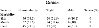
In this cross sectional study of before and after intervention kind, 62 patients with clinical and pathological symptoms of pemphigus vulgaris were studied for one year from July 2011-2012. Written consent was obtained from all the patients. This study was approved by ethic committee of Tabriz university of medical sciences. Before treatment, IgG precipitation along with C3 in skin was traced using direct immunofluorescence (DIF) and titer of IgG levels found in blood circulation using indirect immunofluorescence (IIF) methods. After taking blood samples from the patients, blood serum was isolated, to conduct IIF and added to skin sections resulted from neonates circumcising which were fixed on the slide so that it can connect to desmosomes found among keratinocytes in case of existing of the circulative IgG antibodies. Then, human anti-IgG antibody prepared from rabbits and marked with fluorescent materials were added and finally studied under microscope. To do DIF, the sample resulted from biopsy of the skin located around the lesions at the special immunology laboratory of Imam Reza hospital, Tabriz was sectioned at micron sizes (using cryosection) and was studied under fluorescent microscope using fluorescent conjugation after coating to slide surface and its fixation. Depending on general conditions such as age, records of hypertension and function of the interior organs, the patients were classically treated with prednisolone (1-2 mg kg-1), azathioprine (2-3 mg kg-1) or underwent pulse treatment (using methylprednisolone 1 g day-1 for 4 days and cyclophosphamide 500 mg day-1). After relative recovery and decreasing at least 50% of the clinical symptoms (lasting about 2-3 months) IIF of these patients were reexamined. Changing levels of antibodies found in blood circulation is regarded as a criterion indicating effectiveness of the applied treatment methods. The patients were studied considering variables such as age, gender, disease duration, existence and severity of lesions on skin, mouth and head before and after treatment. Considering disease severity, the patients were divided into four groups: 1) non-morbidity of the desired locus (skin, mouth and head, 2) slight form with morbidity of less than 1/3 of the desired place, 3) mild form with morbidity of 1/3-2/3 of the desired place and 4) severe form with morbidity of more than 1/3 of the desired locus (Table 1). The resulted outcomes were analyzed using SPSS-16 software and stated as mean±SD deviation. The results were regarded meaningful if p>0.05 and highly correlate if r>0.5.
RESULTS
Mean age of these patients was 39.55±12.7 years. The youngest patient was 20 and the oldest one was 79 years old. Mean of the disease starting period was 1.93±1.03 years. The patients was consisted of 28 males (45%) and 34 females (55%). Considering skin morbidity, there were 36 cases (58.3%) of skin non-morbidity and 26 patients (41.7%) of slight, mild, or severe morbidity (Table 1). Considering oral morbidity, 32 cases (51.6%) of oral non-morbidity and 30 cases of oral morbidity with different severities were observed. Regarding head morbidity, there were 45 cases (73.3%) of non-morbidity and only 17 cases of head morbidity as slight, mild, or severe (Table 1). Outcomes resulted from DIF on IgG precipitation in skin before starting treatment demonstrated that DIF skin non-morbidity. In patients with skin lesions, negative DIF were seen in 6 cases (23.7%), IgG+ precipitation in 9 patients (34.6%) and IgG++ precipitation in 11 cases (42.3%). Considering IIF before treatment, it was negative in 10 patients (16.1%) but circulative antibody was observed in 52 cases (83.9%) and its assay varied from 1/20 to 1/160 (Table 2). IIF tests after treatment demonstrated that circulative antibody was not observed in 16 patients (30.2%) but it was seen in 37 cases (69.8%). Repeating IIF test was not possible in 9 patients due to their non-reference to the center. There were no cases of mortality in patients during conducting the study. Evaluating correlation between circulative IgG titer before starting the treatment with its titer after treatment revealed that p = 0.005 and r = 0.415. Significant difference was not observed considering mean of the obtained results on circulative IgG levels before and after treatment (p = 0.122, r = 1.577).
DISCUSSION
There is no standard treating method and different methods are used to treat and control the disease. The final goal is to eradicate or removing circulative antibodies (Yokoyama et al., 2011) the disease severity is controlled by clinical symptoms at acute stages. But, when the new blisters do not appear and the previous lesions recover, changes of circulative antibody levels will be helpful in determining drug dosage (Oberkirchner et al., 2011).
| Table 1: | Prevalence and severity of clinical symptoms in patients suffering from pemphigus vulgaris |
 | |
| Values in brackets one percentage | |
| Table 2: | Levels of circulative IgG antibodies using IIF method before and after treatment in patients suffering from pemphigus vulgaris |
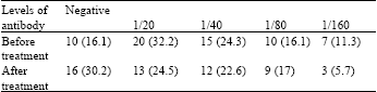 | |
| Values in brackets one percentage | |
In the present study, there was no significant difference between mean of circulative antibody before and after treatment (p = 0.122, t = 1.577). Correlation between circulative antibody levels before and after treatment was positive but lower than moderate level (p = 0.005, r = 0.415). The results demonstrate that decreasing the amount of antibody after treatment was not valuable and the applied treatment methods, in spite of improving the clinical symptoms, were not successful in significantly decreasing the antibody levels. Several studies have been conducted on how titer of these antibodies changes during treatment and different results have been obtained (Lanza et al. 2010) according to the reports, significant decrease of circulative antibodies levels after successfully treating the disease using immunofluorescence test have been observed in some studies (Prado et al., 2011). In a previous study conducted on effects of plasmapheresis in a patient resisting against prednisolone and cyclophosphamide, significant decrease of circulative antibodies in immunofluorescence test associated with recovery of the clinical symptoms was observed about 6 weeks after starting the treatment (Jennings et al., 2011). In another study conducted on effects of intravenous immunoglobulin (IVIg) in 21 patients suffering from severe form of pemphigus vulgaris, significant decrease of serum antibodies levels have been reported 4-6 months after starting the treatment in indirect immunofluorescence (Wada et al., 2011). But, other studies have presented different and, sometimes, contrary results. In a study on effects of oral prednisolone in 46 patients suffering from pemphigus vulgaris, decrease of circulative antibodies titer after treating with immunofluorescence method has only been observed in patients with severe disease. In patients with slight or moderate forms of the disease, changes of the circulative antibodies levels was not considerable (Lanza et al., 2011). In a previous study, effects of replacing plasma and corticosteroid was evaluated in patients suffering from pemphigus vulgaris and decrease of circulative antibodies levels in immunofluorescence was observed in some patients. But, no change was seen in some other patients or it has been increased in spite of improving of the clinical symptoms (Mejri et al., 2011). Another study evaluated 78 patients suffering from pemphigus vulgaris. In this study, circulative antibodies levels were decreased in some patients after treatment using immunofluorescence method. But, no change or even increasing Ab titer was observed in some other understudy patients. In this study, evaluating response to treatment through measuring serial of circulative antibodies levels is not recommended (Muller et al., 2010). As observed, the above studies emphasize unpredictability of changes of circulative antibodies levels in patients suffering from pemphigus vulgaris. In the present study, mean of circulative antibodies levels were not significantly changes (p = 0.1222, t = 1.5777) which may have different reasons. Firstly, type of the applied treatment method is effective in changing circulative antibodies levels (Endo et al., 2010). Simultaneous use of several treatment methods results in more decrease of antibodies levels. Secondly, changes of antibodies levels vary depending on the disease severity (Santiago-et-Sanchez-Mateos et al., 2011). In the present study, the patients suffered from slight and mild forms of the disease and no case of the severe form was observed. The applied treatment methods were not successful in significantly changing antibody levels because the patients received different forms and dosages of corticosteroids prescribed by other physicians in the outpatient ward before hospitalization. On the other hand, the changes observed at circulative antibody levels vary depending on involvement locus of the patients. Like the present study, mouth and head morbidity lead to more stability of antibodies due to having high levels of antigens. Some studies suggested that although special autoantibodies of pemphigus vulgaris are of IgG kind, these antibodies have other subgroups (such as 1,3 kinds) and only some of them are effective in pathogenesis process and common measuring methods do not determine these differences (Aoyama et al., 2010). On the other hand, methods used to measure antibodies levels have different sensitivities, for example, ELISA method is more sensitive than IIF. Additionally, dilution of the prepared samples will lead to different results.
CONCLUSION
In the present study, treatment methods used for patients suffering from pemphigus vulgaris were not successful in significantly decreasing the circulative autoantibodies levels. It is recommended that future studies pay more attention to kind and duration of the applied treatment methods, kind of skin, mucous, or oral morbidity and use of more sensitive evaluation methods such as Elisa to measure circulative antibodies.
REFERENCES
- Aoyama, Y., M. Nagai and Y. Kitajima, 2010. Binding of pemphigus vulgaris IgG to antigens in desmosome core domains excludes immune complexes rather than directly splitting desmosomes. Br. J. Dermatol., 162: 1049-1055.
CrossRef - Behzad, M., C. Mobs, A. Kneisel, M. Moller, J. Hoyer, M. Hertl and R. Eming, 2012. Combined treatment with immunoadsorption and rituximab leads to fast and prolonged clinical remission in difficult-to-treat pemphigus vulgaris. Br. J. Dermatol., 166: 844-852.
CrossRef - Brandt, O., D. Rafei, E. Podstawa, A. Niedermeier and M.F. Jonkman et al., 2012. Differential IgG recognition of desmoglein 3 by paraneoplastic pemphigus and pemphigus vulgaris sera. J. Invest. Dermatol., 132: 1738-1741.
CrossRef - Dworschak, J., A. Recke, M. Freitag, R.J. Ludwig and J. Langenhan et al., 2012. Mapping of B cell epitopes on desmoglein 3 in pemphigus vulgaris patients by the use of overlapping peptides. J. Dermatol. Sci., 65: 102-109.
CrossRef - Endo, Y., K. Tsujioka, M. Tanioka, Y. Minegaki and B. Ohyama et al., 2010. Bullous dermatosis associated with IgG antibodies specific for desmocollins. Eur. J. Dermatol., 20: 620-625.
PubMed - Flores, G., D.A. Culton, P. Prisayanh, B.F. Qaqish and K. James et al., 2012. IgG autoantibody response against keratinocyte cadherins in endemic pemphigus foliaceus (fogo selvagem). J. Invest. Dermatol., 132: 2573-2580.
CrossRef - Funakoshi, T., L. Lunardon, C.T. Ellebrecht, A.R. Nagler, C.E. O'Leary and A.S. Payne, 2012. Enrichment of total serum IgG4 in patients with pemphigus. Br. J. Dermatol., 167: 1245-1253.
CrossRef - Goldust, M., E. Rezaee and S. Hemayat, 2012. Treatment of scabies: Comparison of permethrin 5% versus ivermectin. J. Dermatol., 39: 545-547.
CrossRef - Hosoda, S., M. Suzuki, M. Komine, S. Murata, T. Hashimoto and M. Ohtsuki, 2012. A case of IgG/IgA pemphigus presenting malar rash-like erythema. Acta Dermato-Venereologica, 92: 164-166.
CrossRefPubMedDirect Link - Jennings, J.M., D.K. Tucker, M.D. Kottke, M. Saito and E. Delva et al., 2011. Desmosome disassembly in response to pemphigus vulgaris IgG occurs in distinct phases and can be reversed by expression of exogenous Dsg3. J. Invest. Dermatol., 131: 706-718.
CrossRef - Lanza, A., M. Lanza, R. Santoro, V. Soro, S.S. Prime and N. Cirillo, 2011. Deregulation of PERK in the autoimmune disease pemphigus vulgaris occurs via IgG-independent mechanisms. Br. J. Dermatol., 164: 336-343.
CrossRef - Lanza, A., L. Perillo, C. Landi, F. Femiano, F. Gombos and N. Cirillo, 2010. Controversial role of antibodies against linear epitopes of desmoglein 3 in pemphigus vulgaris, as revealed by semiquantitative living cell immunofluorescence microscopy and in cell elisa. Int. J. Immunopathol. Pharmacol., 23: 1047-1055.
PubMedDirect Link - Lotti, R., A. Marconi and C. Pincelli, 2012. Apoptotic pathways in the pathogenesis of pemphigus: Targets for new therapies. Curr. Pharm. Biotechnol., 13: 1877-1881.
CrossRefDirect Link - Mejri, K., O. Abida, M. Kallel-Sellami, S. Haddouk and L. Laadhar et al., 2011. Spectrum of autoantibodies other than anti-desmoglein in pemphigus patients. J. Eur. Acad. Dermatol. Venereol., 25: 774-781.
CrossRef - Muller, R., N. Hunzelmann, V. Baur, G. Siebenhaar and E. Wenzel et al., 2010. Targeted immunotherapy with rituximab leads to a transient alteration of the IgG autoantibody profile in pemphigus vulgaris. Dermatol. Res. Pract.
CrossRefDirect Link - Oberkirchner, U., K.E. Linder, S. Dunston, P. Bizikova and T. Olivry, 2011. Metaflumizone-amitraz (Promeris)-associated pustular acantholytic dermatitis in 22 dogs: Evidence suggests contact drug-triggered pemphigus foliaceus. Vet. Dermatol., 22: 436-448.
CrossRef - Ohyama, B., K. Nishifuji, P.T. Chan, A. Kawaguchi and T. Yamashita et al., 2012. Epitope spreading is rarely found in pemphigus vulgaris by large-scale longitudinal study using desmoglein 2-based swapped molecules. J. Invest. Dermatol., 132: 1158-1168.
CrossRef - Prado, R., S.L. Brice, S. Fukuda, T. Hashimoto and M. Fujita, 2011. Paraneoplastic pemphigus herpetiformis with IgG antibodies to desmoglein 3 and without mucosal lesions. Arch. Dermatol., 147: 67-71.
CrossRef - Santiago-et-Sanchez-Mateos, D., M.A. Juarez, A.G. De Arriba, D. Jimenez, J. Fraga, T. Hashimoto and A. Garcia-Diez, 2011. IgG/IgA pemphigus with IgA and IgG antidesmoglein 1 antibodies detected by enzyme-linked immunosorbent assay: Presentation of two cases. J. Eur. Acad. Dermatol. Venereol., 25: 110-112.
CrossRef - Wada, N., K. Nishifuji, T. Yamada, J. Kudoh and N. Shimizu et al., 2011. Aire-dependent thymic expression of desmoglein 3, the autoantigen in pemphigus vulgaris and its role in T-cell tolerance. J. Invest. Dermatol., 131: 410-417.
CrossRef - Yokoyama, T., S. Matsuda, Y. Takae, N. Wada, T. Nishikawa, M. Amagai and S. Koyasu, 2011. Antigen-independent development of Foxp3+ regulatory T cells suppressing autoantibody production in experimental pemphigus vulgaris. Int. Immunol., 23: 365-373.
CrossRef