
Shahla Babaei Nejad
Student research committee, Tabriz University of Medical Sciences, Iran
Effat Khodaeiani
Student research committee, Tabriz University of Medical Sciences, Iran
Mehdi Amirinia
Student research committee, Tabriz University of Medical Sciences, Iran
Mohamad Goldust
Student research committee, Tabriz University of Medical Sciences, Iran
Pakistan Journal of Biological Sciences
Year: 2013 | Volume: 16 | Issue: 22 | Page No.: 1609-1611


ABSTRACT
Cicatricle alopecia represents a diverse group of diseases characterized by a lack of follicular ostia and irreversible alopecia. This study aimed at evaluating cicatricial alopecia in Iranian patients. One hundred patients with cicatricial alopecia were studied. Patients disease was pathologically proven. All epidemiologic and clinicopathologic data were obtained through questionnaires. The results were analyzed by means of descriptive statistical methods. One hundred patients were consisted of 52 (52%) males and 48 (48%) females. 30 patients (24 males and 6 females) suffered from folliculate decalvans, 25 cases (10 males and 15 females) from DLE, 18 patients (6 males and 11 females) from brocq pseudopelade, 14 patients (4 males and 10 females) from lichen planopilaris, 8 patients (4 males and 4 females) from morphea and 5 cases (4 males and 1 female) from folliculate colloidalis. Early stage diagnosis by biopsy and proper treatment will reduce further progression and especially alleviate psychosocial disturbances.
PDF Abstract XML References Citation
Received: January 08, 2013;
Accepted: March 02, 2013;
Published: May 08, 2013
How to cite this article
Shahla Babaei Nejad, Effat Khodaeiani, Mehdi Amirinia and Mohamad Goldust, 2013. Evaluation of Cicatricial Alopecia in Iran. Pakistan Journal of Biological Sciences, 16: 1609-1611.
DOI: 10.3923/pjbs.2013.1609.1611
URL: https://scialert.net/abstract/?doi=pjbs.2013.1609.1611
DOI: 10.3923/pjbs.2013.1609.1611
URL: https://scialert.net/abstract/?doi=pjbs.2013.1609.1611
INTRODUCTION
Cicatricial alopecia- a diverse group of rare disorders that destroy the hair follicle, replace it with scar tissue and cause permanent hair loss (Goldust et al., 2013a; Lotti et al., 2013). The hair loss may be accompanied with severe itching, pain and burning and progress rapidly. In other cases the hair loss is gradual,without symptoms and is unnoticed for long periods (Goldust et al., 2013a; Mohebbipour et al., 2012). Cicatricial alopecia, also known as scarring alopecia, occurs in otherwise healthy men and women of all ages and is seen worldwide (Goldust et al., 2013b, c). Cicatricial alopecias are classified as primary or secondary. This discussion is confined to the primary cicatricial alopecias in which the hair follicle is the target of the destructive inflammatory process (Goldust et al., 2013d; Vafaee et al., 2012). In secondary cicatricial alopecias, destruction of the hair follicle is incidental to a non-follicle-directed process or external injury, such as severe infections, burns, radiation, or tumors ( Goldust and Rezaee, 2013; Goldust et al., 2012; Sadighi et al., 2011). Hairs less likely grow after recovery of the disease or primary inflammation (Ohyama, 2012). Discoid lupus erythmatosus (chronic), lichen planopilaris, folliculate decalvans are known as primary factors of cicatricle alopecia (Khumalo, 2010). Burn, radiodermatitis and infections such as favus or skin tuberculosis are regarded as the secondary reasons (Chiu and Lin, 2010). In most cases, a diagnostic biopsy is necessary. Biopsy place should be carefully selected and priority is given to the new lesions (Wu et al., 2008). Primary cicatricle alopecia is pathologically divided into lymphocytic and neutrophilic. Lymphocytic types include discoid lupus erythmatosis, lichen planopilaris, pseudopelade of Brocq, morphea and neutrophilic includes folliculate decalvans and folliculate colloidalis ( De Berker, 2002; Gardani et al., 2007). The study aims at evaluating epidemiological and clinicopathological characteristics of patients suffering from cicatricle alopecia in Tabriz.
MATERIALS AND METHODS
The research was conducted on file of 100 patients suffering from cicatricle alopecia and referring to skin clinic of Tabriz Sina Hospital within a 7 year period. Written consent was obtained from all the patients. This study was approved by ethic committee of Tabriz university of medical sciences. Patients skin lesions were underwent biopsy and pathological diagnosis was confirmed. The obtained data including age, gender, occupation, residence, type of lesion resulting in cicatricle alopecia and type of dermal exudation specified in pathology were analyzed using descriptive statistics methods. SPSSTM, version 16 is the used statistical software program. The results were expressed as Means±standard deviations. The Chi-square test was used for statistical analysis. The level of statistical significance was set at a value of p<0.05.
RESULTS
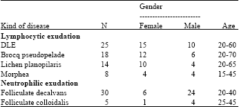
One hundred patients were consisted of 52 (52%) males and 48 (48%) females. Most male patients (50%) were self-employed and most of females (80%) were housewives. In this study, 79 patients were of Tabriz and 21 subjects were of other cities of Azerbaijan. In this study, 30 patients (24 males and 6 females) suffered from folliculate decalvans, 25 cases (10 males and 15 females) from DLE, 18 patients (6 males and 11 females) from brocq pseudopelade, 14 patients (4 males and 10 females) from lichen planopilaris, 8 patients (4 males and 4 females) from morphea and 5 cases (4 males and 1 female) from folliculate colloidalis. DLE was the most common disease with inflammatory lymphocytic exudation. Distribution of kinds of disease with inflammatory lymphocytic exudation based on age, gender and clinical symptoms has been demonstrated in Table 1. There was inflammatory neutrophilic exudation in 30 patients (24 males and 6 females) with age range of 20-40 years old. These patients suffered from folliculate decalvans with pustule and scar plaques in scalp. Such an exudation has been reported in 5 patients (4 males and 1 female) with age range of 25-45 years old suffering from follicular colloid with clinical symptoms of nodule and colloid platelets at lower parts of back of the head posterior.
DISCUSSION
Cicatricle alopecia leading to destruction of hair follicles and hairs permanent disappearing may be a result of some diseases including discoid lupus erythmatosis primarily affecting hair follicles or developed following a secondary disease such as burn, infection or radiotherapy (Messenger, 2002). Significant studies have not been conducted in our country regarding these lesions while most of them indicate diseases which can be controlled regarding their progress and complications through early diagnosis and appropriate treatment. The research was conducted on 100 patients suffering from cicatricle alopecia and their lesions underwent biopsy and type of lesion was diagnosed and confirmed through pathological examination.
| Table 1: | Clinical types of cicatricle alopecia |
 | |
Generally, the patients were consisted of 52 (52%) males and 48 (48%) females with almost equal prevalence. This is while, according to the reports of the researches conducted in other countries, disease in women is more prevalent than men (Tan et al., 2004). It can be attributed to the social-cultural differences of different societies and kind of women coverage-as an appropriate and protective cover against sun light-or delayed referring of women to the clinic. Considering type of lesions cellular exudation, pathological evaluations demonstrated that lymphocytic and neutrophilic exudations are more common in females and males, respectively. Most patients were middle-aged which is in correspondence with other studies (Amato et al., 2002). Lymphocytic and neutrophilic types with exudation were respectively more common in middle-aged women and men which are again in correspondence with other researches (Amato et al., 2002). Considering occupation, most male patients were self-employed and most female ones were housewives. In this case, it can be stated that environmental factors such as sun light and job stress may be effective in developing some lesions including DLE and lichen plan (Sharquie et al., 2002). In this study, 79% of the understudy patients referred to the skin clinic from Tabriz and the rest were of other cities of Azerbaijan. The difference may be attributed to high levels of social, economical, cultural, hygienic factors and awareness of people in big cities and their easy access to therapeutic centers. The lesions were pathologically evaluated to specify (1) type of inflammatory exudation, (2) type of lesion. Generally, the lesions are divided into two categories considering type of inflammatory exudation: (a) Lesions with lymphocytic exudation (b) Lesions with neutrophilic exudation. In this study, 65 patients (65%) suffered from lymphocytic exudation and 35 ones (35%) experienced neutrophilic exudation. High rate of lymphocytic type was in correspondence with researches conducted in other countries (Amato et al., 2002). Lymphocytic exudation lesions included DLE, brocq pseudopelade, lichen planopilaris and morphea. The neutrophilic exudation lesions were folliculate decalvans and folliculate colloidalis. In lymphocytic type, DLE was the most common lesion leading to cicatricle alopecia. It was more common in middle-aged women. The results are in correspondence with other researches (Amato et al., 2002). Considering prevalence, brocq pseudopelade, lichen planopilaris and morphea occupied the next ranks. Again, it corresponded with other studies (Annessi et al., 1999). The present study revealed that folliculate decalvans (30%) was the most prevalent lesion among cicatricle alopecia with neutrophilic exudation and is in correspondence with other studies (Fabbri et al., 2004). Folliculate colloidalis was the next lesion (5%). Considering that the lesion is more common in blacks and it is confirmed by other studies (Amato et al., 2002), it is relatively less common in our country. Generally, primary cicatricle alopecia with neutrophilic exudation is prevalent in middle-aged men such that 24 patients (out of 30 ones) suffered from folliculate decalvans (80%) and 4 patients (out of 5 ones) experienced folliculate colloidalis (80%). High prevalence in males equals with other researches Amato et al. (2002).
CONCLUSION
Cicatricle alopecia is one of the tricologic emergencies and is usually irrevocable. The progress of the lesion and its complications can be prevented through early diagnosis and treatment. The lesions can be diagnosed using an exact clinical-pathological evaluation. Therefore, dermal biopsy should be conducted at initial stages as soon as possible and necessary therapeutic actions should be taken.
REFERENCES
- Amato, L., S. Mei, D. Massi, I. Gallerani and P. Fabbri, 2002. Cicatricial alopecia; a dermatopathologic and immunopathologic study of 33 patients (pseudopelade of brocq is not a specific clinico-pathologic entity). Int. J. Dermatol., 41: 8-15.
CrossRef - Annessi, G., G. Lombardo, T. Gobello and P. Puddu, 1999. A clinicopathologic study of scarring alopecia due to lichen planus: Comparison with scarring alopecia in discoid lupus erythematosus and pseudopelade. Am. J. Dermatopathol., 21: 324-331.
PubMedDirect Link - Chiu, H.Y. and S.J. Lin, 2010. Fibrosing alopecia in a pattern distribution. J. Eur. Acad. Dermatol. Venereol., 24: 1113-1114.
CrossRef - De Berker, D., 2002. Clinical relevance of hair microscopy in alopecia. Clin. Exp. Dermatol., 27: 366-372.
CrossRef - Fabbri, P., L. Amato, C. Chiarini, S. Moretti and D. Massi, 2004. Scarring alopecia in discoid lupus erythematosus: A clinical, histopathologic and immunopathologic study. Lupus, 13: 455-462.
CrossRef - Gardani, G., R. Cerrone, C. Biella, B. Galbiati and E. Proserpio et al., 2007. A case-control study of panicum miliaceum in the treatment of cancer chemotherapy-induced alopecia. Minerva Med., 98: 661-664.
PubMedDirect Link - Goldust, M., S.B. Nejad, E. Rezaee and R. Raghifar, 2013. Comparative trial of permethrin 5% vs. lindane 1% for the treatment of scabies. J. Dermatol. Treat., (In Press).
CrossRefDirect Link - Goldust, M., M.R. Ranjkesh, M. Amirinia, F. Golforoushan, E. Rezaee and M.A.R. Saatlou, 2013. Sertaconazole 2% cream versus hydrocortisone 1% cream in the treatment of seborrheic dermatitis. J. Dermatol. Treat., (In Press).
CrossRefDirect Link - Goldust, M. and E. Rezaee, 2013. The efficacy of topical ivermectin vs. malation 0.5% lotion for the treatment of scabies. J. Dermatol. Treat., (In Press).
CrossRef - Goldust, M., E. Rezaee and S. Hemayat, 2012. Treatment of scabies: Comparison of permethrin 5% versus ivermectin. J. Dermatol., 39: 545-547.
CrossRef - Goldust, M., E. Rezaee and R. Raghifar, 2013. Comparison of oral ivermectin versus crotamiton 10% cream in the treatment of scabies. Cutaneousv Ocul. Toxicol.
CrossRef - Goldust, M., M. Talebi, J. Majidi, M.A.R. Saatlou and E. Rezaee, 2013. Evaluation of antiphospholipid antibodies in youths suffering from cerebral ischemia. Int. J. Neurosci., 123: 209-212.
CrossRefDirect Link - Khumalo, N.P., 2010. Grooming and central centrifugal cicatricial alopecia. J. Am. Acad. Dermatol., 62: 507-508.
CrossRef - Lotti, T., M. Goldust and E. Rezaee, 2013. Treatment of seborrheic dermatitis, comparison of sertaconazole 2% cream vs. ketoconazole 2% cream. J. Dermatol. Treat.
CrossRef - Mohebbipour, A., P. Saleh, M. Goldust, M. Amirnia, Y.J. Zadeh, R.M. Mohamadi and E. Rezaee, 2012. Treatment of scabies: Comparison of ivermectin vs. lindane lotion 1%. Acta Dermatovenerol. Croat, 20: 251-255.
PubMedDirect Link - Ohyama, M., 2012. Primary cicatricial alopecia: Recent advances in understanding and management. J. Dermatol., 39: 18-26.
CrossRef - Sadighi, A., A. Elmi, M.A. Jafari, V. Sadeghifard and M. Goldust, 2011. Comparison study of therapeutic results of closed tibial shaft fracture with intramedullary nails inserted with and without reaming. Pak. J. Biol. Sci., 14: 950-953.
PubMedDirect Link - Sharquie, K.E., J.R. Al-Rawi and H.A. Al-Janabi, 2002. Frictional hair loss in Iraqi patients. J. Dermatol., 29: 419-422.
PubMedDirect Link - Tan, E., M. Martinka, N. Ball and J. Shapiro, 2004. Primary cicatricial alopecias: Clinicopathology of 112 cases. J. Am. Acad. Dermatol., 50: 25-32.
CrossRef - Vafaee, I., M.B. Rahbani Nobar and M. Goldust, 2012. Etiology of ocular trauma: A two years cros sectional study in Tabriz, Iran. J. Coll. Phys. Surg. Pak., 22: 344-344.
PubMedDirect Link - Wu, W.Y., N. Otberg, K.J. McElwee and J. Shapiro, 2008. Diagnosis and management of primary cicatricial alopecia: Part II. SKINmed: Dermatol. Clinician, 7: 78-83.
CrossRef