
Mohammad Shokrzadeh
Pharmaceutical Sciences Research Center, Faculty of Pharmacy, Mazandaran University of Medical Sciences, Sari, Iran
Sepideh Shobi
Department of Biotechnology, Science and Research Branch, Islamic Azad University, Tehran, Iran
Hossein Attar
Department of Biotechnology, Science and Research Branch, Islamic Azad University, Tehran, Iran
Sahel Shayegan
Department of Toxicology and Pharmacology, Faculty of Pharmacy, Mazandaran University of Medical Sciences, Sari, Iran
Sakineh Sadat Hosseini Payam
Department of Toxicology and Pharmacology, Faculty of Pharmacy, Mazandaran University of Medical Sciences, Sari, Iran
Faezeh Ghorbani
Department of Environment, Meybod Branch, Islamic Azad University, Meybod, Iran
Pakistan Journal of Biological Sciences
Year: 2012 | Volume: 15 | Issue: 19 | Page No.: 936-941


ABSTRACT
Diazinon, a commonly used organophosphorus pesticide, has been widely used throughout the world in agriculture and horticulture to control insects that feed on crops, ornamentals, lawns, fruits, vegetables and other food products. The toxicity of the DZN causes adverse effects on many organs. The purpose of this study was to examine the protective effect of vitamins A, E and C on liver enzymes alanine transaminase (ALT), Aspartate aminotransferase (AST) and Lactate Dehydrogenase (LDH) in rats exposed to diazinon. In this study, male wistar rats were randomly divided into 10 different groups. The groups were administered normal saline, soybean oil (as the solvent for diazinon and fat-soluble vitamins), diazinon, (30 mg kg-1), vitamins E, C and A (100, 500 mg kg-1 and 400 IU kg-1, respectively) and a combination of diazinon with the same dose of each vitamin intraperitoneally i.p.daily for 14 days. Seven days after the final injection, the animals were anesthetized and blood samples were taken. The photometric method was used to measure the activity of the enzymes. The activities of ALT and AST in the diazinon group were significantly higher than that observed in the control group; however, the diazinon/vitamin E, A, C group displayed significant reduction in ALT and AST activities compared to the diazinon group. The lowest level of LDH enzyme activity was observed in the dazinon/vitamin C group and this was statistically lower than the diazinon group. The results of this study revealed that vitamin E, A and C have a potent protective effect against diazinon-induced hepatotoxicity in rats, which may be due to the scavenging of free radicals and increased antioxidant status.
PDF Abstract XML References Citation
Received: December 16, 2012;
Accepted: January 18, 2013;
Published: March 04, 2013
How to cite this article
Mohammad Shokrzadeh, Sepideh Shobi, Hossein Attar, Sahel Shayegan, Sakineh Sadat Hosseini Payam and Faezeh Ghorbani, 2012. Effect of Vitamins A, E and C on Liver Enzyme Activity in Rats Exposed to
Organophosphate Pesticide Diazinon. Pakistan Journal of Biological Sciences, 15: 936-941.
DOI: 10.3923/pjbs.2012.936.941
URL: https://scialert.net/abstract/?doi=pjbs.2012.936.941
DOI: 10.3923/pjbs.2012.936.941
URL: https://scialert.net/abstract/?doi=pjbs.2012.936.941
INTRODUCTION
Organophosphate pesticides are the most common cause of poisoning in the world. This group of pesticides is not only used in agricultural fields, gardens and residential areas but is also used for pest control and the protection of public health, as well as in industry and veterinary medicine (Fattahi et al., 2009; Helferich and Winter, 2000). Diazinon is one of the most important organophosphate insecticides and is usually used as an emulsion of 0.1 to 0.2% in farm fields and citrus groves to eliminate pests, particularly the stem borer (Esmaili et al., 1992; Dutta and Maxwell, 2003). The mechanism of action is via the inhibition of the cholinesterase enzyme (Oliveira-Silva et al., 2001; Wu et al., 1996). Typically, diazinon is dissolved in alcohol and acetone and creates an emulsion in water (Coppage and Mattews, 1974). Exposed individuals such as farm workers have reported headaches, neurological complications, skin complications, liver and kidney problems, seizures and even death (Vittozzi et al., 2001). Most organophosphate compounds are converted to toxic metabolites in the liver by the cytochrome P450 system through oxidative dephosphorylation (IPCS, WHO and ILO, 1995). Organophosphates work by inhibiting the enzyme acetylcholinesterase in sympathetic synaptic and parasympathetic synaptic terminals, resulting in the accumulation of the neurotransmitter acetylcholine in the nerve synapses and a consequent excessive stimulation of nicotinic and cholinergic receptors (Civen et al., 1977). The destructive effects of organophosphate compounds are not restricted to enzyme inhibition; non-cholinergic effects, including damage to cell membranes, free radical production and impaired antioxidant activity have also been observed (IPCS, WHO and ILO, 1995). One mechanism that has received much consideration is the production of free radicals by these compounds and subsequent changes in the cell antioxidant system and peroxidation membrane lipids (Sarabia et al., 2009). Many organophosphate insecticides cause oxidative stress and generate Reactive Oxygen Species (ROS) (Pokorny, 2001). ROS, such as superoxide anion and hydrogen peroxide and hydroxyl radicals, are very active and can damage lipids, DNA, nucleic acids and proteins, which may cause genetic mutations. Vitamins E, C and A are known to be antioxidant agents and some studies have shown that these vitamins can inhibit free radicals and mitigate their toxic effects (Kalender et al., 2007). Vitamin E, which belongs to the fat-soluble vitamin family, protects the cell membrane and lipoproteins against peroxidation (Jurczuk et al., 2007; Kalender et al., 2005). Several studies have shown that vitamin E can also act effectively to prevent peroxidation in biological systems via the inhibition of free radicals (Ballantyne et al., 1995). Vitamin C is a low-weight molecular antioxidant and is effective in the aqueous phase in protecting different parts of cells against free radicals of oxygen and nitrogen dissolved in water (IPCS, WHO and ILO, 1995). The combination of vitamins C and E reduces lipid peroxidation and regenerates antioxidant enzyme activity (Akturk et al., 2006). There is some evidence that the major role of vitamin A as an antioxidant is to eliminate oxygen radicals and prevent the formation of lipid hydroperoxidase. Liver enzymes are commonly found in liver cells and when the liver is damaged, liver cells release their enzymes into the blood stream; thus, increased levels of these enzymes are a symptom of liver damage. The first step in the diagnosis of liver damage is a simple blood test that shows the presence of certain liver enzymes in the blood. Aminotransferases are the most sensitive liver enzyme (Dixon and Webb, 1964). Aspartate amino transferase (AST) is in the cytoplasm and mitochondria of heart, liver and muscle cells; if these tissues are damaged, there is a corresponding increase in AST activity in the serum. Alanine transaminase (ALT), like AST, is distributed in most tissues, but at lower levels than AST. This enzyme is primarily used in the diagnosis of liver disease. In evaluating the performance of liver enzymes, ALT is more specific than AST and is known to reach higher levels earlier in the disease process (Pratt and Kaplan, 2005). Lactate dehydrogenase (LDH) is a cytoplasmic enzyme that is used clinically to diagnose cell injury; as such, it is a useful marker for toxic chemical exposure and cell lysis. The activity of this enzyme has a direct relationship with cell mortality. In complete liver failure, blood lactate levels are significantly higher than the reference range (Kalender et al., 2007). Far-reaching effects have been reported for organophosphate compounds. One study showed that under the lethal density of organophosphate pesticides (quinal Foss), the level of liver-specific LDH was reduced but the level of kidney-specific LDH was increased (Das and Mukherjee, 2000). In another study, the hepatotoxic role of diazinon and the preventive role of vitamin E on biochemical markers in the serum (ALT, AST) was explored and vitamin E was shown to reduce hepatotoxicity but not prevent it completely (Kalender et al., 2005). The effects of diazinon on the antioxidation and peroxidation systems of rat kidney lipids was studied and diazinon was found not to produce significant changes compared to a control (Abbasnejad et al., 2009).
The purpose of this study was to examine the effects of the chronic administration of diazinon on the activities of liver enzymes in the presence and absence of antioxidant vitamins.
MATERIALS AND METHODS
Chemicals and systems: Chemical materials include diazinon (technical form: 96% purity; Supelco, United States) which was prepared by the Golsam company in Golestan and dissolved in soybean oil. The diazinon was injected intraperitoneally in a non-lethal dose (1/2 LD50). Vitamins A, E and C were obtained from Sigma company in appropriate doses suspended in soybean oil (as a solvent for vitamins A and E) and were injected intraperitoneally daily to the study groups (after toxin injection with diazinon). Other chemicals that were required in high purities were purchased from Merck or Sigma. This study was performed at the year of 2012.
Animal study: In this study, male Wistar rats weighing 180±10 g at the age of 8 weeks were purchased from the Pasteur Institute of Iran (Amol). Rats were kept in good condition at the university animal section under 12 h light and 12 h darkness and food and water were given.
Animals care: For experimental study, animals were randomly divided into 10 separate groups. Animals were comprised of the following groups:
| • | Group received normal saline |
| • | Group received soybean oil (as the solvent for diazinon and fat-soluble vitamins) |
| • | Group received Diazinon ½ LD50 (approximately 30 mg kg-1 b.wt.) (Fattahi et al., 2007, 2009) |
| • | Group received vitamin E 100 mg kg-1 b.wt. (Kashif et al., 2004) |
| • | Group received vitamin C 500 mg kg-1 b.wt. (Kashif et al., 2004) |
| • | Group given vitamin A 400 IU kg-1 b.wt. (Uboh et al., 2009) |
| • | Group received diazinon/vitamin E |
| • | Group received diazinon/vitamin C |
| • | Group given diazinon/vitamin A |
| • | Group received diazinon/vitamins E and C (at the abovementioned doses) |
Each of abovementioned treatments was administered intraperitoneally for 14 days. Seven days after the final injection, the animals were euthanized using a 3:1 mixture of ketamine and xylazine. Prior to the surgery on the abdomen of mice, through a subcutaneous incision, 1.5-2 cc of blood was drawn straight from the heart of mice and transferred to a centrifuge tube, incubated for 30 m in in vitro and then centrifuged for 15 min with 1500 rpm. Approximately 1 cc of serum was obtained.
Measurement of enzymes level: The Pars Azmon kit was used to photometrically measure the activity levels of all three enzymes (AST, ALT and LDH). These enzymes results were reported in units of IU L-1.
Data analysis: Data analysis was performed using the Prism software and one way ANOVA and Tukey tests were performed with a 5% significant level (Mean±SD).
RESULTS AND DISCUSSION
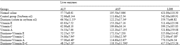
In this study, the protective effect of vitamins C, E and A on rat liver enzyme activity (ALT, AST, LDH) in the presence of diazinon exposure was studied. The results are shown in Table 1. The activity of ALT in the diazinon group was significantly higher (66.5±11.35 IU L-1) than that observed in the control group (52.66±8.144 IU L-1), however, the diazinon/vitamin E, A, C group displayed significant reduction to the diazinon group. The highest ALT activity was observed in the diazinon group (66.5±11.35 IU L-1), while the lowest was observed in the diazinon/vitamin C+E group (48.25±3.2 IU L-1) (p<0.001). A statistically significant AST activity difference was observed in the diazinon group (122.25±17.34 IU L-1) relative to the normal saline treated group (105.5±5.06 IU L-1). The diazinon/vitamin A, C and C+E group displayed significant relative to the diazinon group. The highest levels of AST enzyme activity were unexpectedly observed in the diazinon/vitamin E group (172.33±7.23 IU L-1). Significant reduction in LDH enzyme activity were observed in the diazinon group (359.75±66.82 IU L-1) compared to the control group (542±169.91 IU L-1) and overall, significant changes in the vitamin groups were observed compared to the diazinon group. A Tukey test showed that LDH enzyme activity in the dazinon/vitamin A group (521±259.08 IU L-1) was significantly increased compared to the diazinon group. The lowest level of LDH enzyme activity was observed in the dazinon/vitamin C (272±47.08 IU L-1) group and this was statistically lower than the diazinon group.
This study investigated the activity of liver enzymes in male rats chronically exposed to diazinon and the role of vitamins A, E and C in the reduction of diazinon toxicity. The results showed a significant difference in ALT enzyme activity in the diazinon group compared with the normal control group. The current study was conducted under chronic contact for over 14 days. It is possible that glutathione (GSH) acts as an antioxidant against free radicals at first, but considering the lapse, rats adapted to the various toxin levels, as GSH increased to reduce toxicity; in the diazinon/vitamin groups, diazinon may have promoted the regeneration of GSH, thus, reducing toxin levels and ALT activity.
| Table 1: | Comparison of the effects of diazinon and vitamins A, E and C on rat liver enzymes in the studied groups |
 | |
| ALT: Alanine transaminase, AST: Aspartate aminotransferase, LDH: Lactate dehydrogenase, aValues are the Mean±SD for each group of 5 mice, ap<0.001 compared to the normal control, bp<0.001 compared with the diazinon treated group, cp<0.05 compared with the diazinon treated group | |
Vitamin E/Diazinon significantly increased AST enzyme activity compared with Diazinon (p <0.01). One explanation for this is that when toxic compounds reach the body, the body’s natural antioxidants (GSH) try to clear the poison and repair enzyme activity. It is possible that GSH was unable to completely neutralize diazinon-related toxicity and vitamin E as an antioxidant acted synergistically with GSH, but the toxicities resulting from chronic diazinon contact caused AST levels to increase in the serum even in the presence of vitamin E. The LDH levels observed in the diazinon group were not significantly increased compared to the control group. This may be because chronic contact to diazinon may cause reproduce glutathione, decrease toxin and fix LDH activity. In the diazinon/vitamin A group, LDH activity increases significantly more than that of diazinon group (p<0.001). This may be because Vitamin A decreases diazinon liver damage and because freed liver enzyme in blood stream and liver enzyme produced by gene, too. The results of this study are consistent with the results of other studies (Karakilcik et al., 2004; Yousef et al., 2003; El-Shenawy et al., 2009). However, a few studies have found contradictory results (Gokcimen et al., 2007; Salehi and Jafary, 2010; Kalender et al., 2005; Etim et al., 2006). One study showed the effect of different doses of diazinon on changes in LDH and AST levels (Gokcimen et al., 2007). Another study found that LDH activity was reduced 24 h after the injection of diazinon and paraoxon due to a failure of the antioxidant defense system to protect against free radicals and tissue oxidative damage. (Salehi and Jafary, 2010). Increased serum concentrations of liver enzymes and changes in antioxidant levels were reported to result from chlorpyrifos and sipermetrien in mice (Khan et al., 2005). Lambda-cyhalothrin (LTC) significantly decreased LDH activity in rat brains (Fetoui et al., 2008). After Lindane and diazinon intoxication, LDH levels were shown to increase (Kalender et al., 2005; Etim et al., 2006). Klervos organophosphates created different changes in plasma LDH levels depending on dose and time (Petroianu et al., 2006). Significant plasma AST reductions in carp following acute diazinon toxicity have been reported (Luskova et al., 2002; Sastry and Sharma, 1980). In addition, it has been found that vitamin E partially counteracted the toxic effect of DZN and repaired tissue damage in the liver and kidney (El-Shenawy et al., 2009). The presence of ascorbic acid (vitamin C) may also diminish the adverse effects of Aflatoxin B1 (AFB1) on most hematological and biochemical values and enzymatic activities in rabbits (Yousef et al., 2003). Another study found that AFB1 affected some liver enzymes and other biochemical parameters, but that vitamins C, E and C+E partially prevented an increase in these liver enzymes and biochemical parameters that were induced by AFB1 (Karakilcik et al., 2004).
CONCLUSION
The liver is one of the main tissues affected by the poison. With regard to vitamin levels, changes in the related enzyme levels were observed. The activities of ALT and AST in the diazinon group were significantly higher than that observed in the control group; however, the diazinon/vitamin E, A, C group displayed significant reduction in ALT and AST activities compared to the diazinon group. LDH enzyme activity decreased significantly in the diazinon/vitamin C group compared to the diazinon-exposed group. Differences in chemical composition, the species studied and dose and exposure time vary across the studies. Variations in the chemical structure of organophosphates and their differential effects on various tissues necessitate additional studies to understand the mechanisms of action of this compound.
REFERENCES
- Abbasnejad, M., M. Jafari, A. Asgari, R. Hajihosseini, M. Hjighalamali, M. Salehi and M. Salimian, 2009. Acute toxicity effect of diazinon on antioxidant system and lipid peroxidation in kidney tissues of rats. J. Shahed Univ., 17: 35-42.
Direct Link - Akturk, O., H. Demirin, R. Sutcu, N. Yilmaz, H. Koylu and I. Altuntas, 2006. The effects of diazinon on lipid peroxidation and antioxidant enzymes in rat heart and ameliorating role of vitamin E and vitamin C. Cell Biol. Toxicol., 22: 455-461.
CrossRefPubMedDirect Link - Civen, M., C.B. Brown and R.J. Morin, 1977. Effects of organophosphate insecticides on adrenal cholesteryl ester and steroid metabolism. Biochem. Pharmacol., 26: 1901-1907.
CrossRef - Coppage, D.L. and E. Mattews, 1974. Short-term effects of organophosphate pesticides on cholinesterases of estuarine fishes and pink shrimp. Bull. Environ. Contam. Toxicol., 11: 483-488.
CrossRef - Das, B.K. and S.C. Mukherjee, 2000. Chronic toxic effects of quinalphos on some biochemical parameters in Labeo Rohita. Toxicol. Lett., 114: 11-18.
CrossRef - Dutta, H.M. and L.B. Maxwell, 2003. Histological examination of sub lethal effect of diazinon on ovary of bluegill, Lepomis Macrochirus. Environ. Pollut., 121: 95-102.
PubMed - El-Shenawy, N.S., R.A. Al-Eisa, F. El-Salmy and O. Salah, 2009. Prophylactic effect of vitamin E against hepatotoxicity, nephrotoxicity, haematological indices and histopathology induced by diazinon insecticide in mice. Curr. Zool., 55: 219-226.
Direct Link - Etim, O.E., E.O. Farombi, I.F. Usoh and E.J. Akpan, 2006. The protective effect of aloe vera juice on lindane induced hepatotoxicity and genotoxicity. Pak. J. Pharm. Sci., 19: 337-340.
PubMed - Fattahi, E., K. Parivar, S.G.A. Jorsaraei and A.A. Moghadamnia, 2009. The effects of diazinon on testosterone, FSH and LH levels and testicular tissue in mice. Iran J. Rep. Med., 7: 59-64.
Direct Link - Fetoui, H., E.M. Garoui, F. Makni-Ayadi and N. Zeghal, 2008. Oxidative stress induced by lambda-cyhalothrin (LTC) in rat erythrocytes and brain: Attenuation by vitamin C. Environ. Toxicol. Pharmacol., 26: 225-231.
CrossRefDirect Link - Uboh, F.E., I.S. Ekaidem, P.E. Ebong and I.B. Umoh, 2009. The hepatoprotective effect of vitamin A against gasoline vapor toxicity in rats. Gastroenterol. Res., 2: 162-167.
Direct Link - Gokcimen, A., K. Gulle, H. Demirin, D. Bayram, A. Kocak and I. Altuntas, 2007. Effects of diazinon at different doses on rat liver and pancreas tissues. Pestic. Biochem. Physiol., 87: 103-108.
CrossRefDirect Link - Jurczuk, M., M.M. Brzَska and J. Moniuszko-Jakoniuk, 2007. Hepatic and renal concentrations of Vitamins E and C in lead- and ethanol-exposed rats. An assessment of their involvement in the mechanisms of peroxidative damage. Food Chem. Toxicol., 45: 1478-1486.
Direct Link - Kalender, S., A. Ogutcu, M. Uzunhisarcikli, F. Acikgoz, D. Durak, Y. Ulusoy and Y. Kalender, 2005. Diazinon-induced hepatotoxicity and protective effect of vitamin E on some biochemical indices and ultrastructural changes. Toxicology, 211: 197-206.
CrossRefDirect Link - Kalender, S., Y. Kalender, D. Durak, A. Ogutcu, M. Uzunhisarcikli, B.S. Cevrimli and M. Yildirim, 2007. Methyl parathion induced nephrotoxicity in male rats and protective role of vitamins C and E. Pestic. Biochem. Physiol., 88: 213-218.
CrossRef - Karakilcik, A.Z., M. Zerinm, O. Arslanm, Y. Nazligul and H. Vural, 2004. Effects of vitamin C and E on liver enzymes and biochemical parameters of rabbits exposed to aflatoxin B1. Vet. Hum. Toxicol., 46: 190-192.
PubMedDirect Link - Zaidi, S.M. and N. Banu, 2004. Antioxidant potential of vitamins A, E and C in modulating oxidative stress in rat brain. Clin. Chim. Acta, 340: 229-233.
CrossRefPubMedDirect Link - Khan, S.M., R.C. Sobti and L. Kataria, 2005. Pesticide-induced alteration in mice hepato-oxidative status and protective effects of black tea extract. Clin. Chim. Acta, 358: 131-138.
CrossRefPubMedDirect Link - Luskova, V., M. Svoboda and J. Kolarova, 2002. The effect of diazinon on blood plasma biochemistry in carp (Cyprinus carpio L.). Acta Veterinaria Brno, 71: 117-123.
Direct Link - Oliveira- Silva, J.J., S.R. Alves, A. Meyer, F. Perez, P.N. Sarcinelli, R.C. da Costa Mattos and J.C. Moreira, 2001. Influence of socioeconomic factors on the pesticides poisoning, Brazil. Rev. Saude Publica, 35: 130-135.
PubMed - Sarabia, L., I. Maurer and E. Bustos-Obregon, 2009. Melatonin prevents damage elicited by the organophosphorous pesticide diazinon on mouse sperm DNA. Exotoxicol. Environ. Saf., 72: 663-668.
CrossRefPubMedDirect Link - Sastry, K.V. and S.K. Sharma, 1980. Diazinon effect on the activities of brain enzymes from Ophiocephalus (Channa) punctatus. Bull. Environ. Contam. Toxicol., 24: 326-332.
CrossRef - Vittozzi, L., L. Fabrizi, E. di Consiglio and E. Testai, 2001. Mechanistic aspects of organophosphorothionate toxicity in fish and humans. Environ. Int., 26: 125-129.
CrossRef - Wu, H.X., C. Evreux-Gros and J. Descotes, 1996. Diazinon toxicokinetics, tissue distribution and anticholinesterase activity in the rat. Biomed. Environ. Sci., 9: 359-369.
PubMed - Yousef, M.I., M.H. Salem, K.I. Kamel, G.A. Hassan and F.D. El-Nouty, 2003. Influence of ascorbic acid supplementation on the haematological and clinical biochemistry parameters of male rabbits exposed to aflatoxin B1. J. Environ. Sci. Health B, 38: 193-209.
PubMed