
Mariam A. Abu-Al-Basal
Department of Biological Sciences, Faculty of Science, Al-al-Bayt University, P.O. Box 130040, Mafraq 25113, Jordan
Pakistan Journal of Biological Sciences
Year: 2012 | Volume: 15 | Issue: 7 | Page No.: 306-315


ABSTRACT
Dead Sea (DS) mud and salts are known for their therapeutic and cosmetic properties. Previous studies confirmed their efficacy in treating the more frequent skin diseases such as psoriasis and atopic dermatitis. Therefore, this study aimed to evaluate the wound healing potential of natural and compounded skin-care product (facial mask) of DS black mud in BALB/c mice. Two full-thickness excision round wounds were created on the dorsum region of mouse. Each wound of mice test group were treated topically with 50 μL of 0.1% natural or compounded DS black mud or 50 μL of 0.2% nitrofurazone once a day for 2 consecutive days and the mice control group were left untreated. Healing was assessed by measuring the granulation tissue weight, percentage of wound contraction at day 3, 7, 14 and 21 after wounding. In addition to period of epithelialization and histological evaluation of the regenerated wound area at day 7 and 14 after wounding. Results revealed that DS black mud accelerate wound healing process by enhancing granulation, wound contraction, epithelialization, angiogenesis and collagen deposition. This may be due to high content of minerals and trace elements that possibly act as anti-microbial, anti-inflammatory and antioxidant with enhancement effect on cell proliferation, migration and fibroblast cellular activity. However, the healing property of DS black mud compounded in skin-care product was greater than that of natural black mud, when compared to reference drug, nitrofurazone.
PDF Abstract XML References Citation
Received: March 03, 2012;
Accepted: May 24, 2012;
Published: August 01, 2012
How to cite this article
Mariam A. Abu-Al-Basal, 2012. Histological Evaluation of the Healing Properties of Dead Sea Black Mud on Full-thickness Excision Cutaneous Wounds in BALB/c Mice. Pakistan Journal of Biological Sciences, 15: 306-315.
DOI: 10.3923/pjbs.2012.306.315
URL: https://scialert.net/abstract/?doi=pjbs.2012.306.315
DOI: 10.3923/pjbs.2012.306.315
URL: https://scialert.net/abstract/?doi=pjbs.2012.306.315
INTRODUCTION
Wound is a disruption of cellular, anatomical and functional continuity of living tissue. It may be produced by physical, chemical, thermal, microbial, or immunological insult to the tissue (Ayello, 2005). Wound healing is the interaction of a complex cascade of cellular and biochemical actions aiming to remove damaged tissues and/or invaded pathogen from the body as well as to restore structural and functional integrity of injured tissues (Matsuda et al., 1998; Rozaini et al., 2005). It involves continuous cell-cell and cell-matrix interactions that allow the process of healing to proceed in different overlapping phases including inflammation, wound contraction, epithelialization, granulation, angiogenesis and remodeling of extra-cellular matrix (Eming et al., 2007; Abu-Al-Basal, 2010). However, the normal progress of these interconnected phases may disturb due to exogenous and/or indigenous factors, such as microbial infection, insufficient vascularization, oxidative stress, malnourishment, diabetes and immunocompromised conditions. That may delay or complicate the healing process leading to the development of pathological conditions as ulceration, necrosis and hypertrophic scarring (Martin, 1997; Subramanian et al., 2006; Abu-Al-Basal, 2009).
The Dead Sea (DS) is the lowest point on earth, at 422 m below Sea level. It is located in the Rift Valley between Jordan and Israel and considered the saltiest one among all the hyper saline lakes of the world (Momani et al., 2009). The unique climatic conditions in the DS area make it a famous site in the world for climatotherapy, which is a natural approach in providing cures for many human ailments. These include unusual solar irradiation, oxygen-rich, bromine-rich haze over the sea, exclusive salt composition of the waters, thermal mineral springs and a special natural mud (Oumeish, 1996; Moses et al., 2006). The healing properties of DS have been attributed to its rich salt content and abundance of minerals such as magnesium, calcium, potassium and bromine. Hence, thousands of tourists come every year to the DS not only to enjoy the beauty and quality of the area but more importantly to cover their body with its mud and salts that have been recognized from ancient times in enhancing life and treating rheumatic and skin diseases (Halevy et al., 2001; Wolf et al., 2003).
The DS mud is black in color and rich in salts and minerals that are absorbed from the water. It is mined from the DS shores and extensively used as a raw material in skin care products marketed worldwide including, mud packs, masks, facial and body treatments (Ma’or et al., 2006). The DS black mud and its derivative products are proved to be safe for the consumer with no toxic elements present at elevated levels of concern (Abdel-Fattah and Pingitore, 2009). In addition, several studies verified the therapeutic properties of the DS climatotherapy for the more frequent skin diseases such as psoriasis, eczema and atopic dermatitis (Gambichler et al., 2000; Hodak et al., 2003; Hristakieva, 2005; Ingber, 2006). Therefore, climatotherapy at the DS and its natural products becoming progressively more important as part of alternative modes of therapy among people throughout the world, due to the rising costs of conventional medicine and the public's higher awareness regarding its possible toxic side effects.
The therapeutic effects of the DS climatotherapy on a number of skin diseases and the extensive use of DS natural products in skin care convince searching a new medicinal target as wound healing. Hence, the present study aimed to assess the healing properties of DS black mud at different aspects of cutaneous wound healing process in BALB/c mice.
MATERIALS AND METHODS
Dead Sea black mud: This study concerns in evaluating the healing properties of two samples of DS black mud. The first is a natural sample collected in summer of 2011 from 5 cm deep at the mud/water interface in the eastern shore of the DS region, Jordan. The second sample is compounded in skin-care product (facial mask) made mainly of natural DS black mud in combination with selected plant extracts and vitamin E (Glory Company for Dead Sea quality products, Jordan). The collected natural sample was directly carried to the laboratory and stored at 4°C in a refrigerator for further use.
Animals: Male BALB/c mice six week of age (18-20 g) were obtained from the animal house of the Department of Biological Sciences, Yarmouk University, Irbid, Jordan. Mice were kept under specific pathogenic-free conditions, housed, fed and treated in accordance with the international guidelines principles of laboratory animal use and care (Hedrich and Bullock, 2006). They were maintained on standard pellet diet and water ad libitum for two weeks to be acclimatized prior to the investigation.
Wounding: Mice were anaesthetized with 350 mg kg-1 b.wt. of chloral hydrate (Scharlu chemie, S.A., Barcelona, Spain) via intraperitoneal injection. The dorsal surface of mice was shaved, cleaned with 70% ethanol and 2 full-thickness excision round skin wounds (4 mm diameter) were created on the same mouse along the dorsal middle line using sterile biopsy punch equipment (Revolving punch pliers, Germany). The 2 wounds were separated from each other by at least 1 cm of unwounded skin. The wounds were left open without any dressing material for the duration of the study (Gutierrez-Fernandez et al., 2007).
Mice grouping and treatment: Mice were randomly divided into control and experimental groups of fifteen mice each. Various doses of DS black mud were prepared and preliminary tested for their tolerance in BALB/c mice to select the optimum dose intended for the treatment of experimental animals. Treatments were prepared immediately before use and applied topically at the surface of wound once a day for 2 consecutive days after wounding. Group I: Mice left without treatment, as normal control. Group II: Mice treated with 50 μL of 0.2% nitrofurazone (reference drug)/excision wound, as positive control. Group III: Mice treated with 50 μL of 0.1% natural black mud/excision wound. Group IV: Mice treated with 50 μL of 0.1% compounded black mud/excision wound. Mice were individually housed, maintained on normal food and water ad libitum and those which showed infection signs were separated and excluded from the study.
Granulation tissue: At day 3, 7, 14 and 21, the granulation tissue that was formed on the wounds of mice was excised and the wet weight was recorded.
Wound contraction: Mice were photographed at the time of wounding (0 day) before treatment and again at day 3, 7, 14 and 21 after wounding. The wound surface area was measured from the traced outline of a digital image of the wound by planimetry as described by Flanagan (2003). The percentage of wound contraction was calculated using:
where, A0 is the original wound area and At is the area of wound at specific time period after wounding (Yates et al., 2007).
Epithelialization time: The period of epithelialization was calculated as the number of days required for falling of the dead tissue remnants without any residual raw wound (Nayak et al., 2006).
Histology: Skin samples from each group were obtained at day 7 and 14 after wounding. Samples were dissected, fixed in 10% neutral formalin, dehydrated in ascending grades of alcohol and imbedded in paraffin wax. Five-micrometer thick sections were stained either with hematoxylin and eosin for general histological analysis or Masson's trichrome to assess collagen content and maturation within the dermis. Histological sections of cutaneous wound site were qualitatively evaluated for the following criteria: intensity of inflammation, the extent of epithelialization, maturation and organization of the epidermal layers, regeneration degree of granulation tissue, angiogenesis/neovascularization and amount of collagen deposition and pattern of arrangement in the dermis.
Statistical analysis: Results are expressed as Means±SEM (Standard Error of the Mean). Comparisons between groups were performed by using paired student's t-test on a statistical software package SPSS. Differences were considered significant, if P value is less than 0.05.
RESULTS
Granulation tissue: DS black mud-treated mice showed significant increase in the granulation tissue wet weight at various days after wounding (3, 7, 14 and 21), when compared to those of control groups. The most significant increase in the granulation tissue weight was observed in compounded black mud-treated mice (Group IV: 6.53±0.16, 7.42±0.46), at day 14 and 21, respectively. This proved to be more effective than the reference drug, nitrofurazone (Group II: 5.45±0.31, 6.65±0.51) in enhancing regeneration of granulation tissue (Table 1).
Wound contraction: Significant progress in the percentage of wound contraction was observed in the DS black mud-treated wounds compared with the untreated and nitrofurazone-treated wounds (Table 2). Specifically, at day 7 and 14, both natural and compounded black mud had significant enhancement in the percentage of wound contraction. However, the wound contracting ability of the later (Group IV: 58.50±0.78, 91.23±1.18) was significantly greater than that of the reference drug, nitrofurazone (Group II: 42.25±1.63, 84.20±1.14).
| Table 1: | Influence of Dead Sea black mud on the granulation tissue weight (mg) formed at various days after wounding |
 | |
| Data are Mean±SEM for 6 excision wounds in each group. Values are statistically significant when compared to *Normal control group (I) at p<0.05 and **Nitrofurazone-treated group (II) at p<0.05 | |
| Table 2: | Influence of Dead Sea black mud on the percentage of wound contraction at various days after wounding |
 | |
| Data are Mean±SEM for 6 excision wounds in each group. Values are statistically significant when compared to *Normal control group (I) at p<0.05 and **Nitrofurazone-treated group (II) at p<0.05 | |
 | |
| Fig. 1: | Influence of Dead Sea black mud on epithelialization time of regenerated excision wounds. Data are Mean±SEM for 6 excision wounds in each group. Values are statistically significant when compared to aNormal control group (I) at p<0.05 and bNitrofurazone-treated group (II) at p<0.05 |
Epithelialization time: Faster rate of epithelialization was detected in the DS black mud-treated wounds when compared with the mice control groups. The epithelialization time was significantly reduced from 15.1 days (Group I) to 10.5 (Group III) and 10 days (Group IV) in black mud-treated wounds. DS black mud appeared to accelerate the rate of epithelializiaton more than that of the reference drug, nitrofurazone (Group II: 14.2 day) (Fig. 1).
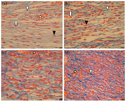
Histological evaluation: Histological analysis of healed wound area, at day 7 and 14 after wounding, revealed marked progress in the healing process of wounds treated with DS black mud compared with those of the controls (Fig. 2-6). At day 7 after wounding, immature and disorganized epidermis with debridement crust overlying the area of the wound was observed in all groups. However, natural and compounded black mud-treated wounds exhibited more advancement in the process of full-thickness epidermal regeneration than that of the controls. In which, a slim layer of dislocate and scantily formed epithelial cells covered the wound area, indicating slow rate of epithelialization (Fig. 2). Cell proliferation, collagen deposition and angiogenesis are important events in the development of granulation tissue. These are clearly detected in black mud-treated wounds that displayed considerable increase in the granulation tissue loaded with fewer inflammatory cells, discernible amount of collagen fibers with fibroblast proliferation and marked dense blood vessels distributed deeply in the tissue. In contrast, scarce fibroblasts, presence of more inflammatory cells, deposition of weak scaffold of collagen fibers and reduced numbers of newly formed blood vessels located mostly at the tissue surface reveal a delay in the progress of granulation tissue development in untreated wounds compared to those treated with the reference drug, nitrofurazone (Fig. 2, 3).
At day 14 after wounding, further progressing in on-going epithelialization and granulation was observed in all treated wounds compared to untreated control (Fig. 4-6). However, both natural and compounded black mud revealed substantial enhancement in the epidermal and dermal architecture of regenerated wounds compared to reference drug. These include: no debridement crust covering the epidermal surface, keratinization, well-developed epidermis with organized tissue layers enclose completely the wound area, full-regenerated granulation tissue loaded with large numbers of blood vessels having various sizes and thick, uniform, compact and regularly arranged collagen fibers. In addition, Masson's trichrome stain revealed that compounded black mud enhance collagen deposition greater than natural black mud due to the increase in activity of fibroblasts (Fig. 6). In which, more distinct, thick, densely associated and well-organized collagen fibers/bands were found with intensely thin-walled blood vessels distributed throughout the granulation tissue.
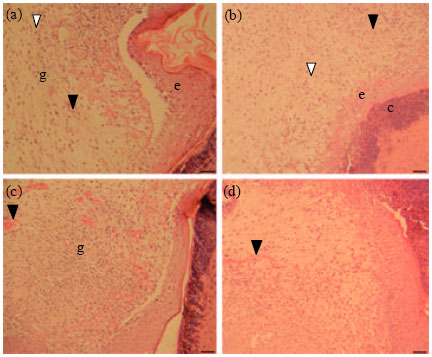 | |
| Fig. 2(a-d): | Hematoxylin and eosin staining histological sections of cutaneous wound site of BALB/c mice, revealing epidermal and dermal architecture of healed wounds at day 7 after wounding. (a) Untreated normal wound; (b) Nitrofurazone-treated wound; (c) Natural black mud-treated wound; (d) Compounded black mud-treated wound. e: Immature epidermis with debridement crust (c) overlying the area of the wound; g: Immature granulation tissue area; Invasion of inflammatory cells (white triangular); Blood vessels (black triangular). Scale bar: 50 μm |
 | |
| Fig. 3(a-d): | Masson’s trichrome staining histological sections of cutaneous wound site of BALB/c mice, revealing dermal architecture of regenerated granulation tissue at day 7 after wounding. (a) Untreated normal wound; (b) Nitrofurazone-treated wound; (c) Natural black mud-treated wound; (d) Compounded black mud-treated wound. Deposition of newly formed collagen fibers (white triangular); Fibroblasts (black triangular); Inflammatory cells (white arrow); Blood vessels (white star). Scale bar: 200 μm |
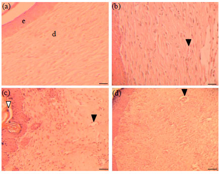 | |
| Fig. 4(a-d): | Hematoxylin and eosin staining histological sections of cutaneous wound site of BALB/c mice, revealing epidermal and dermal architecture of healed wounds at day 14 after wounding. (a) Untreated normal wound; (b) Nitrofurazone-treated wound; (c) Natural black mud-treated wound; (d) Compounded black mud-treated wound. e: Epidermis; d: Dermis rich with newly formed granulation tissue; Blood vessels (black triangular); keratinization (white triangular). Scale bar: 50 µm |
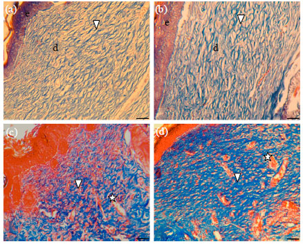 | |
| Fig. 5(a-d): | Masson’s trichrome staining histological sections of cutaneous wound site of BALB/c mice, revealing epidermal and dermal architecture of healed wounds at day 14 after wounding. (a) Untreated normal wound; (b) Nitrofurazone-treated wound; (c) Natural black mud-treated wound; (d) Compounded black mud-treated wound. e: Epidermis; d: Dermis rich with newly formed collagen fibers (white triangular); Blood vessels (white star). Scale bar: 50 µm |
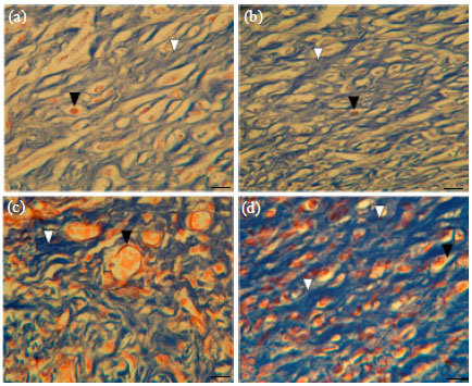 | |
| Fig. 6(a-d): | Masson’s trichrome staining histological sections of cutaneous wound site of BALB/c mice, revealing dermal architecture of regenerated granulation tissue at day 14 after wounding. (a) Untreated wound; (b) Nitrofurazone-treated wound; (c) Natural black mud-treated wound; (d) Compounded black mudtreated wound. Collagen fibers/bands (white triangular); Bood vessels (black triangular). Scale bar: 200 µm |
In contrast, untreated wounds had incomplete maturation of epidermal layers, few fibroblasts, less obvious areas of blood vessels and light stain, thin, less compact, coarse and irregularly arranged collagen fibers (Fig. 5, 6).
DISCUSSION
Results of this study revealed that DS black mud promotes healing of cutaneous full-thickness excision wounds in BALB/c mice by influencing different aspects of the healing process at various days after wounding. This was demonstrated by significant increase in the granulation tissue weight (Table 1), percentage of wound contraction (Table 2), decrease in period of epithelialization (Fig. 1) and marked enhancement of cell and tissue proliferation, collagen deposition and angiogenesis/neovascularization (Fig. 2-6), when compared to control groups. The quicker process of wound healing in black mud-treated wounds could be a function of either individual or synergistic effects of bioactive constituents on one phase or more of the healing process. In fact, the normal subsequent events of healing cutaneous wounds can be classified into three overlapping phases; coagulation and inflammation, proliferation and remodeling (Eming et al., 2007). Coagulation and inflammation begin immediately after the injury and characterized by vasoconstriction, platelet aggregation to induce blood clotting and migration of inflammatory cells to the wound site (Tas et al., 2011). These cells phagocyte necrotic tissue and microorganisms and secrete several cytokines and growth factors to stimulate the development of proliferation phase of healing. That comprises of three events: granulation, contraction and epithelialization (Nagori and Solanki, 2011). However, in the remodeling phase, the wound tensile strength increase due to intermolecular cross-linking of collagen and reorganization, granulation tissue evolve into scar tissue and cells that are no longer needed are removed by apoptosis (Abu-Al-Basal, 2010). Histological evaluation of black mud-treated wounds verified marked progress in the development of proliferation phase of healing, especially at day 14 after wounding, when compared to nitrofurazone-treated and untreated wounds (Fig. 4-6). This may be due to reduced inflammation noticed in the regenerated granulation tissue day 7 after wounding (Fig. 2, 3). Which was high-lighted by fewer inflammatory cells, more fibroblasts, large numbers of new blood vessels and perceptible amount of deposited collagen fibers (Fig. 3). Inflammation is the initial phase of healing and the inflammatory response should occur rapidly to permit the development of subsequent phases of healing damaged tissue (Abu-Al-Basal, 2011). Hence, enhancing early the inflammatory phase in treated wounds may play a key role in speed up regeneration of granulation tissue (Table 1), wound contraction (Table 2) and epithelialization (Fig. 1). That could be attributed to anti-microbial (Ma’or et al., 2006) and/or anti-inflammatory properties (Proksch et al., 2005) of DS salts and minerals.
Granulation tissue is primarily composed of endothelial cells, macrophages fibroblast and the components of a new provisional extracellular matrix (ECM), including collagen (Prasad and Dorle, 2006). High densities of new blood vessels loaded in the granulation tissue of treated wounds (Fig. 3, 5) could be a result of DS mineral's capability to induce proliferation and migration of vascular endothelial cells (Subramanian et al., 2006). Complete wound closure with full epithelialization (Fig. 4, 5) concur with significant increase in the percentage of wound contraction (Table 2) and reduction in epithelialization time (Fig. 1), may be related to enhanced proliferation and migration of epidermal cells and activity of fibroblasts in the regenerated granulation tissue (Ajlia et al., 2010). Myofibroblasts are believed to have a basic role in wound contraction by exerting tension on the surrounding ECM and secreting collagen to stabilize the contraction (Abu-Al-Basal, 2001). Actually, collagen provides strength and integrity to the dermis and hence the synthesis, secretion and subsequent organization of collagen play an integral part in the wound healing process (Yusufoglu and Alqasoumi, 2011). This is clearly demonstrated by marked increase in the amount of collagen fibers deposited, especially in the highly vascular dermis of compounded black mud-treated wounds (Fig. 6). Enhancing quickly vascularization and collagen synthesis and deposition in treated wounds confirmed the synergistic stimulatory effect of DS salts and minerals on angiogenesis, cell proliferation and fibroblast cellular activity.
The DS black mud is a homogeneous mixture of salts, minerals and organic materials retrieved from the shoreline. Chemical analysis of DS black mud samples collected from different locations in Jordan revealed wide range of components including sodium, magnesium, calcium, potassium, chlorides, zinc, iron, iodine, copper, manganese and barium (Khlaifat et al., 2010). These indicate that either natural or compounded black mud accelerate the healing process of excision wounds by providing vital micronutrients required for the regeneration of damaged tissue. Several nutritional factors are known to be essential for sequential events of the healing process, for instance vitamins, building amino acids and minerals (Puratchikody and Nagalakshmi, 2007; Khorshid et al., 2010). The high content of the later in DS black mud might be responsible for the significant stimulatory healing effect observed in treated wounds.
Trace elements especially Zinc (Zn) and Copper (Cu), have important roles in human growth and development, immune function and essential in the wound healing process (Soni et al., 2010). Zinc is a component of hundreds of enzymes, including those involved in collagen synthesis and cell proliferation. Zinc deficiency is linked with delayed healing of wounds and ulcers and lower breaking strength in incisions wounds (Norfarizan-Hanoon et al., 2009). It is required for the production of superoxide dismutase; a powerful skin antioxidant and acts as co-factor of certain metalloproteinases that remove damaged tissue to allow cell migration, proliferation and angiogenesis in the healing process (MacDonald, 2000). Similarly, copper is involved in the metabolism of protein within the body. It is also essential to the absorption of iron and plays a direct role in the growth of new blood vessels by exerting angiogenic effects through inducing proliferation of endothelial cells (Hu, 1998). Furthermore, histological analysis of copper-treated wound margin revealed enhancing in wound closure, more hyper-proliferative epithelial tissue and higher density of cells in the granulation layer of regenerated wound (Sen et al., 2002). Both zinc-dependent and copper-dependent enzymes are proved to be required for cell proliferation and migration along with matrix remodeling, since they induce metallothionein expression at the wound margin that accelerate epithelialization (Lansdown, 2002a).
Calcium (Ca) is an extracellular regulator and an intracellular modulator of epidermal cell proliferation and migration (Khorshid et al., 2010). Experimental studies on animal models confirmed that elevated calcium level improve healing via enhancing blood clotting and aggregation of platelets at the wound site during haemostatic phase of wound healing. It is also evoked a significant increase in the weight of granulation tissue and wound tensile strength (Lansdown, 2002b). In addition, magnesium (Mg) salts exhibited considerable effect in enhancing skin barrier function, influencing epidermal proliferation and differentiation and reducing inflammation in atopic (eczema) dry skin (Proksch et al., 2005). Findings of this study revealed that minerals and trace elements of DS black mud are involved in many of the complicated events and phases of full-thickness excision wound healing process. In reality, the process of healing is complex and requires coordinated interactions between various cell types in the dermis and epidermis, ECM molecules and growth factors to restore structural and functional integrity of damaged tissues (Lim and Yoo, 2010).
The enhanced capacity of wound healing by either natural or compounded black mud could be explained on the basis of anti-microbial, anti-inflammatory, angiogenic and mitogenic effects, along with enhanced level of anti-oxidant enzymes in the regenerated wound. However, the healing property of compounded black mud was observed to be greater than that of natural mud, when compared to reference drug, nitrofurazone. The former is made mainly of DS natural black mud in combination with selected plant extracts and vitamin E. Hence, better healing effect may be owing to the improved antioxidant status at the wound site as a result of combined effect of compounded black mud constituents. Extracts of many medicinal plants have been reported to possess wound healing activity and found effective in the treatment and management of wounds due to the free radical scavenging action and high content of amino acids required for tissue regeneration (Abu-Al-Basal, 2001; Patil et al., 2009).
CONCLUSIONS
According to experimental results, DS black mud accelerate wound healing process of full-thickness excision wounds in BALB/c mice by enhancing granulation, wound contraction, epithelialization, angiogenesis and collagen deposition. This may be due to high content of minerals and trace elements that possibly act as anti-microbial, anti-inflammatory and antioxidant with enhancement effect on cell proliferation, migration and fibroblast cellular activity. However, the healing property of DS black mud compounded in skin-care product was greater than that of natural black mud, when compared to reference drug, nitrofurazone.
ACKNOWLEDGMENTS
The author acknowledge Wasfi Al-Bekearat for his support and technical assistance, Al-al-Bayt University, Department of Biological Sciences, Mafraq, Jordan, Amani Harb for the help in preparing histological sections, Department of Biological Sciences, Jordan University and Al-al-Bayt University, Mafraq, Jordan, for providing necessary facilities to conduct this study.
REFERENCES
- Abdel-Fattah, A. and N.E. Pingitore, 2009. Low levels of toxic elements in Dead Sea black mud and mud-derived cosmetic products. Environ. Geochem. Health, 31: 487-492.
CrossRef - Abu-Al-Basal, M.A., 2009. In vitro and In vivo anti-microbial effects of Nigella sativa linn. seed extracts against clinical isolates from skin wound infections. Am. J. Applied Sci., 6: 1440-1447.
Direct Link - Abu-Al-Basal, M.A., 2010. Healing potential of Rosmarinus officinalis L. on full-thickness excision cutaneous wounds in alloxan-induced-diabetic BALB/c mice. J. Ethnopharmacol., 131: 443-450.
CrossRefDirect Link - Abu-Al-Basal, M.A., 2011. Influence of Nigella sativa fixed oil on some blood parameters and histopathology of skin in staphylococcal-infected BALB/c mice. Pak. J. Biol. Sci., 14: 1038-1046.
CrossRef - Ajlia, S.A.S.H., F.A.A. Majid, A. Suvik, M.A.W. Effendy and H.S. Nouri, 2010. Efficacy of papain-based wound cleanser in promoting wound regeneration. Pak. J. Biol. Sci., 13: 596-603.
CrossRefDirect Link - Ayello, E.A., 2005. What does the wound say? Why determining etiology is essential for appropriate wound care. Adv. Skin Wound Care, 18: 98-109.
PubMed - Eming, S.A., S. Werner, P. Bugnon, C. Wickenhauser and L. Siewe et al., 2007. Accelerated wound closure in mice deficient for interleukin-10. Am. J. Pathol., 170: 188-202.
CrossRef - Flanagan, M., 2003. Improving accuracy of wound measurement in clinical practice. Ostomy Wound Manage., 49: 28-40.
PubMed - Gutierrez-Fernandez, A., M. Inada, M. Balbin, A. Fueyo and A.S. Pitiot et al., 2007. Increased inflammation delays wound healing in mice deficient in collagenase-2 (MMP-8). FASEB J., 21: 2580-2591.
CrossRef - Halevy, S., H. Giryes, M. Friger, N. Grossman, Z. Karpas, B. Sarov and S. Sukenik, 2001. The role of trace elements in psoriatic patients undergoing balneotherapy with Dead Sea bath salt. Isr. Med. Assoc. J., 3: 828-832.
PubMedDirect Link - Hodak, E., A.B. Gottlieb, T. Segal, Y. Politi, L. Maron, J. Sulkes and M. David, 2003. Climatotherapy at the Dead Sea is a remittive therapy for psoriasis: Combined effects on epidermal and immunologic activation. J. Am. Acad. Dermatol., 49: 451-457.
CrossRef - Hristakieva, E., 2005. Climatotherapy in dermatology: Why, how and when? Trakia J. Sci., 3: 27-31.
Direct Link - Hu, G.F., 1998. Copper stimulates proliferation of human endothelial cells under culture. J. Cell Biochem., 69: 326-335.
CrossRefPubMedDirect Link - Khlaifat, A., O. Al-Khashman and H. Qutob, 2010. Physical and chemical characterization of Dead Sea mud. Mater. Charact., 61: 564-568.
CrossRef - Khorshid, F., S.S. Ali, T. Alsofyani and H. Albar, 2010. Plectranthus tenuiflorus (Shara) promotes wound healing: In vitro and in vivo studies. Int. J. Bot., 6: 69-80.
CrossRefDirect Link - Lim, J.S. and G. Yoo, 2010. Effects of adipose-derived stromal cells and of their extract on wound healing in a mouse model. J. Korean Med. Sci., 25: 746-751.
CrossRef - MacDonald, R.S., 2000. The role of zinc in growth and cell proliferation. J. Nutr., 130: 1500S-1508S.
PubMedDirect Link - Ma'or, Z., Y. Henis, Y. Alon, E. Orlov, K.B. Sorensen and A. Oren, 2006. Antimicrobial properties of dead sea black mineral mud. Int. J. Dermatol., 45: 504-511.
CrossRef - Martin, P., 1997. Wound healing: Aiming for perfect skin regeneration. Science, 276: 75-81.
CrossRefPubMedDirect Link - Matsuda, H., H. Koyama, H. Sato, J. Sawada and A. Itakura et al., 1998. Role of nerve growth factor in cutaneous wound healing: Accelerating effects in normal and healing-impaired diabetic mice. J. Exp. Med., 187: 297-306.
CrossRef - Momani, K., T. El-Hasan, S. Auaydeh and K. Al-Nawayseh, 2009. Heavy metals distribution in the dead sea black mud, Jordan. Jordan J. Earth Environ. Sci., 2: 50-59.
Direct Link - Moses, S.W, M. David, E. Goldhammer, A. Tal and S. Sukenik, 2006. The dead sea, a unique natural health resort. Isr. Med. Assoc. J., 8: 483-488.
PubMed - Nagori, B.P. and R. Solanki, 2011. Role of medicinal plants in wound healing. Res. J. Med. Plant, 5: 392-405.
CrossRefDirect Link - Nayak, S., P. Nalabothu, S. Sandiford, V. Bhogadi and A. Adogwa, 2006. Evaluation of wound healing activity of Allamanda cathartica L. and Laurus nobilis L. extracts on rats. BMC Complement. Altern. Med., Vol. 6.
CrossRefDirect Link - Norfarizan-Hanoon, N.A., R. Asmah, M.Y. Rokiah, O. Fauziah and H. Faridah, 2009. Effects of Strobilanthes crispus juice on wound healing and antioxidant enzymes in normal and streptozotocin-induced diabetic rats. J. Biol. Sci., 9: 662-668.
CrossRefDirect Link - Patil, D.N., A.R. Kulkarni, A.A. Shahapurkar and B.C. Hatappakki, 2009. Natural cumin seeds for wound healing activity in albino rats. Int. J. Biol. Chem., 3: 148-152.
CrossRefDirect Link - Prasad, V. and A.K. Dorle, 2006. Evaluation of ghee based formulation for wound healing activity. J. Ethnopharmacol., 107: 38-47.
CrossRef - Proksch, E., H.P. Nissen, M. Bremgartner and C. Urquhart, 2005. Bathing in a magnesium-rich dead sea salt solution improves skin barrier function, enhances skin hydration and reduces inflammation in atopic dry skin. Int. J. Dermatol., 44: 151-157.
CrossRef - Puratchikody, A. and G. Nagalakshmi, 2007. Wound healing activity of Memecylon umbellatum burm. J. Plant Sci., 2: 179-186.
CrossRefDirect Link - Rozaini, M.Z., A.B.Z. Zuki, M.M. Noordin, Y. Norimah and A.N. Hakim, 2005. Macroscopic evaluation of burn wounds healing progress treated with different types of honey. Pak. J. Biol. Sci., 8: 672-678.
CrossRefDirect Link - Sen, C.K., S. Khanna, M. Venojarvi, P. Trikha, E.C. Ellison, T.K. Hunt and S. Roy, 2002. Copper-induced vascular endothelial growth factor expression and wound healing. Am. J. Physiol. Heart Circ. Physiol., 282: H1821-H1827.
CrossRefDirect Link - Soni, A., V.K. Dwivedi, M. Chaudhary, K. Malik, V. Naithani and S.M. Shrivastava, 2010. Plasma cytokines and trace element level in severe burn rat model-with special reference to wound healing potential of ampucare. Res. J. Immunol., 3: 22-30.
CrossRefDirect Link - Subramanian, S., D.S. Kumar and P. Arulselvan, 2006. Wound healing potential of Aloe vera leaf gel studied in experimental rabbits. Asian J. Biochem., 1: 178-185.
CrossRefDirect Link - Tas, A., A. Karasu, B. Comba, D.S. Aksu, E. Duz and P. Tanritanir, 2011. Effects of sildenafil citrate on the hematological parameters in the early phase of wound healing in diabetic rats. Asian J. Anim. Vet. Adv., 6: 290-296.
CrossRefDirect Link - Wolf, R., E. Orion and H. Matz, 2003. Climatotherapy: There is life in the dead sea. Isr. Med. Assoc. J., 5: 124-125.
PubMed - Yates, C.C., D. Whaley, R. Babu, J. Zhang and P. Krishna et al., 2007. The effect of multifunctional polymer-based gels on wound healing in full thickness bacteria contaminated mouse models. Biomaterials, 28: 3977-3986.
CrossRef - Yusufoglu, H.S. and S.I. Alqasoumi, 2011. Anti-inflammatory and wound healing activities of herbal gel containing an antioxidant Tamarix aphylla leaf extract. Int. J. Pharmacol., 7: 829-835.
CrossRefDirect Link