
Abolhassan Shakeri
Department of Radiology, Imam Reza Hospital, Tabriz University of Medical Sciences, Tabriz, Iran
Mohammad Babaei Bazzaz
Department of Radiology, Imam Reza Hospital, Tabriz University of Medical Sciences, Tabriz, Iran
Alireza Khabbazi
Department of Rheumatology, Imam Reza Hospital, Tabriz University of Medical Sciences, Tabriz, Iran
Rohollah Fadaei Fouladi
Department of Radiology, Imam Reza Hospital, Tabriz University of Medical Sciences, Tabriz, Iran
Pakistan Journal of Biological Sciences
Year: 2011 | Volume: 14 | Issue: 16 | Page No.: 812-816


ABSTRACT
This study aimed to evaluate color Doppler sonographic findings in carotid arteries in RA patients under pharmacological treatments and to compare them with normal population. Forty nine patients with late RA and 48 healthy age and sex-matched controls were recruited. The two groups were matched for other known risk factors of atherosclerosis including serum lipid abnormalities, smoking status, diabetes mellitus and hypertension. High resolution B-mode color Doppler ultrasound with a 7 MHz transducer was used for measuring the Common Carotid Intima-Medial Thickness (CCIMT) in both sides in all subjects. Presence of atherosclerotic plaque was also investigated. The mean left and maximum CCIMT was significantly higher in the case group (0.72 vs. 0.62 mm for the left artery; p<0.01; 0.72 vs. 0.64 mm for the maximum reading; p = 0.01). No atherosclerotic plaque was found in common carotid arteries. There were 3 (6.1), 7 (14.3) and 9 (18.4%) plaques in left internal carotid artery, right carotid bulb and left carotid bulb in the case group, respectively with no atherosclerotic plaques in the controls (p = 0.24, 0.01 and <0.001, respectively). Comparing the findings by gender in the case group with the controls, the mentioned significant differences were only between the male patients and the controls. The process of atherosclerosis in RA patients is similar to that in normal population. However, it is apparently accelerated and more advanced in these patients.
PDF Abstract XML References Citation
Received: August 05, 2011;
Accepted: October 14, 2011;
Published: November 24, 2011
How to cite this article
Abolhassan Shakeri, Mohammad Babaei Bazzaz, Alireza Khabbazi and Rohollah Fadaei Fouladi, 2011. Common Carotid Intima-media Thickness in Patients with Late Rheumatoid Arthritis; What Is the Role of Gender?. Pakistan Journal of Biological Sciences, 14: 812-816.
DOI: 10.3923/pjbs.2011.812.816
URL: https://scialert.net/abstract/?doi=pjbs.2011.812.816
DOI: 10.3923/pjbs.2011.812.816
URL: https://scialert.net/abstract/?doi=pjbs.2011.812.816
INTRODUCTION
Rheumatoid Arthritis (RA) is a systemic inflammatory autoimmune disease with an unknown etiology usually presents with small-joint polyarthritis with gradual progression to larger joints. Besides the joints, many other organs may be involved in patients with RA; i.e., it can be called a multisystem disease (Nourmohammadi et al., 2010; Kandil et al., 2007; Shaaban et al., 2006; Haroun, 2004; El-Awady et al., 2007; Ala et al., 2009; Baig et al., 2009; Khan et al., 2011; Deo et al., 2010). It is thought that the prevalence of Coronary Artery Disease (CAD), as well as the cardiovascular mortality may be higher among RA patients comparing with matched normal counterparts (Myllykangas-Luosujarvi et al., 1995; Lowenhoff and Gluszko, 2005). Although, this may be attributed to severity of the disease or long-term pharmacological treatments; the available data are scarce, heterogeneous and inconclusive. Furthermore, possible relationship between these cardiovascular abnormalities and traditional risk factors of the atherosclerosis is not well clarified yet (Wallberg-Jonsson et al., 1997). By the way, majority of available data in this regard are drawn from inappropriate studies due to size and retrospective design. Various studies have proposed that Intima-Media Thickness (IMT) and atherosclerotic plaques in great arteries are good indicators of a more generalized atherosclerosis (Alaee and Khademloo, 2008; Jaarin et al., 2006; Leskinen et al., 2003; Rossi et al., 1996; Mikovanov et al., 2006). This study mainly aimed to compare IMT of common carotid artery (CCIMT) and frequency of atherosclerotic plaques in common carotid artery, internal carotid artery and carotid bulb between patients with late RA and normal controls.
MATERIALS AND METHODS
Study design and patients: In this case-control study, 50 patients with late RA (cases) and 50 healthy individuals (controls) were recruited in Tabriz Educational Imam Reza Hospital in Iran during a 14 month period from April 2010 to June 2011. The study included all patients consecutively admitted to the place of study, if they were fulfilled the 1987 ACR criteria (Arnett et al., 1988) and had a disease duration of more than 2 years and up to 3 years. Finally 49 patients in the case group and 48 subjects in the control group completed this study. Groups were matched for age, gender, menopausal status (in females) and the known risk factors for atherosclerosis, including hypertension, diabetes mellitus, hyperlipidemia and smoking. All patients signed the informed consent form. This study was approved by the ethics committee of Tabriz University of Medical Sciences.
Variables: Hypertension was defined as a systolic blood pressure >140, Diastolic Blood Pressure (DBP) >90 mmHg or a positive history of taking antihypertensive medication. Diabetes mellitus was defined as a fasting plasma glucose >126 mg dL-1 or a positive history of taking diabetic medication. Hypertriglyceridemia was defined as a fasting serum triglyceride level >200 mg dL-1 or a positive history of taking lipid-lowering drugs. Hypercholesterolemia was defined as a fasting total serum cholesterol level >240 mg dL-1 or a positive history of taking cholesterol-lowering drugs. Hypertension was defined as a systolic/diastolic blood pressure >140/90 mmHg. Diabetes mellitus was diagnosed when the fasting blood glucose was >126 mg dL-1. Hypertriglyceridemia and hypercholesterolemia were defined as total serum cholesterol level >250 mg dL-1 and fasting serum triglyceride level >200 mg dL-1, respectively.
Ultrasound evaluation: All subjects were evaluated by a skilled radiologist specialized in Doppler ultrasonography of the vascular system and the CCIMT was calculated. The sonographist was blind to the grouping of the subjects. Sonographic assessment was performed by Aloka ProSound SSD 3500 plus color Doppler machine (Aloka Ltd., Tokyo, Japan) equipped with 7MHz linear array transducer with subjects in the supine position with a slight extension of the neck. All scans were performed by one observer following the method described by Geroulakos et al. (1994). The anterior and lateral projections were employed for imaging the common carotid artery longitudinally. The CCIMT was evaluated at the wall of artery 2 cm proximal to its bifurcation. Internal carotid artery and the bulb of carotid artery were also included in this assessment. The images were zoomed to standard size. The CCIMT was calculated as the mean value of six individual measurements at different points within the region of interest (three for the right and three for the left artery). A plaque was defined as a distinct area with an IMT exceeding twice that of neighboring sites. The CCIMT was calculated in both sides and the maximum reading was also considered as a parameter for evaluation.
Statistical analysis: All descriptive statistics are presented as Mean±Standard deviation and frequency (percent). The differences between the groups regarding the numerical data were assessed using the unpaired t test or Mann-Whitney U-test when appropriate. The differences between the groups regarding the categorical data were assessed using the Chi-square test or the Fisher’s exact test. Correlation was assessed by determining the Pearson’s r. Stepwise logistic regression analysis was used to identify the independent factors among the parameters. Probability values <0.05 were considered statistically significant.
RESULTS
Case and control groups were comparable in terms of characteristics and general data (Table 1). The range of age was 40-86 years in patients and 40-80 years in controls. The duration of RA ranged between 2 and 3 years. History of diabetes mellitus or smoking was negative in all subjects. The mean left and maximum CCIMTs were significantly higher in patients than in controls (0.72±0.21 vs. 0.62±0.10 mm; p<0.01 and 0.72±0.20 vs. 0.64±0.11 mm; p = 0.01, respectively) (Fig. 1). The mean right CCIMT was not significantly different between the two groups (0.69±0.18 mm in the case group vs. 0.64±0.11mm in the control group; p = 0.06) (Fig. 1). There was no atherosclerotic plaque in sonographic evaluation of common carotid and right internal carotid arteries. Atherosclerotic plaques in the left internal carotid artery were present in 3 (6.1%) patients and no controls (p = 0.42). Atherosclerotic plaques in the right and left carotid bulbs were present in 7 (14.3%) and 9 (18.4%) patients and no controls.
 | |
| Fig. 1: | Error bars of mean common carotid intima-media thickness (CCIMT) in patients with rheumatoid arthritis (case) and healthy counterparts (control) |
| Table 1: | Patients’ characteristics, general data and serum laboratory results |
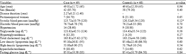 | |
| Data are shown as mean±standard deviation (median) and frequency (percent). *Nonparametric test | |
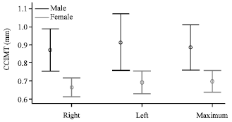 | |
| Fig. 2: | Error bars of mean Common Carotid Intima-Media Thickness (CCIMT) in male and female patients with rheumatoid arthritis |
Frequencies of cases with atherosclerotic plaques in right or left carotid bulbs were significantly higher in patients than controls (p = 0.01 and <0.01, respectively). There was a significant positive correlation between the maximum CCIMT and age of patients (r = 0.47, p<0.01). This correlation was not significant between the CCIMT and duration of RA (r = 0.01, p = 0.67). In the case group, the median left, right and maximum CCIMTs were significantly higher in males than females (0.9 vs. 0.6 mm, p<0.01; 0.9 vs. 0.7 mm, p = 0.01; 0.9 vs. 0.70 mm, p = 0.01, respectively) (Fig. 2). The mean maximum CCIMT was not significantly different between the female patients and the controls (p = 0.08). Frequencies of female patients with atherosclerotic plaques in the right internal carotid artery (2.4%), right carotid bulb (7.1%) or left carotid bulb (7.1%) were not significantly different from those in the controls (p = 0.47, 0.10 and 0.10, respectively). The median maximum CCIMT was significantly higher in the male patients than the controls (nonparametric p<0.001). This difference remained significant after adjusting for other variables including age and the evaluated risk factors for atherosclerosis in Table 1 (p = 0.02). Frequencies of male patients with atherosclerotic plaques in the right internal carotid artery (28.6%), right carotid bulb (57.1%) or left carotid bulb (85.7%) were significantly higher than the controls (p = 0.01, <0.001 and <0.001, respectively).
DISCUSSION
In present study we sonographically determined and compared the mean CCIMT in patients with late RA under appropriate treatment and without symptomatic atherosclerosis and a group of well-matched normal counterparts. Accordingly, the mean maximum CCIMT was significantly higher in the patients (0.72 vs. 0.64 mm). Likewise, frequencies of cases with atherosclerotic plaques in internal carotid artery and bulb of carotid were significantly higher among the patients. These findings are in line with previous reports indicating the mean CCIMT is significantly higher in RA patients comparing with the normal population. The mean CCIMT ranged between 0.63 and 10 mm in RA groups and 0.54 and 0.78 mm in normal population in these reports (Turiel et al., 2009; Kerekes et al., 2008; Cuomo et al., 2004; Hannawi et al., 2007; Carotti et al., 2007). Presence of atherosclerotic plaques has been also reported to be higher in patients with RA (Miasoedova et al., 2009; Pereira et al., 2009). In fact, it is believed by some investigators that the atherosclerosis should be considered as an extra-articular manifestation of RA (Maradit-Kremers et al., 2005; Shoenfeld et al., 2005; Sherer and Shoenfeld, 2006). On the other hand, some studies concluded that there is not a significant difference between RA patients and normal controls in regard to the CCIMT or frequency of atherosclerotic plaques (Diaz et al., 2008; Del Rincon et al., 2003; Jonsson et al., 2001; Park et al., 2002). Summing up the available data, there is not apparently a consensus on this issue in the literature (Van Zanten and Kitas, 2008). This is maybe due to complexity of pathogenesis of atherosclerosis (Georgiadis et al., 2008; Carotti et al., 2007). So for drawing optimal conclusions, we need well-organized and very well-controlled studies in this regard (Sidiropoulos, et al., 2009; Pereira et al., 2008). Although, in the first phase there was a statistically significant preponderance in the mean CCIMT in the patients’ group, reassessment considering the gender of patients showed that this difference is just marginal for the female patients. Schott et al. (2009) also showed that there is no significant difference regarding the CCIMT and the related atherosclerotic plaques between the female RA patients and normal controls. So, it may be concluded that the atherosclerotic process in RA patients is similar to that in normal population but in a more accelerated or at least more progressed fashion. Small sample size of the male patients was the main limitation of current study. Further studies on the male RA patients with enough sample size are recommended.
REFERENCES
- Ala, S., M. Shokrzadeh, A.M.P. Shojah and S.S.S. Saravi, 2009. Zinc and copper plasma concentrations in rheumatoid arthritis patients from a selected population in Iran. Pak. J. Biol. Sci., 12: 1041-1044.
CrossRefPubMedDirect Link - Alaee, A. and M. Khademloo. 2008. Evaluation of correlation between carotid artery intima media wall thickness and coronary artery stenosis in sari, North of Iran. Pak. J. Biol. Sci., 11: 2360-2363.
CrossRefPubMedDirect Link - Arnett, F.C., S.M. Edworthy, D.A. Bloch, D.J. McShane and J.F. Fries et al., 1988. The American rheumatism association 1987 revised criteria for the classification of rheumatoid arthritis. Arthritis Rheum., 31: 315-324.
CrossRefPubMedDirect Link - Baig, M.S., S.M. Humail, S.I. Zaidi, S. Noor, S. Bano, S. Rehman and A. Fawwad, 2009. The efficacy of disease modifying anti-rheumatic drugs in rheumatoid arthritis in local patients of Karachi. Pak. J. Biol. Sci., 12: 339-345.
CrossRefPubMedDirect Link - Carotti, M., F. Salaffi, M. Mangiacotti, A. Cerioni, G.M. Giuseppetti and W. Grassi, 2007. Atherosclerosis in rheumatoid arthritis: The role of high-resolution B mode ultrasound in the measurement of the arterial intima-media thickness. Reumatismo, 59: 38-49.
PubMedDirect Link - Cuomo, G., P. Di Micco, A. Niglio, G. La Montagna and G. Valentini, 2004. Atherosclerosis and rheumatoid arthritis: Relationships between intima-media thickness of the common carotid arteries and disease activity and disabilty. Reumatismo, 56: 242-246.
Direct Link - Deo, S.S., A.R. Chogle, K.J. Mistry, R.R. Shetty and S.B. Londhe, 2010. Pattern of antigen recognition by anti-citrullinated peptide antibodies in undifferentiated arthritis and rheumatoid arthritis. Res. J. Immunol., 3: 157-168.
CrossRef - El-Awady, H.M., A.S. El-Dien El-Wakkad, M.T. Saleh, S.I. Muhammad and E.M. Ghaniema, 2007. Serum melatonin in juvenile rheumatoid arthritis: Correlation with disease activity. Pak. J. Biol. Sci., 10: 1471-1476.
CrossRefPubMedDirect Link - Diaz, Jde D.G., A.L. de Guzman, M.I.D.P. de la Vacas, E.C. Quintana and A.S. Atrio, 2008. Determinants of carotid subclinical atherosclerosis in patients with rheumatoid arthritis. A case-control study. Med. Clin. (Barc), 130: 210-222.
PubMed - Geroulakos, G., D.J. O'Gorman, E. Kalodiki, D.J. Sheridan and S.N. Nicolaides, 1994. The carotid intima-media thickness as a marker of the presence of severe symptomatic coronary artery disease. Eur. Heart J., 15: 781-785.
PubMedDirect Link - Georgiadis, A.N., P.V. Voulgari, M.I. Argyropoulou, Y. Alamanos, M. Elisaf, A.D. Tselepis and A.A. Drosos, 2008. Early treatment reduces the cardiovascular risk factors in newly diagnosed rheumatoid arthritis patients. Semin. Arthritis Rheum., 38: 13-19.
PubMed - Hannawi, S., B. Haluska, T.H. Marwick and R. Thomas, 2007. Atherosclerotic disease is increased in recent-onset rheumatoid arthritis: A critical role for inflammation. Arthritis Res. Ther., Vol. 9.
CrossRef - Haroun, M., 2004. Rheumatoid arthritis patients with acute myeloid leukaemia. J. Medical Sci., 4: 270-275.
CrossRefDirect Link - Jaarin, K., M. Norhayati, G. Norzana, U. Nor Aini and S. Ima-Nirwana, 2006. Effects of heated vegetable oils on serum lipids and aorta of ovariectomized rats. Pak. J. Nutr., 5: 19-29.
CrossRefDirect Link - Jonsson, S.W., C. Backman, O. Johnson, K. Karp, E. Lundstrom, K.G. Sundqvist and S.R. Dahlqvist, 2001. Increased prevalence of atherosclerosis in patients with medium term rheumatoid arthritis. J. Rheumatol., 28: 2597-2602.
Direct Link - Kandil, M.E., A. El Hamshary and N.A.R. Emara, 2007. Seroprevalence of helicobacter pylori in juvenile rheumatoid arthritis and its relation to disease severity. J. Medical Sci., 7: 716-723.
CrossRefDirect Link - Kerekes, G., Z. Szekanecz, H. Der, Z. Sandor and G. Lakos et al., 2008. Endothelial dysfunction and atherosclerosis in rheumatoid arthritis: A multiparametric analysis using imaging techniques and laboratory markers of inflammation and autoimmunity. J. Rheumatol., 35: 398-406.
PubMedDirect Link - Khan, M.O.A., E. Mohiuddin, K. Usmanghani, A. Hannan, M. Akram, S.M.A. Shah and M. Asif, 2011. Clinical evaluation of herbal medicine for the treatment of rheumatoid arthritis. Pak. J. Nutr., 10: 51-53.
Direct Link - Leskinen, Y., T. Lehtimaki, A. Loimaala, V. Lautamatti and T. Kallio et al., 2003. Carotid atherosclerosis in chronic renal failure-the central role of increased plaque burden. Atherosclerosis, 171: 295-302.
PubMedDirect Link - Maradit-Kremers, H., P.J. Nicola, C.S. Crowson, K.V. Ballman and S.E. Gabriel, 2005. Cardiovascular death in rheumatoid arthritis: A population-based study. Arthritis Rheum., 52: 722-732.
CrossRefPubMedDirect Link - Miasoedova, E.E., S.E. Miasoedova and S.V. Obzherina, 2009. A symptomatic carotid atherosclerosis and its predictors in patients with rheumatoid arthritis. Klin. Med. (Mosk), 87: 37-41.
PubMed - Mikovanov, IuS, MIu, Dzitoeva, E.M. Shilov, V.V. Safonov, V.A. Brazhnik and L.N. Savina, 2006. Atherosclerosis/calcinosis of the carotid and peripheral arteries in patients with initial and terminal stages of chronic renal failure. Ter. Arkh., 78: 55-59.
PubMedDirect Link - Myllykangas-Luosujarvi, R., K. Aho, H. Kautiainen and H. Isomaki, 1995. Cardiovascular mortality in women with rheumatoid arthritis. J. Rheumatol., 22: 1065-1067.
PubMed - Nourmohammadi, I., S. Athari-Nikazm, M.R. Vafa, A. Bidari and S. Jazayeri et al., 2010. Effects of antioxidant supplementations on oxidative stress in rheumatoid arthritis patients. J. Biol. Sci., 10: 63-66.
CrossRefDirect Link - Park, Y.B., C.W. Ahn, H.K. Choi, S.H. Lee and B.H. In et al., 2002. Atherosclerosis in rheumatoid arthritis morphologic evidence obtained by carotid ultrasound. Arthritis Rheum., 46: 1714-1719.
CrossRef - Pereira, I., I. Laurindo, R. Burlingame, L. Anjos, V. Viana, E. Leon, M. Vendramini and E. Borba, 2008. Auto-antibodies do not influence development of atherosclerotic plaques in rheumatoid arthritis. Joint Bone Spine, 75: 416-421.
CrossRef - Pereira, I.A., I.M. Laurindo, A.F. Zimmermann, G.R.W. Castro, F. Mello and E.F. Borba, 2009. Single measurements of C-reactive protein and disease activity scores are not predictors of carotid atherosclerosis in rheumatoid arthritis patients. Acta Reumatol. Port., 34: 58-64.
PubMed - Rossi, A., L. Bonfante, A. Giacomini, A. Calabro and G. Rossi et al., 1996. Carotid artery lesions in patients with nondiabetic chronic renal failure. Am. J. Kidney Dis., 27: 58-66.
PubMedDirect Link - Shaaban, F.A., I.M. Metwally, S.M. Samy, I.I. Salama and A.I. Hassanin, 2006. Health related quality of life, disease activity, severity and coping in juvenile rheumatoid arthritis. J. Medical Sci., 6: 561-568.
CrossRefDirect Link - Sherer, Y. and Y. Shoenfeld, 2006. Mechanisms of disease: Atherosclerosis in autoimmune diseases. Nat. Clin. Prac. Rheumatol., 2: 99-106.
CrossRef - Shoenfeld, Y., R. Gerli, A. Doria, E. Matsuura, M.M. Cerinic, 2005. Contemporary reviews in cardiovascular medicine accelerated atherosclerosis in autoimmune rheumatic diseases. Circulation, 112: 3337-3347.
CrossRef - Sidiropoulos, P.I., P. Siakka, K. Pagonidis, A. Raptopoulou, H. Kritikos, D. Tsetis and D.T. Boumpas, 2009. Sustained improvement of vascular endothelial function during anti-TNFα treatment in rheumatoid arthritis patients. Scand. J. Rheumatol., 38: 6-10.
PubMed - Turiel, M., F. Atzeni, L. Tomasoni, S. de Portu and L. Delfino et al., 2009. Non-invasive assessment of coronary flow reserve and ADMA levels: a case-control study of early rheumatoid arthritis patients. Rheumatology (Oxford), 48: 834-839.
PubMed - Van Zanten, J.J.C.S.V. and G.D. Kitas, 2008. Inflammation, carotid intima-media thickness and atherosclerosis in rheumatoid arthritis. Arthritis Res. Ther., Vol. 10.
CrossRef - Wallberg-Jonsson, S., M.L. Ohman and S.R. Dahlqvist, 1997. Cardiovascular morbidity and mortality in patients with seropositive rheumatoid arthritis in Northern Sweden. J. Rheumatol., 24: 445-451.
PubMed