
K.R. N. Reddy
Plant Pathology, School of Biological Sciences, Universiti Sains Malaysia,11800 USM, Penang, Malaysia
N.I. Farhana
Plant Pathology, School of Biological Sciences, Universiti Sains Malaysia,11800 USM, Penang, Malaysia
A. R. Wardah
Plant Pathology, School of Biological Sciences, Universiti Sains Malaysia,11800 USM, Penang, Malaysia
B. Salleh
Plant Pathology, School of Biological Sciences, Universiti Sains Malaysia,11800 USM, Penang, Malaysia
Pakistan Journal of Biological Sciences
Year: 2010 | Volume: 13 | Issue: 16 | Page No.: 794-801


ABSTRACT
The aim of this study was to identify the foodborne pathogens mainly, Aspergillus sp. colonizing rice grains using cultural and microscopic methods. Four differential media (Czapek Dox Agar (CZA), Czapek Yeast Agar (CYA), Malt Extract Agar (MEA) and Czapek yeast 20% sucrose agar (CYA20S)) were used for differentiation of five Aspergillus sp., colonizing rice grains comparing with standard cultures. We studied macroscopic (colony color and diameter, conidia color, exudates, sclerotia and colony texture) and microscopic (conidiophore color, length and breadth, conidia size, shape and surface texture, vesicle diameter and phialides length and breadth) characteristics for identification of 110 isolates of Aspergillus sp. isolated from 65 rice grain samples collected from various countries in South Asia (Cambodia, India, Indonesia, Malaysia and Thailand). According to morphological characters, all these isolates were belonging to Aspergillus flavus (45), A. fumigatus (8), A. ochraceus (7), A. niger (42) and A. tamarii (8). This is the first report on identification of large number of Aspergillus strains isolated from rice grains in South Asia.
PDF Abstract XML References Citation
Received: March 16, 2010;
Accepted: May 23, 2010;
Published: August 18, 2010
How to cite this article
K.R. N. Reddy, N.I. Farhana, A. R. Wardah and B. Salleh, 2010. Morphological Identification of Foodborne Pathogens Colonizing Rice Grains in South Asia. Pakistan Journal of Biological Sciences, 13: 794-801.
DOI: 10.3923/pjbs.2010.794.801
URL: https://scialert.net/abstract/?doi=pjbs.2010.794.801
DOI: 10.3923/pjbs.2010.794.801
URL: https://scialert.net/abstract/?doi=pjbs.2010.794.801
INTRODUCTION
Rice is one of the important staple foods in the world and in the South Asia where high amounts of rice are consumed per capita per year. The main rice producing countries are Bangladesh, China, India, Indonesia, Myanmar, Thailand and Vietnam (Reiter et al., 2010). The environmental conditions in South Asian countries are characterized by high temperatures (26-39°C) coupled with high relative humidity (67-98%) and are hence conducive for the growth of mycotoxigenic fungi and the production of mycotoxins in nearly all agricultural crops throughout the year (Sales and Yoshizawa, 2005). Generally cereal crops usually are harvested during the rainy season resulting in high moisture content of the grains, sub-optimal conditions for processing and storage and potentially rapid accumulation of toxigenic fungi (Sales and Yoshizawa, 2005). Mycotoxigenic fungi are well-known to invade the rice grains under storage conditions and produce mycotoxins (Reddy et al., 2009, 2010;). Several reports are available on colonization of rice grains with Aspergillus sp., from South Asia (Pitt et al., 1994; Sales and Yoshizawa, 2005; Park et al., 2005; Makun et al., 2007; Reddy et al., 2009; Lampak et al., 2009).
Mycotoxigenic fungal contamination not only causes deterioration of foods, but also causes food borne intoxicants in humans and animals as they may produce toxic secondary metabolites called mycotoxin (Murthy et al., 2009). Most human diseases caused by Aspergilli (aspergilloses) are associated with immunosupression. They are frequently fatal. As the number of immunosupressed people in the population has risen, so has the importance of infection by Aspergillus. A. fumigatus is involved in about 90% of human aspergilloses, followed by A. flavus, A. terreus, A. niger, A. nidulans and A. ochraceus (Bertout et al., 2001). Interest in Aspergillus sp., is increasing world wide due to the discovery of a growing number of naturally occurring Aspergillus toxins that have proved to be threat to the human and animal health (Bhat et al., 2010). Aspergillus sp. is known to produce aflatoxins, ochratoxins and gliotoxin (Reddy et al., 2009; Lanier et al., 2009). Aflatoxin B1 (AFB1) has been classified as a class 1 human carcinogen and OTA as class 2B by the International Agency for Research on Cancer (IARC, 1993). Gliotoxin is one of the most abundantly produced epithiodioxopiperazine metabolites from A. fumigatus. Toxicological studies showed that gliotoxin can exacerbate the pathogenesis of aspergillosis (Sutton et al., 1996). A gardener developed a fatal aspergillosis and died after inhalation of decayed plant matter contaminated with A. fumigatus spores (Russel et al., 2008).Generally identification of the Aspergillus sp. is based on the morphological characteristics of the colony and microscopic examinations (McClenny, 2005; Diba et al., 2007). Though molecular methods continue to improve and become more rapidly available, microscopy and cultural methods remain commonly used and essential tools for identification of Aspergillus sp. (Diba et al., 2007). However, the aim of this study was to identify large number of Aspergillus isolates through macroscopical and microscopical characters isolated from 65 rice grain samples collected from South Asian countries. As far as we know this is the first study of its kind for identification of Aspergillus species in South Asia through morphological characters.
MATERIALS AND METHODS
Collection of rice grain samples and isolation of Aspergillus sp.: A total of 65 rice grain samples were randomly collected from sundry markets and supermarkets in South Asia during the year 2009. The samples were mainly from white rice, basmati, black glutinous rice, brown rice, fragrant rice and white glutinous rice originating from Cambodia, India, Indonesia, Malaysia and Thailand. All samples were analyzed for Aspergillus sp. using the agar-plate method according to Reddy et al. (2009). Hundred seeds were placed on one-half strength Potato Dextrose Agar (PDA) added with rose bengal at a concentration of 50 ppm. The plates were incubated at 25±2°C for 7 days and then the Aspergillus colonies emerging out from rice grains were transferred onto fresh potato dextrose agar plates for further studies.
Purification of cultures through single spore isolation: All Aspergillus sp. strains were purified through single spore isolation technique (Samapundo et al., 2007). The single conidial isolates were maintained on low nutrient medium for further studies.
Identification of Aspergillus sp. through morphological characters: All Aspergillus sp. isolates were identified to the species level using taxonomic systems of Aspergillus by Klich (2002). All Aspergillus sp. were cultured on Czapeks yeast agar (CYA; Czapek concentration 10.0 mL, K2HPO4 1.0 g, powdered yeast extract 5 g, sucrose 30 g, agar 15 g, distilled water 1 L), Czapeks yeast agar with 20% sucrose (CYA20S; Czapek concentration 10.0 mL, K2HPO4 1.0 g, powdered yeast extract 5 g, sucrose 200 g, agar 15 g, distilled water 1 L), Malt extract agar (MEA; powdered malt extract 20 g, peptone 10 g, glucose 20 g, agar 20 g, distilled water 1 L) and Czapeks Dox agar (CZA; Czapek concentration 10 mL, K2HPO4 1 g, sucrose 30 g, agar 17.5 g, distilled water 1 L) at 25±2°C for 7 days. Some of the CYA plates were also incubated at 37°C for 7 days. Macroscopical characters included colony color and diameter, conidia color, exudates, sclerotia, colony texture and shape. Microscopic characteristics for the identification were color and length of conidial heads, stipes, vesicles shape and seriation, conidia size, shape and roughness and phialides length and breadth (Diba et al., 2007). To confirm our identification, we compared the morphological characteristics of tested Aspergillus isolates with those of the standard species obtained from Microbial Type Culture Collection (MTCC), Chandigarh, India.
RESULTS
A total of 110 isolates of Aspergillus spp. which included A. flavus (45), A. fumigatus (8), A. ochraceus (7), A. niger (42) and A. tamari (8) were obtained from 65 rice samples collected from South Asian countries. Morphological characters were studied for identification of all these isolates along with standard cultures using four differential culture media.
Morphological characters of A. flavus isolates: Conidia of all isolates were light sparse grey green to pale blue green or parrot green, mycelium fluffy creamy white to dull white color and exudates were present on surface, reverse uncolored to yellowish or orange and wrinkled mycelial growth; soluble pigments were absent; very few sclerotia were present in wheat brown color (Fig. 1a-h). On reverse side of MEA plates no wrinkles were observed. Conidia very sparse in dull blue green color reverse yellowish orange to light peach on CZA (Table 1). Macroscopical and microscopical characters of A. flavus are presented in Table 1.
Morphological characters of A. fumigatus isolates: Conidial colors on CYA25 grayish, mycelium white, inconspicuous to florescence; exudates were absent; reverse uncolored to yellowish, red brown or green, soluble pigments were absent; sclerotia were absent in all media (Table 2). On MEA, conidia colored as on CYA25, mycelium white, reverse uncolored to dull yellow or grey, soluble pigments were absent. Macroscopical and microscopical characters of A. fumigatus are presented in Table 2.
Morphological characters of A. niger isolates: Conidia are black and densely packed on CYA; hyphae inconspicuous, white to dull yellow; exudates were absent; reverse uncolored to florescent yellow and wrinkled mycelial growth; soluble pigments were absent; sclerotia were absent in all media.
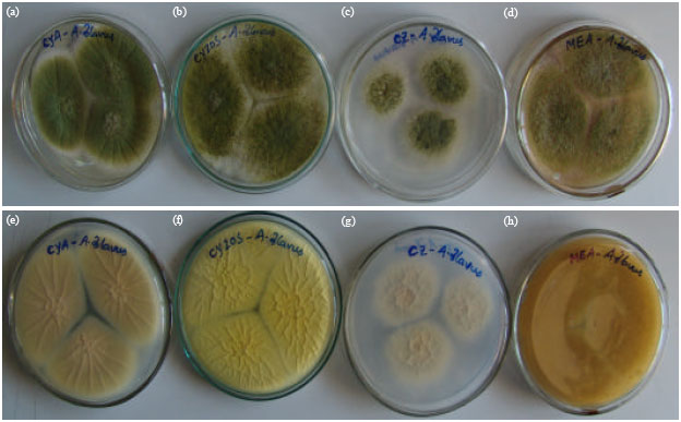 | |
| Fig. 1: | Macroscopical characters of A. flavus on different agar media. (a-d) Front view and (e-h) Reverse view. (a, e) Mycelia growth on CYA; (b, f) Growth on CYA20S; (c, g) Growth on CZA; (d, h) Growth on MEA |
| Table 1: | Macroscopic and microscopic characters of A. flavus isolates |
 | |
| ± = Standard deviation | |
On MEA, reverse uncolored to light brown and no wrinkles were present. On CZA, reverse florescent yellow to light white, wrinkled. Macroscopical and microscopical characters of A. niger are presented in Table 3.
Morphological characters of A. ochraceus isolates: Conidial color on CYA25 near wheat to light yellow; fluffy mycelial growth, white or creamy white, inconspicuous to florescence; exudates were absent; reverse dull yellow to dark yellow or some times brown and wrinkled mycelial growth; soluble pigments were absent; sclerotia were absent in all media.
| Table 2: | Macroscopic and microscopic characters of A. fumigatus isolates |
 | |
| ± = Standard deviation | |
| Table 3: | Macroscopic and microscopic characters of A. niger isolates |
 | |
| ± = Standard deviation | |
On MEA, conidia not dense usually pale to light yellow; reverse light yellow, pale orange to grayish gold shades; and no wrinkles were present; colonies not densely sporulating, variable in appearance (Table 4). Macroscopical and microscopical characters of A. ochraceus are presented in Table 4.
Morphological characters of A. tamarii isolates: Colony and conidia color on CYA25 dark olive green; mycelium white to dull white, usually inconspicuous, reverse uncolored to red tinge with chocolate brown or brown and straight wrinkled mycelial growth; the outer surface of the colony was circular; colonies usually quite low, velutinous; sclerotia, exudates and soluble pigments were absent in all media (Table 5). On MEA, mycelium usually inconspicuous, reverse brown color with normal, sparse, loose, not dense and floccose growth and in all other media wrinkled growth; on CY20S generally puffy growth with slightly more yellow green than on CYA25; reverse reddish brown with wrinkled mycelial growth. On CZA, conidia very sparse in dark bluish green or olive green colors reverse reddish tinge to dark brown shade; on CYA37 dark blackish brown and concentric rings were observed on reverse (Table 5). Macroscopical and microscopical characters of A. tamarii are presented in Table 5.
| Table 4: | Macroscopic and microscopic characters of A. ochraceus isolates |
 | |
| ± = Standard deviation | |
| Table 5: | Macroscopic and microscopic characters of A. tamarii isolates |
 | |
| ± = Standard deviation | |
DISCUSSION
When rice grains with moisture content higher than the desired level enter the storage system, invasion of both field and storage fungi take place. The harmful effects of such fungal invasion are glume or grain discoloration, loss in viability, quality and toxin contamination (Reddy et al., 2008). A. flavus is of ubiquitous occurrence in nature. Since the discovery of aflatoxins, it has become the most widely reported food-borne fungus, reflecting its economic and medical importance and ease of recognition, as well as its universal occurrence. A. parasiticus is less common, but the extent of its occurrence is obscured by the tendency for A. flavus and A. parasiticus to be reported only as A. flavus (Reddy et al., 2009). In this study we observed low frequency of A. fumigatus in some of the rice grains. A. fumigatus is an airborne fungus and infection occurs by inhaling conidia which may colonize airways prior to invasion. It is an opportunistic fungal pathogen responsible for most cases of Invasive Aspergillosis (IA), the most common systemic filamentous fungal infection worldwide (Bertout et al., 2001).
Several reports are available on contamination of rice grains with Aspergillus sp. A high incidence of A. flavus was found in the seed mycoflora of rice (Reddy et al., 2008). The rice crop exposed to frequent and heavy rainfall and flood is subjected to infection by Aspergillus sp. (Reddy et al., 2009). Begum and Samajpati (2000) isolated mycotoxin-producing fungi from contaminated grains of rice sold in the local markets of Calcutta, India. Recently, Jayaraman and Kalyanasundaram (2009) reported Aspergillus sp. contamination of rice bran oils. Sales and Yoshizawa (2005) described the incidence of A. flavus and A. parasiticus in rice bran (14%) and rough rice (78%). Abdullah et al. (1998) reported the incidence of aflatoxingenic fungi in rice grains from Malaysia. Recently, Lampak et al. (2009) reported various fungal mycoflora (Acremonium, Aspergillus, Bipolaris, Colletotrichum, Curvularia, Drechslera, Fusarium, Geotrichum, Nigrospora and Penicillium) in brown rice from Thailand. A study by Purwoko et al. (1991) revealed that A. flavus was the dominant fungi in broken rice and rice bran samples in Indonesia. Still today there are no reports on occurrence of Aspergillus sp. in rice from Cambodia. In this we made attempts to isolate and identify Aspergillus sp. in rice samples collected from Cambodia.
Many Aspergillus strains are very close in their morphological characters and chances are very high to misidentify them. Therefore, accurate identification of Aspergillus sp. is important to develop proper management practices to control these toxigenic fungi and their mycotoxins in food grains. Recently, Kim et al. (2009) and Diba et al. (2007) studied the morphological characters for identification of clinical Aspergillus sp. isolates. Alwakeel (2007) identified Aspergillus sp. isolated from kitchen samples in Riyadh, Saudi Arabia using morphological methods. Morya et al. (2009) used morphology based methods for identification of Aspergillus sp. isolated from soil of teak forest. But none of them studied morphological characters for Aspergillus isolates from rice grains. This is the first report on identification of huge number of Aspergillus sp. isolates obtained from rice grain samples collected from various countries in South Asia through morphological characters.
Askun (2006) used three (CZ, MEA and PDA) differential media for identification of Aspergillus sp. using morphological characteristics isolated from maize kernels. Similarly, Khosravi et al. (2007) used only two differential media (PDA and CZ) for identification of Aspergilli isolated from nut products in Iran. In this study we used morphological method with four differential culture media for identification of five important Aspergillus species isolated from rice grains. Using this method, all standard strains were identified successfully. For the identification of Aspergilli based on morphological methods requires adequate growth for evaluation of colony characteristics and microscopic features. Diba et al. (2007) reported that use of potato dextrose, potato flake, malt extract, inhibitory mould agar, or similar sporulation agars as primary isolation media for Aspergillus may accelerate growth rate and the production of conidia (Diba et al., 2007). In our study, using four differential media including CZA, CYA, CYA20S and MEA with macroscopic and microscopic characteristics of fungal growth on this culture media enabled us to discriminate five Aspergillus species isolated from rice grains. We preserved all 110 isolates of Aspergillus sp. at Universiti Sains Malaysia, Malaysia culture collection centre for future studies.
CONCLUSION
This study concludes that Aspergillus sp. can contaminate rice grains under storage conditions. Though molecular methods are well developed for identification of Aspergillus sp., the developing countries are still highly depending on morphological identification. In this study we have identified five Aspergillus sp. isolated from rice grains based on their morphological characters. Most important human pathogen, A. fumigatus were also observed in few rice grain samples destined for human consumption. In our view, morphological method using the differential media is the most reliable and sensitive assay to identify important Aspergillus sp. isolated from rice grains or some other sources. This study may be a basis for those who are interested to study morphological characters for Aspergillus sp. in developing countries. Mycotoxin profiles produced by these Aspergillus sp. isolates are under progress.
ACKNOWLEDGMENTS
Dr. K.R.N. Reddy acknowledges the Universiti Sains Malaysia, Penang, Malaysia for providing a postdoctoral fellowship. We are grateful for the research grants 1001/PBIOLOGI/811009 and 1001/PBIOLOGI/811147 provided by USM.
REFERENCES
- Alwakeel, S.S., 2007. Bacterial and Aspergillus spp. contamination of domestic kitchens in Riyadh, Saudi Arabia. Saudi J. Biol. Sci., 14: 1-5.
Direct Link - Askun, T., 2006. Investigation of fungal species diversity of maize kernels. J. Biol. Sci., 6: 275-281.
CrossRefDirect Link - Bertout, S., F. Renaud, R. Barton, F. Symoens and J. Burnod et al., 2001. Genetic polymorphism of Aspergillus fumigatus in clinical samples from patients with invasive aspergillosis: Investigation using multipl typing methods. J. Clin. Microbiol., 39: 1731-1737.
PubMed - Bhat, R., R.V. Rai and A.A. Karim, 2010. Mycotoxins in food and feed: Present status and future concerns. Comprehen. Rev. Food Sci. Food Safety, 9: 57-81.
CrossRefDirect Link - Diba, K., P. Kordbacheh, S.H. Mirhendi, S. Rezaie and M. Mahmoudi, 2007. Identification of Aspergillus species using morphological characters. Pak. J. Med. Sci., 23: 867-872.
Direct Link - Begum, F. and N. Samajpati, 2000. Mycotoxin production on rice, pulses and oil seeds. Naturwissenschaffen, 87: 275-277.
CrossRefPubMedDirect Link - Jayaraman, P. and I. Kalyanasundaram, 2009. Natural occurrence of aflatoxins and toxigenic fungi in rice bran oil and de-oiled bran. Indian J. Sci. Technol., 2: 35-37.
Direct Link - Khosravi, A.R., S. Hojjatollah and Z. Tahereh, 2007. Evaluation of fungal flora in some important nut products (pistachio, peanut, hazelnut and almond) in Tehran, Iran. Pak. J. Nutr., 6: 460-462.
CrossRefDirect Link - Kim, D.H., S.H. Kim, Y.K. Kim, S.O. Kim, S.J. Kim and S.B. Hong, 2009. Reidentification of Aspergillus spp. isolated from clinical specimens of patients suspected as pulmonary Aspergillosis in Korea. Korean J. Med. Mycol., 3: 133-144.
Direct Link - Lampak, K., S. Lumyong, R. Wangspa and U. Sardsud, 2009. Diversity of filamentous fungi on brown rice from Pattalung province. Thailand J. Agric. Technol., 5: 129-142.
Direct Link - Lanier, C., N. Heutte, E. Richard, V. Bouchart, P. Lebailly and D. Garon, 2009. Mycoflora and mycotoxin production in oilseed cakes during farm storage. J. Agric. Food Chem., 57: 1640-1645.
PubMed - Makun, H.A., T.A. Gbodi, O.H. Akanya, E.A. Salako and G.H. Ogbadu, 2007. Fungi and some mycotoxins contaminating rice (Oryza sativa) in Niger State, Nigeria. Afr. J. Biotechnol., 6: 99-108.
Direct Link - McClenny, N., 2005. Laboratory detection and identification of Aspergillus species by microscopic observation and culture: The traditional approach. Med. Mycol., 43: S125-S128.
CrossRefDirect Link - Morya, V.K., Kamal and D. Yadav, 2009. Diversity of indigenously isolated Aspergilli from soil of monoculture teak forest. Res. J. Soil Biol., 1: 77-83.
CrossRef - Murthy, K.K., E.R. Rati and H.K. Manonmani, 2009. Incidence of Fusarium toxins in rice from Karnataka, India. Res. J. Toxins, 1: 1-7.
Direct Link - Park, J.W., S.Y. Choi, H.J. Hwang and Y.B. Kim, 2005. Fungal mycoflora and mycotoxins in Korean polished rice destined for human. Int. J. Food Microbiol., 103: 305-314.
CrossRefDirect Link - Pitt, J.I., A.D. Hocking, K. Budhasamai, B.F. Miscamble, K.A. Wheeler and P. Tanboon-Ek, 1994. The normal mycoflora of commodities from Thailand.2. Beans, rice, small grains and other commodities. Int. J. Food Microbiol., 23: 35-53.
CrossRef - Purwoko, H.M., B. Hald and J. Wolstrup, 1991. Aflatoxin content and number of fungi in poultry feed stuffs from Indonesia. Lett Applied Microbiol., 12: 212-215.
Direct Link - Reddy, K.R.N., C.S. Reddy, H.K. Abbas, C.A. Abel and K. Muralidharan, 2008. Mycotoxigenic fungi, mycotoxins and management of rice grains. J. Toxicol. Toxin Rev., 27: 287-317.
CrossRef - Reddy, K.R.N., C.S. Reddy and K. Muralidharan, 2009. Detection of Aspergillus spp. and aflatoxin B1 in rice in India. Food Microbiol., 26: 27-31.
CrossRef - Reddy, K.R.N., B. Salleh, B. Saad, H.K. Abbas, C.A. Abel and W.T. Shier, 2010. An overview of mycotoxin contamination in foods and its implications for human health. Toxin Rev., 29: 3-26.
CrossRefDirect Link - Reiter, E.V., F. Vouk, J. Bohm and E. Razzazi-Fazeli, 2010. Aflatoxins in rice: A limited survey of products marketed in Austria. Food Control, 21: 988-991.
CrossRef - Russel, K., C. Broadbridge, S. Murray, D. Waghorn and A. Mahoney, 2008. Gardening can seriously damage your health. Lancet, 371: 2056-2056.
PubMed - Samapundo, S., F. Devlieghere, B.D. Meulenaer and J. Debevere, 2007. Growth kinetics of cultures from single spores of Aspergillus flavus and Fusarium verticillioides on yellow dent corn meal. Food Microbiol., 24: 336-345.
PubMed - Sales, A. and T. Yoshizawa, 2005. Updated profile of aflatoxin and Aspergillus flavi contamination in rice and its by products from the Philippines. Food Addit. Contam., 22: 429-436.
CrossRef - Sutton, P., P. Waring and A. Mullbacher, 1996. Exacerbation of invasive aspergillosis by the immunosuppressive fungal metabolite, gliotoxin. Immunol. Cell Biol., 74: 318-322.
CrossRef - Abdullah, N., A. Nawawi and I. Othman, 1998. Survey of fungal counts and natural occurrence of aflatoxins in Malaysian starch-based foods. Mycopathology, 143: 53-58.
CrossRefDirect Link