
Leila Shabahang
Department of Obstetrics and Gynecology, Tabriz University of Medical Sciences, Bostanabad, Iran
Pakistan Journal of Biological Sciences
Year: 2010 | Volume: 13 | Issue: 22 | Page No.: 1106-1109


ABSTRACT
Erythema Multiforme (EM) is a type of allergic reaction that occurs in response to medications, infections, or different illnesses. In many cases, a definite underlying cause may not be identified. This study aimed to evaluate adult EM outpatients with regard to patients’ characteristics, the disease and the underlying contributors. In this cross-sectional study, 61 adult EA outpatients referred to the Dermatology Clinic of Tabriz Sina Hospital were recruited during a 12-month period (January 2009-January 2010). The diagnosis was made based on clinical and morphologic grounds. Age, sex, types of EA, location and type of the lesions and the underlying causes were documents. An infectious etiology was suspected when a preceding illness was noticed without drug ingestion within 1 week prior to the onset of the rash. A drug related to the condition was defined as every drug that has been taken during 21 days prior to the onset of any symptoms. There were 34 males and 27 females with a mean age of 26.8±15.3 (18-57) years enrolled. The EM was minor in 62.3% and major in 37.7% of patients. The upper limb was involved in all patients. The lesions were maculo-papular, vesiculobullous and bullous in 83.6, 13.1 and 3.3% cases, respectively. Drugs and herpes simplex were the main causes in 49.2 and 16.4% of patients, respectively. The disease was idiopathic in 34.4%. The underlying drugs were sulfonamides in 12 cases (19.7%), penicillin in 5 cases (8.2%), salicylic acid, aspirin, cimetidine and amoxicillin each one in 3 cases (4.9%) and barbiturate in 1 case (1.6%). Five cases (8.2%) were recurrent EM including 4 males and 1 female, 3 idiopathic and 2 cases due to sulfonamides.
PDF Abstract XML References Citation
Received: July 09, 2010;
Accepted: September 15, 2010;
Published: November 02, 2010
How to cite this article
Leila Shabahang, 2010. Characteristics of Adult Outpatients with Erythema Multiforme. Pakistan Journal of Biological Sciences, 13: 1106-1109.
DOI: 10.3923/pjbs.2010.1106.1109
URL: https://scialert.net/abstract/?doi=pjbs.2010.1106.1109
DOI: 10.3923/pjbs.2010.1106.1109
URL: https://scialert.net/abstract/?doi=pjbs.2010.1106.1109
INTRODUCTION
EM is an acute and self-limited skin condition which may be sometimes recurring. It is believed that the EM is a type IV hypersensitivity reaction associated with various triggers such as infections and certain medications (Freedberg et al., 2003). The characteristic skin manifestations include eruption of annular, maculo-papular lesions with dark raised, erythematous, or vesiculobullous center surrounded by a pale zone (Patel and Patel, 2009). These polymorphous eruptions are composed of symmetrically distributed macules, papules and bullae, with an edematous, petechial, vesicular, or bullous dusky violet center (Ayangco and Rogers, 2003; Fauci et al., 2008). The classification is in two ways; morphological and clinical. In morphological classification, the erythema multiforme lesions are of two types: Iris type (target lesions) which include annular lesions with a raised and cyanotic center due to vascular damage; surrounded by a halo and erythematous rim; and the vesiculo-bullous type which includes annular lesions with a vesicular or even bullous center due to greater skin damage (Du Vivier, 2002). Clinically, erythema multiforme is classified into three groups: erythema multiforme minor; a mild recurrent benign self-limited variant localized to the face and acral parts with one or no mucosal involvement or systemic symptoms. The major subtype is acute self-limited nonrecurrent condition with prominent prodrome, mucosal involvement and systemic symptoms such as fever and prostration. Mucosal lesions are ocular (conjunctivitis, keratitis), oral (stomatitis, cheilitis), nasal, pharyngeal, tracheal and genital (balanitis and valvulitis). Maximal variant or Stevens-Johnson Syndrome (SJS) is characterized by features of erythema major with atypical, confluent target lesions distributed in the face, trunk or both associated with systemic complications (lung, gastrointestinal system, renal and central nervous systems, lymphadenitis, hairs and nail involvement) (Farthing et al., 2005). The epidemiological data of the EM are scarce, many out-of-date and retrospectively conducted in majority (Kamaliah et al., 1998; Lam et al., 2004; Li and Ma, 2006; Leaute-Labreze et al., 2000; Chang et al., 2007; Wetter and Davis, 2009). In addition, majority of studies with appropriate methodology have only dealt with severe cases (Khoo and Foo, 1996; Pushker et al., 2000). To our knowledge, this is the first study ever in the literature assessing the characteristics of adult EM outpatients. The result of current investigation may be considered as an epidemiological source in the region, as well.
MATERIALS AND METHODS
In this cross-sectional study, 61 adult outpatients (18 years≤) with diagnosis of EM presenting in the Dermatology Clinic of Tabriz Teaching Sina Hospital during a one-year period from January 2009 to January 2010 were enrolled. The diagnosis was made by an experienced dermatologist based on clinical and morphologic grounds. The minor subtype was diagnosed when the typical targets or raised edematous papules were acrally distributed. The major type was diagnosed as the minor type plus involvement of one or more mucous membranes (Leaute-Labreze et al., 2000). Skin biopsy was taken when there was an undetermined case. The recurrent case was assigned based on the presence of a symmetrically distributed, fixed eruption, including target lesions, with or without mucous membrane involvement, occurring on at least three occasions. (Schofield et al., 1993). An infectious etiology was suspected when a preceding illness was noticed without drug ingestion within 1 week prior to the onset of the rash (Lam et al., 2004). A drug related to the condition was defined as every drug that has been taken during 21 days prior to the onset of any symptoms (Kamaliah et al., 1998). The age, sex, distribution and types of lesions, type of the disease, underlying causes and recurrence were determined. Data were analyzed by SPSS software version 15.0 and are shown as Mean±SD (range) or frequency (percentage).
RESULTS
Sixty one outpatients with EM, 34 males and 27 females (Fig. 1) with a male to female ratio of 1.3 to 1 and a mean age of 26.8±15.3 (18-57) years were recruited. The age groups are shown in Fig. 2. As seen, the most frequent age group is 18 to 30 years. The EM was minor in 38 cases (62.3%) and major in 23 cases (37.7%). Lesions were present in upper limb in 61 patients (100%), lower limb in 56 patients (91.8%), forehand in 21 patients (34.4%), face in 11 patients (18.0%), penis in 2 patients (3.3%) and neck and oral mucosa each one in 1 patient (1.6%). The lesions were in majority maculo-papular in 51 cases (83.6%), vesiculobullous in 8 cases (13.1%) and bullous in 2 cases (3.3%). Twenty one cases were idiopathic, 10 cases due to herpes simplex infection and 30 cases due to various drugs (Fig. 3). The underlying drugs were sulfonamides in 12 cases (19.7%), penicillin in 5 cases (8.2%), salicylic acid, aspirin, cimetidine and amoxicillin each one in 3 cases (4.9%) and barbiturate in 1 case (1.6%).
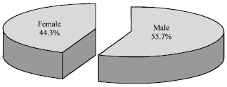 | |
| Fig. 1: | Gender of the studied patients |
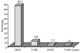 | |
| Fig. 2: | Age-groups of the studied patients |
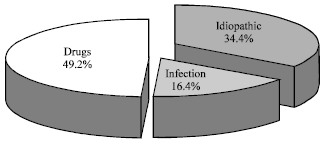 | |
| Fig. 3: | Etiological causes of erythema multiforme in the studied patients |
Five cases (8.2%) were recurrent EM including 4 males and 1 female, 3 idiopathic and 2 cases due to sulfonamides.
DISCUSSION
In present study 61 adult outpatients with EM were evaluated. The male to female ratio was 1.3 to 1, 55.7% and 44.3% of patients, respectively. The mean age was 26.8±15.3 (18-57) years and the most frequent age group was 18-30 years. Kamaliah et al. (1998) recruited 4 Malaysian patients with EM, 2 males and 2 females aged from 15 to 65 years. In this study, the male to female ratio is 1 to 1, almost similar to our finding. However, the sample size is too small. Chang et al. (2007) assessed 65 EM patients in Taiwan. The male to female ratio was 1.3 to 1, 57.6 and 42.4% of patients, respectively. The mean age was 23.5±27.2 years in this study. The result of this study regarding the male to female ratio is exactly similar to ours. The mean age is also very near to that in present study. Apparently, the EM is a little more frequent in the males and majority are young adults aged between 20 and 30 years. The gender preponderance, however is quite opposite in pediatric population. Lam et al. (2004) studied 19 Taiwanese children with EM. The male to female ratio was 0.7 to 1 in this study. Leaute-Labreze et al. (2000) also studied 22 children with EM in France. The male to female ratio was 0.6 to 1. Lifetime longitudinal studies could better describe the change of gender preponderance in EM from childhood to adulthood. The underlying cause of EM was unknown in 34.4% of our patients. The disease was attributed to viral infection (herpes simplex) in 16.4% and various drugs in the remaining 49.2% of the patients. The reports in this regard are very heterogeneous. In retrospective study of 4 hospitalized EM cases by Kamaliah et al. (1998), drugs as a definitive cause was observed in one case (antiepileptic) (25%). Two cases (50%) were secondary to infection (viral illness and tonsillitis) and one case (25%) had an unknown cause. These rates are similar to our finding with regard to the underlying etiologies. Very small sample size and a retrospective method were the major limitations of this study. In addition, only hospitalized cases were recruited. In another series by Stewart et al. (1994), antecedent medication use was identified in 59% of EM patients. This rate is also similar to ours. Chang et al. (2007) reported drugs as underlying causes of EM in 28.8%, viral infection (including herpes simplex) in 27.3% and unknown etiology in the remaining. It seems that geographical variations have a key role regarding the underlying etiologies of RM. The most common etiology in EM was infection (84.2%) in pediatric sample evaluated by Lam et al. (2004). Auquier-Dunant et al. (2002) concluded that there appears to be an association between the type of etiological agent and the severity of the disease. Thus viral infections appear to trigger EM minor or major but drug ingestion tends to trigger more severe forms of the disease. However they also emphasized that this is not absolute and a small but significant proportion of EM minor and major cases are precipitated by drugs, while likewise some cases of severe forms of the disease are virally associated. Based on our finding, we may conclude that restricting the etiological causes to some especial type of the EM disease is not correct. In a study by Leaute-Labreze et al. (2000), they concluded that the childhood EM was mostly related to herpes infection. As mentioned earlier, the only infectious agent in our series was also herpes simplex. So the same conclusion might be drawn in adult population, as well. EM can be triggered by a number of factors. It is previously shown that the best documented triggering agent of EM is preceding infection with herpes simplex virus, the lesions resulting from a cell mediated immune reaction triggered by simplex virus-DNA (Farthing et al., 2005). Drugs in the list of contributing factors of EM in our series were sulfonamides, penicillin, salicylic acid, aspirin, cimetidine, amoxicillin and barbiturate in decreasing order of prevalence. This list also varies greatly in different studies including antibiotics, non-steroidal anti-inflammatory drugs, anticonvulsants, anti-gout agents, etc. (Kamaliah et al., 1998; Li and Ma, 2006; Chang et al., 2007). As mentioned earlier, it is thought that the epidemiology and underlying causes of EM varies greatly from region to region (Kamaliah et al., 1998). Present findings also support this justification. This also necessitates appropriate epidemiological studies in this regard in different regions. The recurrent cases comprised 8.2% of patients (5 cases) in our study. These patients were 4 males and 1 female, 3 idiopathic and 2 cases due to sulfonamides. To the best of our knowledge, there is not any similar report. Wetter and Davis (2009) conducted a retrospective review of patients with recurrent EM. Of 48 patients, 58% were female. Herpes simplex virus caused recurrent EM in 11 (23%) patients and the cause remained unknown in 28 (58%). In contrary to this study, no recurrent case was due to herpes simplex infection in our patients. Small sample size does not let us a definite conclusion. Regional differences may also play a role in this regard. In conclusion, some features of adult patients with EM were discussed in current study. A prospective approach, assessment of outpatient adult cases and appropriate methodology in determining the underlying causes of the disease were the main advantages. Based on our findings, it should be noticed that considerable differences exists between the patients with EM in different regions.
REFERENCES
- Auquier-Dunant, A., M. Mockenhaupt, L. Naldi, O. Correia, W. Schroder, J.C. Roujeau and SCAR Study Group, 2002. Severe cutaneous adverse reactions. Correlations between clinical patterns and causes of erythema multiforme majus, Stevens-Johnson syndrome, and toxic epidermal necrolysis: Results of an international prospective study. Arch. Dermatol., 138: 1019-1024.
PubMedDirect Link - Ayangco, L. and R.S. Rogers, 2003. Oral manifestations of erythema multiforme. Dermatol. Clin., 21: 195-205.
PubMedDirect Link - Chang, Y.S., F.C. Huang, S.H. Tseng, C.K. Hsu, C.L. Ho and H.M. Sheu, 2007. Erythema multiforme, Stevens-Johnson syndrome and toxic epidermal necrolysis: Acute ocular manifestations, causes and management. Cornea, 26: 123-129.
PubMed - Farthing, P., J.V. Bagan and C. Scully, 2005. Mucosal disease series. Number IV. Erythema multiforme. Oral. Dis., 11: 261-267.
PubMedDirect Link - Kamaliah, M.D., D. Zainal, N. Mokhtar and N. Nazmi, 1998. Erythema multiforme, Stevens-Johnson syndrome and toxic epidermal necrolysis in northeastern Malaysia. Int. J. Dermatol., 37: 520-523.
PubMedDirect Link - Khoo, A.K. and C.L. Foo, 1996. Toxic epidermal necrolysis in a burns centre: A 6-year review. Burns, 22: 275-278.
PubMedDirect Link - Lam, N.S., Y.H. Yang, L.C. Wang, Y.T. Lin and B.L. Chiang, 2004. Clinical characteristics of childhood erythema multiforme, Stevens-Johnson syndrome and toxic epidermal necrolysis in Taiwanese children. J. Microbiol. Immunol. Infect., 37: 366-370.
PubMedDirect Link - Leaute-Labreze, C., T. Lamireau, D. Chawki, J. Maleville and A. Taieb, 2000. Diagnosis, classification, and management of erythema multiforme and Stevens-Johnson syndrome. Arch. Dis. Child., 83: 347-352.
PubMedDirect Link - Li, L.F. and C. Ma, 2006. Epidemiological study of severe cutaneous adverse drug reactions in a city district of China. Clin. Exp. Dermatol., 31: 642-647.
PubMedDirect Link - Patel, N.N. and D.N. Patel, 2009. Erythema multiforme syndrome. Am. J. Med., 122: 623-625.
PubMedDirect Link - Pushker, N., R. Tandon and R.B., Vajpayee, 2000. Stevens-Johnson syndrome in India-risk factors, ocular manifestations and management. Ophthalmologica, 214: 285-288.
PubMed - Schofield, J.K., F.M. Tatnall and I.M. Leigh, 1993. Recurrent erythema multiforme: Clinical features and treatment in a large series of patients. Br. J. Dermatol., 128: 542-545.
PubMedDirect Link - Stewart, M.G., N.O. Duncan, D.J. Franklin, E.M. Friedman and M. Sulek, 1994. Head and neck manifestations of erythema multiforme in children. Otolaryngol. Head. Neck. Surg., 111: 236-242.
PubMedDirect Link - Wetter, D.A. and M.D. Davis, 2009. Recurrent erythema multiforme: Clinical characteristics, etiologic associations, and treatment in a series of 48 patients at Mayo Clinic, 2000 to 2007. J. Am. Acad. Dermatol., 62: 45-53.
PubMedDirect Link