
A.R. Fathilah
Department of Oral Biology, Faculty of Dentistry, University of Malaya, 50603 Kuala Lumpur, Malaysia
Z.H.A. Rahim
Department of Oral Biology, Faculty of Dentistry, University of Malaya, 50603 Kuala Lumpur, Malaysia
Y. Othman
Institute of Biological Sciences, Faculty of Science, University of Malaya, 50603 Kuala Lumpur, Malaysia
M. Yusoff
Institute of Biological Sciences, Faculty of Science, University of Malaya, 50603 Kuala Lumpur, Malaysia
Pakistan Journal of Biological Sciences
Year: 2009 | Volume: 12 | Issue: 6 | Page No.: 518-521


ABSTRACT
In this study, the bacteriostatic effect of Piper betle and Psidium guajava extracs on selected early dental plaque bacteria was investigated based on changes in the doubling time (g) and specific growth rates (μ). Streptococcus sanguinis, Streptococcus mitis and Actinomyces sp. were cultured in Brain Heart Infusion (BHI) in the presence and absence of the extracts. The growth of bacteria was monitored periodically every 15 min over a period of 9 h to allow for a complete growth cycle. Growth profiles of the bacteria in the presence of the extracts were compared to those in the absence and deviation in the g and μ were determined and analyzed. It was found that the g and μ were affected by both extracts. At 4 mg mL-1 of P. betle the g-values for S. sanguinis and S. mitis were increased by 12.0- and 10.4-fold, respectively (p<0.05). At similar concentration P. guajava increased the g-value by 1.8- and 2.6 -fold, respectively (p<0.05). The effect on Actinomyces sp. was observed at a much lower magnitude. It appears that P. betle and P. guajava extracts have bacteriostatic effect on the plaque bacteria by creating a stressed environment that had suppressed the growth and propagation of the cells. Within the context of the dental plaque, this would ensure the attainment of thin and healthy plaque. Thus, decoctions of these plants would be suitable if used in the control of dental plaque.
PDF Abstract XML References Citation
How to cite this article
A.R. Fathilah, Z.H.A. Rahim, Y. Othman and M. Yusoff, 2009. Bacteriostatic Effect of Piper betle and Psidium guajava Extracts on Dental Plaque Bacteria. Pakistan Journal of Biological Sciences, 12: 518-521.
DOI: 10.3923/pjbs.2009.518.521
URL: https://scialert.net/abstract/?doi=pjbs.2009.518.521
DOI: 10.3923/pjbs.2009.518.521
URL: https://scialert.net/abstract/?doi=pjbs.2009.518.521
INTRODUCTION
The extracts of the Piper betle (L.) and Psidium guajava (L.) plants have been reported to posses many biological activities that have contributed to their role in the development of therapeutic products (Nair and Chanda, 2008; Wirotesangthong et al., 2007; Kamath et al., 2008). Piper betle is popularly used in traditional medicine as it possesses antioxidant, antibacterial, antifungal, antidiabetic and radioprotective activities (Wirotesangthong et al., 2007). Psidium guajava is often used as astringent for skin diseases (Ponglux et al., 1987) and also showed antidiarrheal, hepatoprotective, hypoglycemic, lipid lowering, antibacterial and antioxidant activities (Kamath et al., 2008). The methanolic extract of P. betle and P. guajava have been shown to exhibit antimicrobial effects on various Gram positive and negative food borne pathogens (Francis Parillon, 2006). While the effect of P. betle was on both the Gram-positive and Gram-negative bacteria, the effect of P. guajava was specific to the Gram-negatives. Membrane damage that causes loss of cell viability and leakage of intracellular constituents was suggested to be the main mechanism of action of these extracts.
In the perspective of oral health maintenance, the aqueous extracts of P. betle and P. guajava have showed positive antiplaque activities that act on dental plaque bacteria at the early phase of plaque formation. These extracts were reported to act by first reducing the adhering capacity of the acquired pellicle which forms on the tooth surface at the early phase of plaque formation, to receive and bind the bacteria (Fathilah and Rahim, 2003) and second by diminishing the cell-surface hydrophobicity of the bacteria which are required to assist the adherence process (Fathilah et al., 2006). Study by Nalina and Rahim (2006) has also shown that the crude extract of P. betle inhibited the activity of glucosyltransferase (GTF) which is required for glucan synthesis by the cariogenic bacteria Streptococcus mutans. The Minimal Inhibitory Concentration (MIC) and Minimal Bactericidal Concentration (MBC) of aqueous extracts of P. betle and P. guajava were reported within the range of 2.16-4.69 and 5.21-10.42 mg mL-1, respectively (Fathilah and Rahim, 2003).
The objective of this study was to investigate the bacteriostatic effect of the aqueous crude extracts of P. betle and P. guajava on early dental plaque bacteria, S. sanguinis, S. mitis and Actinomyces sp. Bacteriostatic effect will be determined based on deviations in the doubling time and specific growth rate of the growth profiles produced in the presence and absence of the extracts.
MATERIALS AND METHODS
Preparation of plant extracts: Fresh leaves of P. betle and P. guajava were obtained from the university’s botanical garden. Aqueous extracts of P. betle and P. guajava were prepared by concentrating decoctions of the fresh leaves of the plants using a speed-vacuum concentrator (HETO/HS-1-110, Denmark). The dried extracts were then kept refrigerated at -80°C (Hetofrig, Denmark) prior to use in the experiment. The dried extracts of P. betle and P. guajava were weighed into sterile microfuge vials and prepared into stocks of 20 mg mL-1 using sterile distilled water as the diluents. The extracts were dissolved by sonicating the microfuge vials in a sonicator (Ultrasonic sonicator, Selecta CE95).
Preparation of bacterial suspensions: Streptococcus sanguinis, S. mitis and Actinomyces sp. used in the investigation were pure cultures obtained from frozen (-80°C) stocks isolated from dental plaque specimens collected from volunteers visiting the Dental Clinic at the Faculty of Dentistry, University of Malaya (Fathilah and Rahim, 2003). Each bacteria species was revived in Brain Heart Infusion (BHI, Oxoid) broth at 37°C overnight. Following incubation the bacteria cells were harvested by centrifugation at 10,000 rpm for 10 min. The cells were then resuspended in BHI broth and the concentration was standardized at 106 cells mL-1 [Optical Density (OD) of 0.014] by using a spectrophotometer read at 550 nm.
The effect of P. betle and P. guajava aqueous extracts on bacterial growth profiles: The antibacterial activity of P. betle and P. guajava was determined using an assay procedure which involved the changes in the optical absorbance as an indication of changes in the bacterial growth profile. Metal capped borosilicate glass tubes (13x75 mm) were sterilized and used as culture tubes in the experiments. The growth of S. sanguinis, S. mitis and Actinomyces sp. under four different conditions was monitored by measuring the increased in the OD of the growing cells every 15 min. The four growth conditions were: (a) in 5 mL BHI broth to represent the control, (b) in 5 mL BHI added with P. betle extract at 4 mg mL-1, (c) in 5 mL BHI added with P. guajava extract at 4 mg mL-1 and (d) in 5 mL BHI added with chlorhexidine (CHX)-containing mouth rinse at neat concentration of 0.12 mg mL-1. Each of the test tubes was then inoculated with 50 μL of the respective bacterial cells suspension. The concentration of extracts was selected at 4 mg mL-1 to be within the range of the MIC. This would ensure that the addition of the extract would not kill the bacteria cells but instead would allow the growth of cells to be at its minimum. CHX-containing mouth rinse in the test was used to represent a positive control for the study as CHX is considered the standard antimicrobial agent in the dental and hospital arena (Jones, 1997; Kornman, 1986). All tests were carried out in triplicate and repeated three times for reproducibility of results.
The content of each culture tubes were mixed well using a vortex mixer. The OD readings of each tube were set to zero with a test tube containing everything else except for the bacterial cells. This was to accommodate differences in the OD of the mixture caused by the varying color intensities of the plant extracts and the CHX-containing mouth rinse. The cultures were then incubated at 37°C in a shaking water bath and changes in the OD readings of each tube were periodically monitored and recorded at every 15 min intervals over a period of 9 h. The growth curves of each bacterium under the four growth conditions were plotted and compared with the profile of the CHX-containing mouth rinse. The growth rate (μ) and doubling time (g) of S. sanguinis, S. mitis and Actinomyces sp. under the different growth conditions were then determined using the following equations (Gerhardt et al., 1981; Cappuccino and Sherman, 2005):
μ = [(log10 N-log10 N0) (2.303/(t-t0)) ] |
g = (log10 N - log10 N0)/log10 2 |
where, N is No. of cells at log phase, N0 is No. of cells at zero time and t is time to reach, t0 is zero time log phase.
Statistical analysis: The effect of the extracts on the growth of the bacteria was illustrated by comparative analysis of their growth profiles under the various growth conditions. Statistical analysis was carried out using the one way analysis of variance (ANOVA). The MINITAB 13 for Windows statistical program was used to determine the Mean, Standard Deviation and evaluate the significance of the data in the experiments. Results were expressed as Mean±SD from one nine determinations (n = 9) set at a significance level of p<0.05.
RESULTS AND DISCUSSION
Throughout the study, the concentration of P. betle and P. guajava was set within the sub-MIC concentration of 4 mg mL-1 and not exceeding the MBC concentration.
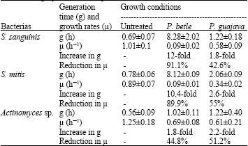
This is important as the aim of a good antiplaque agent is not to kill all but to allow some of the plaque bacteria especially those of the normal species, to grow at a minimal rate. Within the sub-MIC range the bacteria exist in a condition where the biological cell functions are not disrupted. The growth profiles in Fig. 1a-c strongly suggested that the antimicrobial activity of P. betle and P. guajava towards S. sanguinis, S. mitis and Actinomyces sp. was bacteriostatic and may have been targeted at the early lag phase of the growth cycle. P. betle and P. guajava extracts seem to have created a stressed environment for the cells to perform their normal biological functions. This explains for the extended g-values and reduction in the μ-values in Table 1. The attainment of minimal population size as the bacteria enters the stationary phase indicated the bacteriostatic activities of P. betle and P. guajava towards S. sanguinis, S. mitis and Actinomyces sp. Under the stressed growth environment the bacteria were unable to perform normal biological function and eventually ceased to propagate. Such growth inhibiting mechanism has also been reported when the requirement for nutrient was restricted for S. sanguinis growth (Fathilah et al., 2007).
The search for new alternative agents to be used as adjuncts in oral health care products has spurred due to the unfavorable staining effect caused by the prolonged usage of CHX (Cummins, 1992; Baehni and Takeuchi, 2003). In this study CHX-containing mouth rinse at 0.12 mg mL-1 was found to be bactericidal to S. sanguinis, S. mitis and Actinomyces sp. as no growth profile can be generated following the 9 h incubation period. This finding confirmed the reputation of CHX as the most effective antimicrobial agent for the oral microorganisms. Alternative to CHX, plant based bioactive compounds such as sanguinarine, gallotannins and catechins have been isolated using solvent extraction procedures from Sanguinaria canadensis (Kopczyk et al., 1991; Harper et al., 1990), Melaphis chinensis (Wu-Yuan et al., 1988) and Japanese green tea (Hirasawa et al., 2006; Otake et al., 1991), respectively. Exhibiting significant antiplaque activities, these compounds have been incorporated as adjuncts in dental dentifrices. In other plants like Azarachdita indica (Neem) (Wollinsky et al., 1996), P. betle (Fathilah and Rahim, 2003; Fathilah et al., 2006; Nalina and Rahim, 2006, 2007) and P. guajava (Fathilah and Rahim, 2003; Fathilah et al., 2006), aqueous extraction procedure were employed and the crude extracts have been used. The exploration of antimicrobial activities of these plants has been based on their effective used in folklore medicines which often make use of simple decoction using water. Aqueous extraction procedure was employed in this study as it is environment friendly and would also avoid any possibility of side effect that could arise from the exposure to solvents if these extracts are to be used in the development of oral health care product.
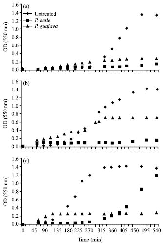 | |
| Fig. 1: | The growth profiles of (a) S. sanguinis, (b) S. mitis and (c) Actinomyces sp. in the absence of extract, presence of P. betle and presence of P. guajava. Deviation of profiles from the untreated growth condition indicated the bacteriostatic effect of the extracts |
| Table 1: | Changes in the generation times (g) and specific growth rates (μ) of S. sanguinis, S. mitis and Actinomyces sp. when cultured in the absence (untreated) and presence (extract treated) of P. betle and P. guajava were compared |
 | |
| p<0.05 | |
CONCLUSION
The aqueous extracts of P. betle and P. guajava exhibited bacteriostatic effect on early dental plaque bacteria S. sanguinis, S. mitis and Actinomyces sp. under the stressed growth environment the bacteria appear to be unable to perform normal biological function and eventually ceased to propagate. Such events would ecologically control the development of dental plaque. Thus, both the plant extracts may have potential to be used as an active ingredient in the development of oral health care products.
ACKNOWLEDGMENTS
This research was financially supported by the IRPA 09-02-03-0197-EA197 Grant from the Malaysian Government and Vote F0392/2004B from the University of Malaya.
REFERENCES
- Baehni, P.C. and Y. Takeuchi, 2003. Anti-plaque agents in the prevention of biofilm-associated oral disease. Oral Dis., 9: 23-29.
Direct Link - Razak, F.A. and Z.H. Rahim, 2003. The anti-adherence effect of Piper betle and Psidium guajava extracts on the adhesion of early settlers in dental plaque to saliva-coated glass surfaces. J. Oral Sci., 45: 201-206.
PubMedDirect Link - Razak, F.A., Y. Othman and Z.H. Abd Rahim, 2006. The effect of Piper betle and Psidium guajava extracts on the cell-surface hydrophobicity of selected early settlers of dental plaque. J. Oral Sci., 48: 71-75.
CrossRefPubMedDirect Link - Harper, D.S., L.F. Mueller, J.B. Fine, J. Gordon and L.L. Laster, 1990. Effect of 6 months use of a dentifrice and oral rinse containing sanguinaria extract and zinc chloride upon the microflora of the dental plaque and oral soft tissues. J. Periodontol., 61: 359-363.
CrossRefDirect Link - Hirasawa, M., K. Takada and S. Otake, 2006. Inhibition of acid production in dental plaque bacteria by green tea catechins. Caries Res., 40: 265-270.
PubMedDirect Link - Jones, C.G., 1997. Chlorhexidine: Is it still the gold standard? Periodontology, 15: 55-62.
CrossRefDirect Link - Kopczyk, R.A., J. Abrams, A.T. Brown, J.L. Matheny and A.L. Kaplan, 1991. Clinical and microbiological effects of a sanguinaria-containing mouthrinse and dentifrice with and without fluoride during 6 months of use. J. Periodontol., 62: 617-622.
CrossRefDirect Link - Nalina, T. and Z.H.A. Rahim, 2006. Effect of Piper betle L. leaf extract on the virulence acticity of Streptococcus mutans: An in vitro study. Pak. J. Biol. Sci., 9: 1470-1475.
CrossRefDirect Link - Nalina, T. and Z.H.A. Rahim, 2007. The crude aqueous extract of Piper betle L. and its antibacterial effect towards Streptococcus mutans. Am. J. Biotechnol. Biochem., 3: 10-15.
Direct Link - Otake, S., M. Makimura, T. Kuroki, Y. Nishihara and M. Hirasawa, 1991. Anticaries effects of polyphenolic compounds from Japanese green tea. Caries Res., 25: 438-443.
PubMed - Wollinsky, L.E., S. Mania, S. Nachnani and S. Ling, 1996. The inhibiting effect of aqueous Azadirachta indica (neem) extract upon bacterial properties influencing in vitro plaque formation. J. Dent. Res., 75: 816-822.
CrossRefDirect Link - Wu-Yuan, C.D., C.Y. Chen and R.T. Wu, 1988. Gallotannins inhibit growth, water-insoluble glucan synthesis and aggregation of mutans streptococci. J. Dent. Res., 67: 51-55.
CrossRefDirect Link - Kamath, J.V., N. Rahul, C.K.A. Kumar and S.M. Lakshmi, 2008. Psidium guajava L.: A review. Int. J. Green Pharm., 2: 9-12.
Direct Link - Nair, R. and S. Chanda, 2008. Antimicrobial activity of Terminalia catappa, Manilkara zapota and Piper betel leaf extract. Indian J. Pharmaceut. Sci., 70: 390-393.
PubMedDirect Link - Wirotesangthong, M., N. Inagaki, H. Tanaka, W. Thanakijcharoenpath and H. Nagai, 2008. Inhibitory effects of Piper betle on production of allergic mediators by bone marrow-derived mast cells and lung epithelial cells. Int. Immunopharm., 8: 453-457.
CrossRef