
P. Tajik
Department of Clinical Sciences, Faculty of Veterinary Medicine, University of Tehran, P.O. Box 14155-6453, Tehran, Iran
R. Beheshti-Govij
Faculty of Veterinary Medicine, Islamic Azad University, Science and Research Branch, Tehran, Iran
J. Soleimani-Rad
Deparment of Anatomy and Embryology, Faculty of Medicine, Tabriz University of Medical Sciences, Tabriz, Iran
H. Ghasemzadeh-Nava
Department of Clinical Sciences, Faculty of Veterinary Medicine, University of Tehran, P.O. Box 14155-6453, Tehran, Iran
A. Mohamadi-Roshandeh
Deparment of Anatomy and Embryology, Faculty of Medicine, Tabriz University of Medical Sciences, Tabriz, Iran
Pakistan Journal of Biological Sciences
Year: 2008 | Volume: 11 | Issue: 4 | Page No.: 628-632


ABSTRACT
Murine 2-cells embryos were isolated from murine oviducts at laboratory and transferred into Ham's F-10 medium containing 0.1 mg mL-1 streptomycin and 100 IU mL-1 penicillin G and supplemented with 3 mg mL-1 bovine serum albumin (BSA) or different concentrations of bovine follicular fluid (bFF) and estrous cow serum (ECS). Significantly higher (p<0.05) >=4-cell embryos were developed when embryos were cultured 20% bFF (84.33%) comparing to 10 and 15% bFF (48.33 and 69.33%) as well as 3 mg mL-1 BSA (65.66%). Morula rates were also lower in 10% bFF (22.33%) comparing to the other groups and were similar in 15 and 20% bFF (62.66 and 72.33% morula rates) as well as BSA containing media (55.33%). The highest (p<0.05) blastocyst rates were obtained in medium containing 20% bFF (64.33%) and the lowest belonged to 10% bFF (15%) comparing to 15% bFF (33.66%) or 3 mg mL-1 BSA. When embryos were cultured in ECS, no significant different was observed in different culture media (76.66, 72.33, 82.5 and 65.66% >=4-cell embryos in 10, 15 and 20% bFF and 3 mg mL-1 BSA, respectively). Morula and blastocyst rates were also similar in all groups (32.33, 41.66 and 66.25 and 55.33% morula rates and 15.33, 27, 44.50 and 29.66% blastocyst rates for 10, 15 and 20% bFF and 3 mg mL-1 BSA, respectively). The results of the present study demonstrated that 20% bFF could be substituted for BSA when in vitro culture of murine embryos is carried.
PDF Abstract XML References Citation
How to cite this article
P. Tajik, R. Beheshti-Govij, J. Soleimani-Rad, H. Ghasemzadeh-Nava and A. Mohamadi-Roshandeh, 2008. Effects of Different Concentrations of Bovine Follicular
Fluid and Estrous Cow Serum on Development of Murine 2-Cell Embryos. Pakistan Journal of Biological Sciences, 11: 628-632.
DOI: 10.3923/pjbs.2008.628.632
URL: https://scialert.net/abstract/?doi=pjbs.2008.628.632
DOI: 10.3923/pjbs.2008.628.632
URL: https://scialert.net/abstract/?doi=pjbs.2008.628.632
INTRODUCTION
It is now well understood that the development of preimplantation mammalian embryos in vitro is less than optimal. Early attempts to develop fertilized mouse ova in vitro demonstrated that murine morula developed into blastocysts in complex medium or in simple medium consisting mainly of Kreb`s-Ringer bicarbonate (Loutradis et al., 1987). Studies on culture of preimplantation embryos advanced considerably after the development of biological medium containing egg white egg yolk and a chemically semi-defined medium with BSA for mouse embryos (Han and Niwa, 2003).
Mammalian embryos are generally cultured in medium supplemented with serum as protein supplement (Babaei et al., 2006; De Santis et al., 2007; Banwell et al., 2007). Any use of serum involves the addition of wide range of proteins, hormones and other elements which may vary widely from batch to batch (Barnes and Sato, 1980). Serum is an extremely complex fluid containing a variety of energy substrates, amino acids, vitamins and growth factors that may support survival and growth of mammalian cells in culture (Han and Niwa, 2003). However, many reports indicate that exposure of 2- to 8-cell embryos to FBS is detrimental to their development to blastocyst in vitro (Bavister, 1995; Maurer, 1992), indicating that sera contain toxic factors. In our previous study (Tajik, 2006) bovine fertilized eggs could reach to blastocyst stages in protein-free medium. On the other hands, Pinyopummintr and Bavister (1991) reported that serum had a biphasic effect on bovine embryo development, inhibiting the first cleavage of 1-cell embryos and having no beneficial effect from the 2-cell to the morula stage, but subsequently enhancing development of morula to blastocyst. The beneficial effect of serum on advanced-stage embryos also been reported in pigs (Pollard et al., 1995) and mice (Torensi and Archer, 1996).
It has been hypothesized that protein in embryo culture medium may function as a fixed nitrogen source (Fissore et al., 1989). Fetal cord serum (FCoS) has been used as protein source for embryo culture media since 1988 because it was easily available and was considered a good substitute for maternal serum (Lavarge et al., 1997).
Serum albumin, on the other hand, is a relatively pure fraction, although its content can also be very variable (Bavister, 1995). The varying conditions present current embryo culture systems may contribute to the poor cultured embryos (Racowsky, 2002). On the other hand, it is well documented that the follicular fluid plays an important biological role in folliculogenesis, oocyte maturation, granulosa cells luteinization and ovulation (Gotting et al., 2002).
Follicular fluid is primarily the transudation of plasma that contains specific constituents such as steroids, glycosaminoglycans and many other metabolites synthesized by the cells of the follicle wall (Choi et al., 1998). Moreover, it has been reported that the concentrations of vitamins (Schweigert and Kucker, 1988), insulin-like growth factors (Einspanier et al., 1993) and other factors vary with follicular size and degree of atresia (Kruip et al., 2000).
Bovine Serum Albumin (BSA) is the most common protein added to culture media as a fixed nitrogen source for embryos but it is very expensive and hard to prepare. However, we (Tajik and Niwa, 1998) have shown that bovine oocytes can be fertilized and embryos will develop to blastocyst in the complete absence of any exogenous fixed nitrogen source, although a high molecular weight such as polyvinylpyrrolidone was added to the culture media as a replacement for BSA. The aim of the present study was to examine development of mouse 2-cell embryos in the presence different concentrations of follicular fluid and estrus cow serum (of bovine source) as substitutions for BSA.
MATERIALS AND METHODS
Animals and embryo collection: This study was conducted during 1 year almost from spring to winter 2006. Randomly bred female mice (BALB/c), 8-10 weeks old, were superovulated by an intraperitoneal injection of 5 IU of human menopausal gonadotrophin (hMG, Humegun, Zaran, Iran), followed 48 h later by 5 IU of human chorionic gonadotrophin (hCG; Oregone, Holland). After the injection human chorionic gonadotropin (hCG) (purchased from Darou Pakhsh, Iran). Superovulated females were caged overnight with males. Insemination was verified the following morning finding a copulation plug in the vagina. Embryos were recovered 40h after hCG injection by flushing uterine. Any embryos appearing degenerate or abnormal were discarded. Normal 2-cell embryos different litters were pooled, washed 3 times and transferred to Ham`s F10 medium. Groups of 15 embryos were placed in drops with different culture media (Ham`s F10) in 35mm Petri dishes of each protein supplemented media and three replicates were done.
Follicular fluid collection and preparation: Mixed bovine follicular fluid was retrieved from slaughterhouse ovaries. Follicles with diameters between 3 and 15mm were aspirated by an 18 gauge needle attached to a 10 mL syringe the pooled follicular fluid was then centrifuged at 4000rpm, the supernatant stored at -20°C, until use. All experiments were performed with the same batch.
Estrus cow serum collection: Serum used in this experiment was obtained from six estrous cows, heat-inactivated (56°C, 30min), pooled, filtered with a 0.22 µm membrane (Millipore, Brussels, Belgium) and frozen in 1.5mL vials until used.
Culture treatments: For embryo culture, Ham`s F-10 medium was supplemented with either estrous cow serum (ECS) at three concentrations of 10, 15 and 20% (v/v) or bovine follicular fluid (bFF) at three concentrations of 10, 15 and 20% (v/v). Each 10-15 2-cell harvested embryos were randomly allocated into 50 µL media droplets of the mentioned culture media in a polystyrene culture dish (35x10 mm). The dishes were kept in a CO2 incubator (5% CO2 in air at 37°C) for about 2 h before embryos were added. After 24, 48 and 72 h culture of embryos in CO2 incubator embryos were observed under a stereomicroscope (Nikon co. ltd, Japan). Embryonic development was scored every 24 h and the proportion the 4 to 8-cell, morula and blastocyst stages were recorded.
Statistical analysis: The proportions of total embryos in each stages of development were subjected to an arc-sin transformation and the transformed values were analyzed using a mathematical model that included fixed effect due to treatment (serum concentrations) and residual error. When the analysis revealed a significant effect, the values were compared by Duncan`s multiple range test.
RESULTS
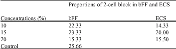
When murine 2-cell embryos were cultured in Ham`s F-10 medium supplemented with different concentrations of sera, 22.33, 23.33 and 15.33% were blocked and did not
| Table 1: | The 2-cell block in different concentrations of bovine follicular fluid (bFF) and estrous cow serum (ECS) in Ham`s F-10 medium 48 h post-culture |
 | |
| Table 2: | The proportion of >=4-cell in different concentrations of bovine follicular fluid (bFF) and estrous cow serum (ECS) in Ham`s F-10 medium 24 h post-culture |
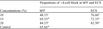 | |
| a-bValues in rows, columns and in control group with different superscript are significantly different (p<0.05) | |
developed to higher stages in !0, 15 and 20% bFF. These values were 14.33, 20 and 15.5% for 10, 15 and 20% ECS respectively. More embryos were blocked in medium containing BSA. However, the different was not significant (Table 1).
Significantly (p<0.05) lower development rat (48.33%) was observed in 10% bFF comparing to 20% bFF (84.33%) or different concentrations of ECS studied (76.66, 72.33 and 82.5% development rates for 10, 15 and 20% ECS respectively). This value was not either significantly lower than the medium supplemented with BSA (65.66%) (Table 2).
Forty hour post-culture only 22.33% of embryos in 10% bFF developed to morula stage. However, these values were significantly (p<0.05) higher in 15 and 20% bFF (with 62.66 and 72.33% development rates). The development rates in different concentrations of ECS are also increasing in a dose dependency (Table 3).
Observation for detection of blastocyst on 72 h post-culture showed that 64.33% of embryos reached to blastocyst stage in 20% bFF. This value was significantly higher than blastocyst rates in all other groups except in 20% ECS in which 44.50% embryos reached to blastocyst (Table 4).
DISCUSSION
Follicular fluid is instrumental in the nutritional and developmental support of the oocyte. Follicular maturation and the maturation of its oocyte are parallel events and also functionally related. Malekshah et al. (1996) have reported that developments of 2-cell mouse embryos are possible in human heat inactivated follicular fluid (hFF). In their study, the development of
| Table 3: | The proportion of morula in different concentrations of bovine follicular fluid (bFF) and estrous cow serum (ECS) in Ham`s F-10 medium 48 h post-culture |
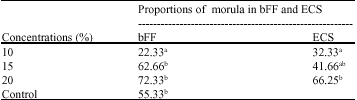 | |
| a-bValues in rows, columns and in control group with different superscript are significantly different (p<0.05) | |
| Table 4: | The proportion of blastocyst in different concentrations of bovine follicular fluid (bFF) and estrous cow serum (ECS) in Ham`s F-10 medium 72 h post-culture |
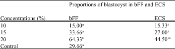 | |
| a-bValues in rows, columns and in control group with different superscript are significantly different (p<0.05) | |
2-cell embryos to 4 cells or beyond is likely supported in 15% FF comparing to the control group. In the present study regarding development of embryos to >=4-cell stage, no significant difference was observed between 10 and 15% bFF with control. However, the medium with 20% bFF, significantly (p<0.05) supported embryonic development to >=4-cell.
It has been reported that there is a significant improved development of ICR mouse 2-cell embryos to 8-cell and morula by the addition of amino acids and in the presence of 10% hFF comparing to the medium supplemented with BSA (Cho et al., 2002).
Recent studies have shown that the follicular fluid derived from small, medium, large and pre-ovulatory follicles supplemented to the maturation medium at the range of 10% (Carolan et al., 1996; Elmileik et al., 1995; Sirard et al., 1995), 20% (Romero-Arredondo and Seidel, 1996) and 100% (Choi et al., 1998) improved the developmental capacity of bovine oocytes. In swine, also a medium of 100% follicular fluid supplemented with FSH 0.12 IU mL-1 was used for oocyte maturation, markedly improved male pronucleus formation (Naito et al., 1988, 1989).
However, 60% of bovine follicular fluid derived from small or large follicles had a detrimental effect on embryonic development (Elmileik et al., 1995; Kim et al., 1996). In present, development of mice 2-cell embryos to and beyond the 4-cell stage was not inhibited when they were cultured in HF-10+20% ECS or HF-10+20% FF. It was also observed that, protein supplementation has a beneficial effect on embryo development from 2-cell embryos to blastocyst stages.
The BSA used was 98% pure and we consider that the 2% of uncharacterized impurities are the probable source of the variability between batches of BSA and may also be important essential factors for the hatching process.
Albumin, on the other hand, has the advantage of being a single protein that is commercially available. The source of albumin can be human or bovine. Ashwood-Smith et al. (1989) reported on the outcome of embryo development comparing embryo culture in Earle`s medium with either Albuminar-5 or patient`s serum as protein source. No significant differences were found regarding fertilization rate and implantation rates. In the present study the proportions of >=4-cell in BSA 65.66% was not significantly different with dose in different concentrations of bFF and ECS. The proportion of morula (55.33%) and blastocyst (29.66%) in control medium containing BSA were also similar to those of cultured in 15 and 20% of bFF and ECS, which is in agreement with Ashwood-Smith et al. (1989). However, Staessen et al. (1990) found that the morphological appearance and the pregnancy rate were significantly higher in the Albuminar group.
In conclusion, media supplemented with concentration of 20% bFF and ECS is likely to support murine 2-cell embryos development to blastocyst stage. These observations could have important implications for human in vitro fertilization and development
REFERENCES
- Ashwood-Smith, M.J., P. Hollands and R.G. Edwards, 1989. The use of Albuminar 5 (TM) as a medium supplement in clinical FVF. Hum. Reprod., 4: 702-705.
Direct Link - Babaei, H., S.N. Nematallahi and A. Kheradmand, 2006. The effects of vitamin A administration on the development of verified-warmed mouse blastocyst. Anim. Reprod. Sci., 95: 125-133.
Direct Link - Banwell, K.M., M. Lane, D.L. Russell, K.L. Kind and J.G. Thompson, 2007. Oxygen concentration during mouse oocyte in vitro maturation affects embryo and fetal development. Hum. Reprod., 10: 2768-2775.
Direct Link - Barnes, D. and G. Sato, 1980. Serum-free cell culture: A unifying approach. Cell, 22: 649-655.
CrossRefDirect Link - Bavister, B.D., 1995. Culture of preimplantation embryos: Facts and artifacts. Hum. Reprod. Update, 1: 91-148.
CrossRefDirect Link - Carolan, C., P. Lonergan, P. Monget, D. Monniaux and P. Mermillod, 1996. Effect of follicle size and quality on the ability of follicular fluid to support cytoplasmic maturation of bovine oocytes. Mol. Reprod. Dev., 43: 477-483.
CrossRefDirect Link - Cho, J., S. Park, H. Chung, H. Shim and B. Lee et al., 2002. Improved development of ICR mouse 2-cell embryos by the addition of amino acids to a serum-phosphate- and glucose-free medium. J. Vet. Med. Sci., 64: 797-801.
Direct Link - Choi, Y.H., M. Takagi, H Kamishita, M.P.B. Wijayagunawardane, T.J. Acosta, K. Miyazawa and K. Sato, 1998. Developmental capacity of bovine oocytes matured in two kinds of follicular fluid and fertilized in vitro. Anim. Reprod. Sci., 50: 27-33.
CrossRefDirect Link - De Santis, L., G. Coticchio, S. Paynter, D. Albertini and K. Hutt et al., 2007. Permeability of human oocytes to ethylene glycol and their survival and spindle configuration after slow cooling cryopreservation. Hum. Reprod., 10: 2776-2783.
Direct Link - Einspanier, R., H. Schuster and D. Schams, 1993. A comparison of hormone levels in follicle-lutein-cysts and in normal bovine ovarian follicles. Theriogenology, 40: 181-188.
CrossRefDirect Link - Elmileik, A.M.A., T. Maeda and T. Terada, 1995. Higher rates of development into blastocyst following the in vitro fertilization of bovine oocytes matured in a medium supplemented with the fluid from large bovine follicles. Anim. Reprod. Sci., 38: 85-96.
CrossRefDirect Link - Fissore, R.A., K.V. Jackson and A.A. Kiessling, 1989. Mouse zygote development in culture medium without protein in the presence of ethylenediaminetetraacetic acid. Biol. Reprod., 41: 835-841.
CrossRefDirect Link - Han, M.S. and K. Niwa, 2003. Effect of BSA and fetal bovine serum in culture medium on development of rat embryos. J. Reprod. Dev., 49: 235-242.
Direct Link - Kim, K.S., N. Mitsumizo, K. Fujita and K. Utsumi, 1996. The effects of follicular fluid on in vitro maturation, oocyte fertilization and the development of bovine embryos. Theriogenology, 45: 787-799.
CrossRefDirect Link - Kruip, T.A.M., M.M. Bevers and B. Kempt, 2000. Environment of oocyte and embryo determines health of IVP offspring. Theriogenology, 53: 611-618.
Direct Link - Laverge, H., P. de Sutter, R. Desmet, J. van der Elst and M. Dhont, 1997. Prospective randomized study comparing human serum albumin with fetal cord serum as protein supplement in culture medium for in-vitro fertilization. Hum. Reprod., 12: 2263-2266.
CrossRefDirect Link - Loutradis, D.K., D. John and A.A. Kiessling, 1987. Hypoxanthine causes a 2-cell block in random-bred mouse embryos. Biol. Reprod., 37: 311-316.
CrossRefDirect Link - Naito, K., Y. Fukuda and Y. Toyoda, 1988. Effects of porcine follicular fluid on male pronucleus formation in porcine oocytes matured in vitro. Gamete Res., 21: 289-295.
CrossRefDirect Link - Naito, K., Y. Fukuda and I. Ishibashi, 1989. Developmental ability of porcine ova matured in porcine follicular fluid in vitro and fertilized in vitro. Theriogenology, 31: 1049-1057.
CrossRefDirect Link - Pollard, J.W., C. Plante and S.P. Leibo, 1995. Comparison of development of pig zygotes and embryos in simple and complex culture media. J. Repord. Fert., 103: 331-337.
PubMed - Racowsky, C., 2002. High rates of embryonic loss, yet high incidence of multiple births in human art: Is this paradoxical? Theriogenology, 57: 87-96.
PubMed - Romero-Arredondo, A. and G.E. Seidel, 1996. Effects of follicular fluid during in vitro maturation of bovine oocytes on in vitro fertilization and early embryonic development. Biol. Reprod., 55: 1012-1016.
PubMed - Schweigert, F.J. and H. Kucker, 1988. Concentrations of vitamin A, b-carotene and vitamin E in individual bovine follicles of different quality. J. Reprod. Fert., 52: 575-579.
PubMed - Sirard, M.A., F. Roy, B. Patrick, P. Mermillod and L.A. Guilbault, 1995. Origin of the follicular fluid added to the media during bovine IVM influences embryonic development. Theriogenology, 44: 85-94.
Direct Link - Staessen, C., E. van den Abbeel, M. Carle, I. Khan, P. Devroey and A.C. van Steirteghem, 1990. Comparison between human serum and Albuminar-20 (TM) supplement for in-vitro fertilization. Hum. Reprod., 5: 336-341.
PubMedDirect Link - Torensi, M.B. and J. Archer, 1996. The early development of mouse embryos in vitro in medium supplemented with different batches of serum and bovine serum. Vet. Res. Commun., 20: 15-19.
PubMedDirect Link