
O .S. Udengwu
Department of Botany, University of Nigeria, Nsukka, Nigeria
J. C. Chukwujekwu
Research Centre for Plant Growth and Development, University of Kwa-Zulu Natal, Pietermaritzburg, Private Bag X01, Scottville 3209, South Africa
Pakistan Journal of Biological Sciences
Year: 2008 | Volume: 11 | Issue: 18 | Page No.: 2184-2192


ABSTRACT
The Allium test was used to study the cytotoxic effects of five commonly abused skin toning creams-Ikb, Tura, Top gel, Dorot and Mililo. These creams are commonly used by some black skinned people (especially the females) as skin lightening (bleaching) agents. The results showed that all the five bleaching creams were mito-depressive in action. They exhibited both chromatoclassic and mitoclassic effects. Their depressive effects were found to increase with duration of treatment. The induced abnormalities included chromosome contraction, spindle breakages, c-metaphase, star anaphase, chromosome stickiness and sticky bridges, precocious chromosome movement as well as endomitosis. It is suggested that since all eukaryotic cells are basically the same, these observed abnormalities could be similar to the effects these chemicals have on human skin when they are applied. Some of these are known to cause alteration in melanin formation as well as the biosynthesis of the enzyme tyrosinase. Furthermore, since certain points on the chromosomes called fragile sites have been implicated in oncogenesis, the observed abnormalities may be part of (or include) the switching on mechanisms of such genes, which could be responsible for the transformation of normal skin cells to malignant cells in those who abuse these creams.
PDF Abstract XML References Citation
How to cite this article
O .S. Udengwu and J. C. Chukwujekwu, 2008. Cytotoxic Effects of Five Commonly Abused Skin Toning (Bleaching)
Creams on Allium cepa Root Tip Mitosis. Pakistan Journal of Biological Sciences, 11: 2184-2192.
DOI: 10.3923/pjbs.2008.2184.2192
URL: https://scialert.net/abstract/?doi=pjbs.2008.2184.2192
DOI: 10.3923/pjbs.2008.2184.2192
URL: https://scialert.net/abstract/?doi=pjbs.2008.2184.2192
INTRODUCTION
A practice persists where some dark skinned people, especially the women folk, abuse skin toning creams in their bid to bleach their skin for better look and acceptance, despite the fact that many researchers like Barsh (2003), Fitzpatrick (1988), Lin and Fisher (2007) and Radhakrishnan et al. (2007) have written about the protective functions of the black skin against the damaging effects of UV irradiation; which could cause sunburn and skin cancer for exposed vulnerable skins. The black skin is able to perform this protective function due to the physical barrier imposed by the epidermal melanin.
Despite the fact that several researchers, like Bernstein et al. (1970), Forbes et al. (1970), Elmett et al. (1977), Parrish et al. (1978), Bergstresser (1989), Cohn and Emmett (1978), Harber et al. (1982), Bickers (1988) and Krutman and Emmett (1988) have written about the inherent health hazards which the use of bleaching creams pose for the users, not much research study has been carried out to throw adequate light on the nature of the possible cytotoxic and genetic complications they could cause those who use them.
Available literature indicate that related studies done in this area had to do with the study of the general effects of some of the chemical compounds used in the making of some of these bleaching creams on human skin. Such studies revealed that chemicals like, hydrogen peroxide preparations, ammoniated mercury, phenols and catechols including monobenzyl ether of hydroquinone, monomethyl ether of hydroquinone, (p-hydroxyanisole), p-tertiary butyl phenol, p-tertiary amylphenol and 4 -tertiary butyl catechol could act as demelanizing agents (Parrish et al., 1978; Douglas, 1980; Cohn and Emmett, 1978, Marzulli and Maibach, 1980; Anderson and Parrish, 1981; Krutman and Emmett, 1988).
As a result of its critical role in melanin biosynthesis, the enzyme tyrosinase has become a major target for inhibition in skin-lightening cosmetics. Hydroquinone is one of the most popular depigmenting agents and is used extensively to treat several hyper pigmentation disorders. Depigmentation by hydroquinone is because of its ability to inhibit tyrosinase as well as its cytotoxicity to melanocytes. However, because of its carcinogenic properties, use of hydroquinone is banned or limited in cosmetic products in many countries (Radhakrishnan et al., 2007).
The health and social problems caused by the use of bleaching creams were compounded following the introduction of topical steroid in 1951 and super potent steroid in 1974 (Frumess and Lewis, 1957; Mihan and Ayres, 1964; Sneddon, 1969; Leyden et al., 1974; Ljubojeviae et al., 2002; Rathi, 2006).
Apart from the above reported research on the effects of some of the chemicals on human skin, there are also some cytological studies done by past researchers that revealed many cytological effects of some other chemicals on dividing cells of plants. Deysson (1968), Torkowska (1971), Gulati et al. (1975), El-Bayoumi et al. (1979), Shehab (1979), Amer and Enaam (1980), Kabirity and Malallah (1980) and Ene-Obong and Amadi (1987) reported that many glycosides, plant alkaloids, pesticides and some other chemicals have some cytological effects on root tip mitosis of experimental plants like Allium cepa, Vicia faba, Lens esculenta and some other plants. Their effects could generally be described as mitodepressive, mitopromotive, mitoclassic and chromatoclassic.
The cytological abnormalities they induced include, accumulation of prophase at the expense of other phases, stickiness of chromosomes, contraction of chromosomes and sticky bridges, lagging of chromosomes (Raj and Shubba, 1971; Torkowska, 1971; Shehab, 1979; Kabarity and Malallah, 1980; Ene-Obong and Amadi, 1987). Other effects include spindle disturbances, polyploidy, chromosome breakages, pycnosis, binucleate cells, chromosome denaturation (Raj and Shubba, 1971; Torkowska, 1971; Shantharmurthy and Rangaswamy, 1979; Shehab, 1979; Ene-Obong and Amadi, 1987).
Similar studies with some extracted known alkaloids like Colchicine, Podophy-lotoxin, Coumarin, Vinblastine, Vincristine and Mimosine exhibited remarkable cytological effects which include sticky bridges by colchicine spindle breakage by Vinblastine spindle damage, stickiness and sticky bridges, laggards, precocious chromosomes by podophyllin and mimosine (Deysson, 1968); spindle formation inhibition by colchicines (Inone , 1981).
The aim of this present study was to ascertain the nature of the cytotoxic effects of these bleaching creams on the building blocks of life- the cells, which contain the chromosomes and which in turn contain the genes that control all the biological activities of an organism. It is believed that since the structure of the cells of all eukaryotic cells are basically the same, coupled with the fact that it is easier to study with plant materials, the observed cytological effects of these bleaching creams studied, with the Allium test, may throw some light on the possible mode of action of the chemical constituents of these creams on the cells of the human skin in changing melanin present in melanosomes from the dark- coloured oxidized form to the lighter coloured reduced form, interference with the biosynthesis of melanin, prevention of the biosynthesis of tyrosinase, premature skin aging, dermatitis and carcinogenesis as noted by Douglas (1980), Lin and Fisher (2007) and Radhakrishnan et al. (2007). The Allium test is acclaimed to be a very cheap and sensitive tool for the detection of potentially genotoxic substances (Fiskesjo, 1985; Sabti, 1989; Smaka-Kinkl et al., 1996; Chang et al., 1997; Rank and Nielson, 1998; Cotelle et al., 1999; Moraes and Jordao, 2001). The findings may also help to caution the users of such creams about the potential dangers they might be exposing themselves to.
MATERIALS AND METHODS
This research was initiated in 1994 and concluded in 2007 in the Department of Botany, University of Nigeria, Nsukka. Rooted onions Allium cepa (2 n = 16) bulbs were used to investigate the cytotoxic effects of Top gel, Ikb, Tura, Dorot and Mililo which are among the commonly abused commercial bleaching creams. Table 1 gives the chemical composition of the creams as indicated on the cream containers by the manufacturers. One hundred and eight medium sized, fresh red onion bulbs, each weighing approximately 85 g, bought from the Nsukka market, were grown in water soaked, well cured, Gmelina wood saw dust, in wooden germination boxes in the Botanical garden, University of Nigeria, Nsukka. When the roots were about 2-5 cm long, after about 4-6 days of planting, the rooted bulbs were transferred to 100 mL beakers containing distilled water and left for 24 h in order to allow enough time for recovery, in case there were any abnormalities caused by the sawdust culture.
| Table 1: | Chemical composition of the five bleaching creams |
 | |
| Table 2: | Time range and duration of treatments (h) |
 | |
Treatment procedure and duration of treatment: The one hundred and eight bulbs were divided into 6 groups, with three bulbs set for each of the six treatment durations for each of the creams and the water control. The onion roots were evenly rubbed with each of the bleaching creams, for the different treatments, just like humans rub creams on their skin. The roots were covered with moistened cotton wool to enhance absorption of the creams by the roots, before suspending the bulbs in 150 mL beakers. Three bulbs, for each treatment (duration), were retained in distilled water, in a 150 mL beaker, without any cream, to serve as the control. Table 2 gives a summary of the durations and times of treatment.
Fixation and hydrolysis of the root tips: At the end of each treatment period, four healthy roots were cut off from each of the three bulbs for each of the treatment durations and for each of the creams as well as the water control. The roots were washed in distilled water for 2-3 times and then fixed in Carnoy`s solution (1:3 acetic acid:absolute alcohol). The fixed materials were kept in the refrigerator for at least 24 h after which they were stored in 70% alcohol before usage. The root tips were hydrolysed in I N HCl at an acid temperature of 60oC for 5-7 min using a Gallenkamp water bath.
Slide preparation and study: Hydrolysed root tips were then washed 2-3 times in tap water, sliced and squashed on a clean glass slide in Lacto Proprionic Orcein (LPO) and left for 5 min so, that the stain will be absorbed by the chromosomes. The temporary slides were then studied under the microscope. Good preparations were sealed off using nail varnish. Good plates were photographed with Leitz Ortholux II microscope at 1000 magnification and the prints were done at 4x negative enlargements.
For making the cell counts, three slides were prepared for each treatment duration and for each cream and the water control. Different fields were picked at random with the three different slides and the views of interests scored. The number of cells counted ranged between 3,000 and 3,200.
Data collections and analysis: After observing, counting and recording dividing cells and total number of cells from 12 different fields for each cream for each treatment duration, the mitotic indexes were calculated using the formula below. Thereafter the means and standard errors of the mitotic indexes for each of the creams as well as the treatment durations were determined:
Other computations that were done based on classification of cytological abnormalities were:
 |
For all analysis of variance the Randomized Complete Block Design (RCBD) was used. Three main effects and three first order interactions were analyzed for the analysis of variance of the dividing cells.
RESULTS AND DISCUSSION
The Allium cepa root tips treated with the five bleaching creams (Ikb, Tura, Dorot, Top-gel and Mililo) exhibited many types of abnormalities. These abnormalities involved all stages of mitosis. Generally, all the five bleaching creams induced mitodepressive effects i.e., the reduction in number of dividing cells. The bleaching creams showed different degrees of depression based on duration of treatment. On the average, the highest degree of depression was scored by Tura followed by Dorot, Top-gel, Mililo and the least by IKB. It was found that increase in duration of treatment affected this depression. For all the bleaching creams the trend of depression showed highest reduction of mitotic indexes at the longest durations of treatment of 24 h (Table 3). The reason for Tura being the most mitodepressive may not be unconnected with the fact that essentially it contains hydroquinione mixed with allantoin. The effects of hydroquinone on human skin have engaged the attention of dermatologists for a long time. For Ikb, which showed the least mitodepression, the presence of vitamin E as one of its constituents, may have mitigated the effect of hydroquinone and allantoin on the root tip cells. Vitamin E is known to play vital roles in the formation of red blood cells, muscles and other tissues and in preventing the oxidation of vitamin A and fats. It is also popularly advocated for a wide range of diseases.
| Table 3: | Effect of treatment duration on the mitotic index of Allium cepa root tips treated with five bleaching creams |
 | |
| Table 4: | Percentage mean No. of dividing cells at different phases of mitosis |
 | |
| Means followed by the same letter(s) in each column or row are not significantly different at 5% level using LSD | |
| Table 5: | Analysis of variance of dividing cells |
 | |
| **: Significant at p<0.01, ***: Significant at p<0.001 | |
The effect on the mitotic index also affected the mean percentage number of cells at different phases. Significant increase in percentage of prophase phase with significant corresponding decrease in other phases was observed (Table 4).
This was found to be highest with Top-gel followed by Tura, Ikb, Mililo and least with Dorot. A possible reason for the accumulation of prophase at the expense of the other phases could be due to the ability of the constituents of these bleaching creams to attack and disrupt the spindle apparatus that are normally formed prior to the cell transiting into metaphase. Analysis of variance of the dividing cells as given in Table 5 indicate that there was a very highly significant difference between the treatments (p<0.001).
The differences between the mitotic stages on one hand and the durations of treatment on the other were very highly significant (p<0.001). The first order interaction on Table 5 (i.e., Treatment x Mitotic stage showed a very highly significant difference (p<0.001), the last interaction (i.e., Mitotic stages x Duration) also showed a highly significance difference (p<0.01). In the case of mitodepression, the degree of depression varied with the creams and this can be attributed to their chemical components as shown in Table 1. Similar depressions were also observed by some researchers using chemicals like 2,4,- D, Amitrols, Phenols, Isoprophyl and some plant alkaloids like colchicine, podophylotoxin, Coumarin, Vinblastine etc. (Amer and Farah, 1974; Amer and Enaam, 1980; Kabarity and Malallah, 1980; Ene-Obong and Amadi, 1987). They attributed the phenomenon to the inhibition of DNA replication at interphase during the S-phase leading to inhibition of other mitotic stages.
It is worth noting that the observed mitodepressive actions of these creams can be largely attributed to hyroquinone, which is the major chemical component of almost all the creams (Table 1). It has been reported that hydroquinone inhibits the synthesis of melanin that protects the skin from UV radiation in a process known as de-pigmentation (Douglas, 1980; Lin and Fisher, 2007; Radhakrishnan et al., 2007). Its mitodepressive ability, as the major component of the five creams suggests that one of the possible processes through which it causes de-pigmentation in human skin could be through the suppression of the mitotic process needed for the replacement of worn out melanocytes.
| Table 6: | Percentage mean No. of dividing cells showing abnormalities after treatment with five bleaching creams for different time durations |
 | |
| Means followed by the same superscript letters in each column or row are not significantly different at 5% level using LSD | |
| Table 7: | Percentage mean No. of abnormal cells at different phases of mitosis |
 | |
Means followed by the same superscript letter(s) in each column or row are not significantly different at 5% level using LSD | |
With serious reduction in the amount of new melanocytes being formed (mitodepression), the skin may gradually loose its characteristic black colour resulting to a fair skin appearance for the user with the attendant exposure of the skin to ultraviolet radiations and other potential infections. Table 3 shows that mitodepression increased with duration of treatment and this may account for the fact that the longer people used these creams, the lighter and more tender their skin become and in cases of excessive use the skin may loose its ability to perform its primary protective functions. Additionally, it has been observed that once users stop the application of these creams, nature fights back to restore the status quo by stimulating rapid cell division among the melanocytes. Unfortunately such natural response does not restore the status quo, but rather the skin of the individual becomes darker and less attractive, hence the tendency to continue usage or even look for stronger brands which may eventually lead to malignancy of the melanocytes.
Table 6 indicates that there were no significant differences between the induced abnormalities by the five creams over the different time durations. A possible reason for this could be that the creams being powerful de-pigmenting agents were able to cause different damages to the genetic materials in the cells shortly after their application. These quick damages however appear not be influenced by duration of treatment, unlike the situation with mitodepression. This observation may account for the frequent application of these creams by their users as well as the observable lightening of the skin a few days after commencement of their application.
Table 7 shows that most of the abnormalities were scored at the prophase and metaphase stages and least at anaphase and telophase stages.
The different types of abnormalities observed were mostly of the sticky (chromotoclassic) and disturbed (mitoclassic types). Figure 1a-f shows that some of the abnormalities observed included disturbed prophase, sticky metaphase and anaphase, c-metaphase, star anaphase and anaphase with precocious chromosomes. Table 8 indicates that disturbed phases, stickiness, anaphase bridges and to a less extent precocious chromosome movements at anaphase and star anaphase were the most frequent abnormalities encountered. The stickiness types occurred in the form of sticky metaphase, anaphase and telophase. It was also found that the degree of stickiness varied with the treatments (creams) for example Ikb gave the highest degree of stickiness followed by Top-gel, Tura and the least by Dorot and Mililo. This type of anomaly has been interpreted to be due to the depolymerization of DNA, partial dissolution of nucleoproteins, breakage and exchange of the basic folder fibre unit of chromatids and the stripping of the protein covering of DNA in chromosomes (Mercykutty and Stephen, 1980). This anomaly was also reported to be induced by phosphonothioate insecticide, leptophos, on Vicia faba (Ali and Amer, 1974; Amer and Farah, 1974; Shehab, 1979).
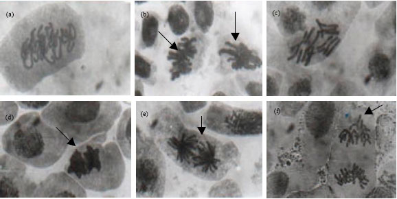 | |
| Fig. 1: | (a) Disturbed prophase (b) Mildly sticky metaphase (left arrow) and Early anaphase (right arrow) (c) C-metaphase (d) Very sticky anaphase (arrow) (e) Star anaphase and (f) Anaphase with precocious chromosomes (arrow) |
| Table 8: | Percentages of the main types of abnormalities observed at different phases |
 | |
| Distur: Disturbed, Endo: Endomitosis, Preco: Precocious chromosome movement | |
The disturbed types of abnormalities included; disturbed prophase, metaphase, anaphase and telophase. The degree of disturbance varied with the creams, for example Mililo gave the highest degree of mitoclassic effect followed by Ikb, Tura, Dorot and the least was scored with Top-gel. The disturbed prophase observed by earlier researchers was attributed to irregular arrangement of chromatin threads (Shehab, 1979). However disturbed metaphase and the later phases are believed to be as a result of spindle formation inhibition or damage (Borisy and Taylor, 1967a, b; Deysson, 1968; Pritchard and Court, 1968). This spindle disturbance during the metaphase stage resulted to the scattering of the chromosomes within the cell, thus preventing the chromosomes from moving towards the opposite poles during anaphase leading to what is referred to as c-metaphase (Fig. 1c) (Weisenberg et al., 1967; Torkowska, 1971; Artvinli, 1987).
The consequences of these observed abnormalities are often interrelated. According to Fiskesjo (1985), c-mitosis is regarded as indicative of a weak toxic effect which may be reversible, a vagrant chromosome, a weak c-mitotic effect indicating risk of aneuploidy, while sticky chromosomes indicate a highly toxic, irreversible effect, probably leading to cell death. Anueploidy has been reported to probably be the only mutation that can explain all aspects of carcinogenesis (Duesberg and Rasnick, 2000).
Endomitosis or endopolyploidy was another abnormality observed. It was observed only in metaphase stage. It occurred in almost all the treatments with highest frequency occurrence in Dorot followed by Top-gel, Ikb, Mililo and least by Tura. This abnormality could have arisen as a result of inhibition of spindle mechanism leading to the arrested phase reverting to the interphase stage (Nelson, 1972; Ene-Obong and Amadi, 1987). Single and multiple bridges were observed in nearly all the treatments. They occurred only in anaphase and telophase stages. The degree of this anomaly also varied with different treatments (creams). Top-gel gave the highest degree of bridges followed by Ikb, Dorot, Tura and the least by Mililo. The incidence of bridges have been attributed to the sticky nature of chromosomes that brought about non-synchronization at separation of chromatids during the movement of chromosomes towards the poles, such chromosomal bridges were also reported by Amer and Farah (1974) and Kabarity and Malallah (1980).
Other rare abnormalities observed include precocious chromosomes movements at anaphase, which is believed to be caused by the non-synchronization of the spindles in their poleward movement during anaphase or due to early disjunction of a pair of chromatids such that one starts off its poleward journey earlier than others (Shehab, 1979; Sarbhoy, 1980). Star anaphase (Fig. 1e), could be attributed to disorientation of the chromosome spindles with the result that the centromeres all point towards a centre in a circular form instead of in the direction of the poles. Few cases of chromosomal breakages were also observed with Tura which could be linked to the actions of hydroquinone, the major component of the cream, on the chromosomes. Hydroquinone according to Radhakrishhnan et al. (2007) has carcinogenic properties and hence its use is banned or limited in cosmetic products in many countries. Breakages on certain locations on the chromosomes called fragile sites that contain oncogenes have been linked with cancer (Yunis and Soreng, 1984; De Braekeleer et al., 1985; LeBeau, 1986; Pellicia and Rocchi, 1986; Yunis et al., 1987).
Incidences of skin cancer in those who use these creams may therefore not be unconnected with the actions of hydroquinone on fragile sites of some chromosomes.
It has been shown that these creams apart from being mitodepressive also exhibit both mitoclassic and chromatoclassic effects. These effects were found to depend on individual creams and duration of treatment. This duration dependent mitodepressive actions could be attributed to the different chemical compositions (i.e., hydroquinone, fluocinonide, cetrimide, allantoin, propylene glycol, irgassan etc) of the creams. It is suspected that these chromatoclassic and mitoclassic effects could be close to their mode of action in the alteration of melanin formation and inactivation or the prevention of the biosynthesis of the enzyme tyrosinase in humans.
It is postulated that since all eukaryotic cells are basically the same, these observed anomalies with plant cells are expected to be similar, if not more pronounced, with the animal (human) cells which lack rigid cell walls. Studies by different researchers indicate that the Allium test is a very sensitive tool for the detection of potentially genotoxic substances (Fiskesjo, 1985; Sabti, 1989; Smaka-Kinkl et al., 1996; Chang et al., 1997; Rank and Nielson, 1998; Cotelle et al., 1999; Moraes and Jordao, 2001). The protocol of rubbing the creams on the roots the way humans rub these creams on their bodies, as well as the observed cytotoxic effects even after short durations of application, suggests that the Allium test could be a pertinent tool for a better understanding of the cytotoxic problems and complications the abuse of these creams could cause.
Finally, since this study has shown that the degree of mitodepression increased with duration of treatment as well as with the nature of the chemical constituents of these creams; this could be related to the observed fact that the skins of the abusers of these creams show severe deterioration with prolonged usage. Incidentally, to shorten the action time of these creams, some manufacturers now produce more powerful creams whose bleaching effects, together with its concomitant degradation of the skin, manifests after shorter periods of time. From the cytological point of view, such a development may further complicate the social and medical problems the abuses of these products are likely to cause.
REFERENCES
- Amer, S.M and O.R. Farah, 1974. Cytological effects of pesticides VI. Effects of insecticide Rogor on the mitosis of Vicia faba and Gossypium bar-badense. Cytologia, 39: 507-514.
PubMed - Barsh, G.S., 2003. Unsolved mystery: What controls variation in human skin colour?. PLOS Biol., 1: 19-23.
CrossRefDirect Link - Bernstein, H.N., J. Curtis and F.L. Earl, 1970. Phototoxic corneal and lens opacitiesin dogs receiving a fungicide: 2,6-diebloro-4-nitro-analine. Arch. Ophthalmol., 83: 336-348.
PubMed - Bickers, D.R., 1988. Metabolic activation of carcino-gens by keratinocytes. Ann. N. Y. Acad. Sci., 548: 102-107.
CrossRef - Borisy, G.G. and E.W. Taylor, 1967. The mechan-ism of action of colchicine-3H to cellular protein. J. Cell Biol., 34: 525-533.
PubMed - Borisy, G.G. and E.W. Taylor, 1967. The mechanism of action of colchici-ne. Colchicine binding tosea urchin eggs and mitotic apparatus. J. Cell Biol., 34: 535-548.
PubMed - Chang L.W., J.R. Meier and M.K. Smith, 1997. Application of plant and earthworm bioessays to evaluate remediation of a lead contaminated soil. Arch. Environ. Contam. Toxicol., 32: 166-171.
Direct Link - Cotelle, S., J.F. Masfaraud and G.F. Ferard, 1999. Assessment of the genotoxicity of contaminated soil with the Allium/Vicia micronucleus and the Tradescantia-micronucleus assays. Mutation Res., 426: 167-171.
CrossRefDirect Link - Duesberg, P. and D. Rasnick, 2000. Aneuploidy, the somatic mutation that makes cancer a specie of its own. Cell Motil. Cytoskeleton, 47: 81-107.
CrossRefPubMedDirect Link - Fiskesjo, G., 1985. The Allium test as a standard in environmental monitoring. Hereditas, 102: 99-112.
CrossRefDirect Link - Fitzpatrick, T.B., 1988. The validity and practicality of sun-reactive skin types I through VI. Arch. Dermatol., 124: 869-871.
PubMed - Lin, J.Y. and D.E. Fisher, 2007. Melanocyte biology and skin pigmentation. Nature, 445: 843-850.
CrossRefDirect Link - Ljubojeviae, S., A. Basta-Juzbasiae and J. Lipozeneiae, 2002. Steroid dermatitis resembling rosacea: Aetiopathogenesis and treatment. J. Eur. Acad. Dermatol. Venereal., 16: 121-126.
PubMedDirect Link - Mercykutty, V.C. and J. Stephen, 1980. Adriamycia induced genetic toxicity as demonstrated by Allium cepa test. Cytologia, 45: 769-777.
Direct Link - Moraes, D. and B. Jordao, 2001. Evaluation of the genotoxic potential of municipal waste water discharge into the Paraguay River during periods of food and drought. Environ. Toxicol., 16: 113-116.
CrossRefDirect Link - Radhakrishnan, N., K. Vijayachandra and S. Ranganathan, 2007. Changing skin colour: Evolution and modern trends. Indian J. Dermatol., 52: 71-77.
Direct Link - Rathi, S., 2006. Abuse of topical steroid as cosmetic cream: A social background of steroid dermatitis. Indian J. Dermatol., 51: 154-155.
Direct Link - Sabti, K., 1989. Allium test for air and water borne pollution control. Cytobios, 58: 71-78.
PubMedDirect Link - Smaka-Kincl, V., P. Stegnar, M. Lovka and M.J. Toman, 1996. The evaluation of waste, surface and ground water quality using the Allium test procedure. Mutat. Res./Genet. Toxicol., 368: 171-179.
CrossRefPubMedDirect Link - Yunis, J.J. and A.L. Soreng, 1984. Constitutive fragile sites and cancer. Science, 266: 1199-1204.
CrossRef