
Arash Khaki
Department of Veterinary Pathology, Islamic Azad University Tabriz Branch, Iran
Marefat Ghaffari Novin
Cellular and Molecular Biology Research Center, Shaheed Beheshti University MC, Tehran, Iran
Amir Afshin Khaki
National Management Center for Health (NPMC), Department of Anatomical Science,Tabriz University of Medicine Science, Tabriz, Iran
Mohammad Nouri
Department of Biochemistry Science, Tabriz University of Medicine Science, Tabriz, Iran
Ehsan Sanati
Department of Veterinary Pathology, Islamic Azad University Tabriz Branch, Iran
Mahdad Nikmanesh
Department of Veterinary Pathology, Islamic Azad University Tabriz Branch, Iran
Pakistan Journal of Biological Sciences
Year: 2008 | Volume: 11 | Issue: 13 | Page No.: 1683-1689


ABSTRACT
The aim of this study was to investigate the comparative effects of aminoglycosides and fluoroquinolones on testis apoptosis and sperm parameters in rats. Fifty male Wistar rats were randomly divided into control (n = 10) and experimental (n = 40) groups. The experimental groups subdivided into four groups of ten. Each received 5 mg kg-1 (IP) gentamicin, 50 mg kg-1 (IP) neomycin, 40 mg kg-1 (IP) streptomycin and 72 mg kg-1 (IP) ofloxacin daily for 14 days, respectively; however, the control group just received vehicle (IP). In the fourteenth day, rats were killed and sperm analyzed for sperm parameters. Testis tissues were also prepared for TUNEL assay for detection of apoptosis. There was a significant decrease in sperm count, viability and motility in all of experimental groups when compared with control group. Although in streptomycin group these parameters were less decreased than in the other experimental groups. The apoptotic cells were significantly increased in all experimental groups when compared with those seen in the controlled group. Gentamicin, neomycin and streptomycin and ofloxacin have negative effects on sperm parameters and testis apoptosis in rats. However, these side effects are less seen in the streptomycin group. Therefore, it is recommended that usage of this drug have fewer side effects on male fertility.
PDF Abstract XML References Citation
How to cite this article
Arash Khaki, Marefat Ghaffari Novin, Amir Afshin Khaki, Mohammad Nouri, Ehsan Sanati and Mahdad Nikmanesh, 2008. Comparative Study of the Effects of Gentamicin, Neomycin, Streptomycin and Ofloxacin Antibiotics on Sperm Parameters and Testis Apoptosis in Rats. Pakistan Journal of Biological Sciences, 11: 1683-1689.
DOI: 10.3923/pjbs.2008.1683.1689
URL: https://scialert.net/abstract/?doi=pjbs.2008.1683.1689
DOI: 10.3923/pjbs.2008.1683.1689
URL: https://scialert.net/abstract/?doi=pjbs.2008.1683.1689
INTRODUCTION
In the last few years, a marked decrease in the quality of semen has been reported (Carlsen et al., 1992). These changes in semen quality are more likely to be due to environmental factors. Chemicals and drugs which add are particularly misused are among these environmental factors (Oliva et al., 2002). Antibiotics are commonly prescribed for a multitude of everyday condition. Not surprisingly, a proportion of male patients attending fertility clinics may have been prescribed antibiotics by their general practitioner to treat these unrelated infections. In addition, some patients requiring assisted conception occasionally show evidence of infection of the male reproductive tract. The antibiotic aminoglycosides (gentamicin, neomycin, streptomycin) and fluoroquinolones (ofloxacin) are routinely used by urologists, andrologist and fertility specialists to treat such bacterial infections occurring prior to in vitro fertilization treatment, or when high concentration of leukocytes are present in the semen of these patients, irrespective of microbial evidence of infection. They have a broad spectrum against of microbial pathogens, especially Gram-negative infectious diseases, that has been approved in more than 100 countries worldwide. They are among the most effective antibacterial for the treatment of bacterial genitourinary tract (Herbold et al., 2001; Schlegel et al., 1991). In the laboratory, aminoglycosides are commonly used in embryo culture, sperm wash and cryopreservation media (King et al., 1997; Magli et al., 1996) for controlling growth of bacteria and fungi. The most frequently used antibiotics are streptomycin sulfate and penicillin (Magli et al., 1996; Erbach et al., 1994), which are usually used in combination. Some laboratories use gentamycin to maintain the sterility of culture (Takahashi and First, 1992). In vivo and in vitro genotoxicity studies suggest these antibiotics as safe for therapeutic use (Herbold et al., 2001). However, other studies have demonstrated they impair significantly both testicular function and structure (Schlegel et al., 1991; Abd-Allah et al., 2000; Demir et al., 2007). The aim of the present study was to compare the effect of gentamicin, neomycin, streptomycin and ofloxacin on sperm parameters and testis apoptosis in rat.
MATERIALS AND METHODS
Animals: Fifty adult Wistar albino male rats were 8 weeks old and weighing 250±10 g, they were obtained from animal facility of pasture institute of Iran. Male rats were housed in temperature controlled rooms (25°C) with constant humidity (40-70%) and 12/12 h light/dark cycle prior to use in experimental protocols. All animals were treated in accordance to the Principles of Laboratory Animal Care. All Rats were fed a standard diet and water. The daily intake of animal water was monitored at least one week prior to start of treatments in order to determine the amount of water needed per experimental animal. Thereafter, the rats were randomly selected and divided into control (n = 10) and experimental (n = 40) groups. The experimental groups subdivide four groups of ten. Each received 5 mg kg-1 (IP) gentamicin, 50 mg kg-1 (IP) neomycin, 40 mg kg-1 (IP) streptomycin and 72 mg kg-1 (IP) ofloxacin daily for fourteenth days, respectively; however, the control group just received vehicle (IP). Body weight daily intake of food and water were determined several times per week throughout the study. All chemicals used in the present study were purchased from Sigma chemical.
Surgical procedure: In the fourteenth day, the Pentobarbital sodium (40 mg kg-1) was administered intra peritoneal for anesthesia and the peritoneal cavity was opened through a lower transverse abdominal incision. Then testis and epididymis were immediately removed. The weights of testis in both groups were recorded. At the end of the experiment; the animals were anesthetized with diethyl ether and killed by decapitation between 9:00 and 11:00 am.
Sperm analysis: Epididymal sperm was collected by cutting the cauda region of the epididymis into small pieces in 5 mL of Ringer`s medium at 32°C. A sperm viability test was done by the method described by World Health Organization (WHO). Epididymal sperm count and motility were evaluated and the methods used have been detailed elsewhere (Brooks, 1976).
Histopathology of testis: The testis was fixed in 10% formalin and embedded in paraffin. Five-micron thick sections were prepared and stained with hematoxylin and eosin (H and E). The specimens were examined under Olympus/3H light microscope. The diameter of seminiferous tubules was measured in 20 round tubular sections per animal at 100x magnification and the digitized images were analyzed for morphometric study. Then D = mean of diameter, a = high diameter = low diameter and put a and b in this formula: D =![]() axb. the software for measurement diameter of seminiferous tubules was image toll 2007.
axb. the software for measurement diameter of seminiferous tubules was image toll 2007.
TUNEL analysis of apoptosis: The in situ DNA fragmentation was visualized by TUNEL method (Xiaozhong et al., 2001). Briefly, dewaxed tissue sections were predigested with 20 mg mL-1 proteinase K for 20 min and incubated in phosphate Buffered Saline Solution (PBS) containing 3% H2O2 for 10 min to block the endogenous peroxidase activity. The sections were incubated with the TUNEL reaction mixture, fluorescein-dUTP (in situ Cell Death Detection, POD kit, Roche, Germany), for 60 min at 37°C. The slides were then rinsed three times with PBS and incubated with secondary antifluorescein-POD-conjugate for 30 min. After washing three times in PBS, diaminobenzidine-H2O2 (DAB, Roche, Germany) chromogenic reaction was added on sections and was counterstained with hematoxylin. As a control for method specificity, the step using the TUNEL reaction mixture was omitted in negative control serial sections andnucleotide mixture in reaction buffer was used instead. Apoptotic germ cells were quantified by counting the number of TUNEL stained nuclei per seminiferous tubular cross section. Cross sections of 100 tubules per specimen were assessed and the mean number of TUNEL positive germ cells per tubule cross- section was calculated.
Statistical analysis: Statistical analysis was made using the ANOVA and Chi-square tests for comparison of data in the control group with the experiment groups. The results were expressed as mean±SEM (standard error of means). p-value less than 0.05 were considered significant.
RESULTS
Weight of individual male reproductive organs: Weights of the testis, epididymis and seminal vesicle were significantly lower in animals treated with gentamicin and ofloxacin relative to the control group (p<0.05), which suggests that these antibiotics have the toxicity to male reproductive organs. However, these decreases for testis weight were not seen in the animals receiving streptomycin and neomycin and this decrease for seminal vesicle weight was not seen in the animals receiving streptomycin (Table 1).
| Table 1: | Male reproductive organ weights of control and treated rats |
 | |
| Values are mean±SE, *Significant different at p<0.05 level, (compared with the control group) | |
| Table 2: | The effect of gentamicin, neomycin, streptomycin and ofloxacin on sperm count, motility and viability |
 | |
| Data are presented as mean±SE, *Significant different at p<0.05 level, (compared with the control group), ***Significant different in viability at p<0.001, (compared with the control group) | |
| Table 3: | The effect of gentamicin, neomycin, streptomycin and ofloxacin on diameter of seminiferous tubules |
 | |
| Data are presented as mean±SE, *Significant different at p<0.05 level, (compared with the control group) | |
Sperm analysis: Administration of 5 mg kg-1 (IP) gentamicin, 50 mg kg-1 (IP) neomycin, 40 mg kg-1 (IP) streptomycin and 72 mg kg-1 (IP) ofloxacin daily for consecutive 14 days significantly reduced sperm count in all experimental groups as compared with the control group. Sperm motility was significantly decreased in gentamicin and neomycin groups as compared with the control group. Moreover, sperm viability was significantly declined in the experimental groups when compared with the control group (Table 2) (p<0.001).
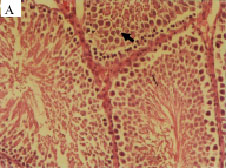
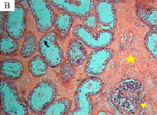
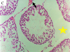
Histopathology of testis: Histopathological study showed that the cycle of spermatogenesis was regular in the control group (Fig. 1A). However, in all animals exposed to drugs were seen a depletion of germ cells, germinal cells necrosis, especially in spermatogonia, evidence of cell debris in lumen and present of lymphocyte and plasmocyte (except streptomycin group) (Fig. 1B). Expansion of interstitial space and intertubular space with vacuolization was developed and congestion in veins were increased in all experimental groups as compared with those seen in the control group (Fig. 1C). Diameter of seminiferous tubules was decreased in all experimental groups as compared with those measured in the control group (p<0.05) (Table 3).
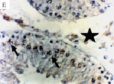
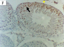
Results of apoptosis: In this study, the number of Tunel positive apoptotic germ cells per tubule cross-section at stages 1-4 and 5-8 and stages 9-13 increased following gentamicin, neomycin, ofloxacin treatment compared to the control group. The apoptotic spermatogonia were present in stages 1-4 and stages 9-13 but rarely observed in stages 5-8 in all groups except control group. The spermatocytes especially, leptotene-zygotene spermatocytes and pachytene spermatocytes were the main germ cells undergoing apoptosis in stages 9-13. Figures Tunel assay demonstrate the typical histograms of tunnel assay analyzed by light microscope in all groups (Fig. 1D-H). Using the in situ detection method by which the apoptotic cells can be identified by their darkly stained nuclei, we observed a low incidence of spontaneous apoptosis in normal rat testis from the control group. Although in low numbers, apoptotic germ cells were also observed in the control group when compared with the experimental groups (p<0.05). The rate of total Tunel positive apoptotic germ cells (spermatogonia and spermatocytes) were (24.15±3.216), (25.15±2.881), (34.15±2.584) and (15.15±3.523), respectively for gentamicin, neomycin, ofloxacin and streptomycin groups and (7.3±0.762) for the control group (Table 4). The incidence of Tunel positive apoptotic germ cells/100 tubules were higher in stages 1-4 and 9-13 in the experimental group when compared with those seen in the control group, according to the definition of Clermont-method, according this method the generations of developing germ cells within the cycle of the seminiferous epithelium in fourteenth day of study in all type of cells type A spermatogonia, intermediate spermatogonia, type B spermatogonia, Pereleptotene sparmatocytes, leptotene sparmatocytes, Zygotene sparmatocytes, Diplotene Spermatocytes in all stages were recorded (Clermont, 1972).
 |  |
 |  |
 |  |
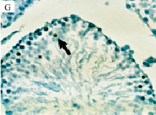 | 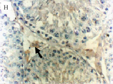 |
| Fig. 1: | Photomicrograph of testis in control group show regular epithelium seminiferous layers (arrow), H and E (A), X40, Photomicrograph of testis in ofloxacin group show depletion of germ cells, germinal cells necrosis (arrow) and present of lymphocyte and plasmocyte (triangle), Expansion of interstitial space with fibrosis were seen (star) (H and E) method (B), X40, Photomicrograph of testis in gentamicin group show expansion of interstitial space (star) with congestion in vein (arrow) (H and E) method (C), X40, Photomicrograph of testis in the control group shows apoptosis in primary spermatocyte that dark brown apoptotic body (arrow) detecting by Tunel assay (D), X40, Photomicrograph of testis in gentamicin group show Expansion of interstitial and intertubular space (star) and more dark brown apoptotic body (arrow) detecting by Tunel assay, (E), X40, Photomicrograph of testis in ofloxacin group shows Tunel positive apoptotic germ cells in spermatogonia cells (triangle) and more dark brown apoptotic body (arrow) detecting by Tunel assay (arrow) (F), X40 Photomicrograph of testis in neomycin group show that dark brown Tunel positive apoptotic germ cells in spermatocyte (arrow) detecting by Tunel assay (G), X40, Photomicrograph of testis in streptomycin group show dark brown Tunel positive apoptotic body in leydig cells (arrow) detecting by Tunel assay and (H), X40 |
| Table 4: | The effect of gentamicin, neomycin, streptomycin and ofloxacin on germinal cells apoptosis |
 | |
| Data are presented as mean±SE, *Significant different at p<0.05 level, (compared with the control group) | |
DISCUSSION
Antibiotics are used in the treatment of infection and as anticancer, antiprotozoal andantihelminthic agents. However, antimicrobial therapy has been shown to significantly affect semen parameters in human and animal models (Schlegel, 1991). This effect on spermatogenesis may have a significant impact on patients treated with these agents who desire fertility. In the present study, all four different antibiotics were tested, ofloxacin and three forms of aminoglycosides, neomycin, gentamycin and streptomycin, had adverse effect on spermatogenesis in rats. The present results indicated that, administration of ofloxacin for 14 consecutive days, results in a marked reduction in sperm count and viability as compared to respective controls. This is in agreement with that of Abd-Allah et al. (2000) who reported that ofloxacin at a dose of 72 mg kg-1, showed almost the highest potential in terms of impairment of the rat testicular functions. In addition, AndreeBen et al. (1993) reported that sperm was significantly decreased after 50 days when ofloxacin was administered to patients in a dose of 200 mg two times daily for 20 days. On the other hand, Crotty et al. (1995) have shown that ofloxacin in a dose of 10 mg/kg/day for 10 consecutive days in rats revealed testicular impairment, indicated by decreased haploid cells at days 11 and 56 from starting the treatment using flow cytometric analysis of testicular aspirate. In present study, sperm motility was not affected by ofloxacin. This is in agreement with Erhart et al. (1998) study. They showed that at physiologic or higher concentration, ofloxacin appears to be safe and does not affect sperm kinematic parameters when compared with controls. Present study is also comparable to the study by of King et al. (1997), who obtained similar results with a slightly different concentration of ofloxacin. In their study, no difference in motility, kinematic parameters, or hyperactivation were noted between the control and ofloxacin-exposed sperm. Moreover, Schramm (1986) showed that sperm motility was not affected by ofloxacin in a concentration up to 4.5 mg L-1 as tested on fresh ejaculate of andrologic patients. We have shown that male genital organ weight was decreased by ofloxacin. Degenerative changes in the seminiferous tubules and decrease of spermatozoa in the testis, epididymis and vas deferens are the evidence for genotoxicity. In the testis, a large component of the tissue weight is associated with spermatogenic function; hence, suppression of testicular weight could be due to large changes in the content of spermatids and spermatozoa. Therefore, the marked reduction of spermatozoa and spermatid in the seminiferous tubules, epididymis is most likely account for the effects on genital organ weights. The present study is demonstrated that apoptosis in male rat germ cells is induced by ofloxacin. These results indicate that ofloxacin look like as other chemical agents may directly interfere in the process of spermatogenesis. This increase in germ cell apoptosis is possibly due to an increased peroxide radical generation in the testis following ofloxacin treatment (Weyers et al., 2002) which then induces DNA single-strand breaks and chromosomal aberrations as demonstrated by in vitro genotoxicity studies (Sanchez et al., 2005; Itoh et al., 2006). In addition, ofloxacin could active Caspases 3 and induce apoptotic pathways (Aranha et al., 2002). The aminoglycosides widely used for treating many gram negative bacterial infections. Present results showed that aminoglycosides (gentamicin, neomycin, streptomycin) had an adverse effect on spermatogenesis. The effects of gentamicin treatment on spermatogenesis were investigated by Timmermans (1989) who found that men treated with gentamicin before prostatic surgery developed a cessation of meiosis at the stage of primary spermatocystes with an increase of normal and abnormal primary spermatocytes on testis biopsy. Studies using rats treated with therapeutic doses of gentamicin confirmed the observations in humans regarding the adverse effects of aminoglycosides on spermatogenesis (Narayana, 2008). These animals were found to have spermatogenic arrest with cessation of spermatogonial division and interruption of meiosis in primary spermatocytes. This could be due to decrease of the protein synthesis in the nuclei of the spermatogonia and of the spermatocytes and second a decrease of the glucose metabolism in the spermatocytes layers (Timmermans, 1989). In addition, we found that apoptosis in male rat germ cells is induced by gentamicin which is in agreement with other study (Servais et al., 2006). Servais et al. (2006) showed that gentamicin causes apoptosis at low concentrations in renal cells. The present study indicated that administration of neomycin resulted in a marked reduction in sperm count, motility and viability and increased in germ cell apoptosis as compared to respective controls. This result in agreement with earlier studies who reported that neomycin have an adverse effect on sperm concentration, total sperm count and spermatozoal motility in men with chronic inflammatory urologic conditions (Itoh et al., 2006). All antibiotics used in the present study had negative effect on spermatogenesis. However, these negative effects are less seen in the streptomycin group. Streptomycin and penicillin are the most common antibiotics used to maintain sterility in embryo and sperm culture media (Magli et al., 1996; Erbach et al., 1994; Gardner et al., 1998). Some laboratories use gentamycin to maintain the sterility of culture (Takahashi and First, 1992). However, little attention has been paid to their possible effect on sperm and embryo development in vitro. Previous study has reported an adverse effect of penicillin and streptomycin or streptomycin plus penicillin retarded or depressed protein and DNA synthesis (Moss et al., 1984). It has been reported that individually, penicillin, streptomycin and gentamicin did not affect embryo development in vitro. However, when penicillin and streptomycin were used together, the percentages of both 8-cell embryos at 58 h and blastocysts at 82 h were significantly lower than the control (Zhou et al., 2000). In conclusion, aminoglycosides (gentamicin, neomycin and streptomycin) and fluoroquinolone (ofloxacin) antibiotics have negative effect on sperm parameters and testis apoptosis in rats. However, these side effects are less seen in the streptomycin group. Therefore, it is recommended that usage of this drug have fewer side effects on male fertility.
ACKNOWLEDGMENTS
We thank the staff at Islamic Azad University of Tabriz for their help and support in the preparation of this research.
REFERENCES
- Abd-Allah, A.R., H.A. Aly, A.M. Moustafa, A.A. Abdel-Aziz and F.M. Hamada, 2000. Adverse testicular effects of some quinolone members in rats. Pharmacol. Res., 41: 211-219.
PubMed - AndreeBen, R., F. Sudhoff, V. Borgmann and R. Nagel, 1993. Results of ofloxacin therapy in andrologic patients suffering from therapy-requiring asymptomatic infections. Andrologia, 25: 377-383.
PubMed - Aranha, O., L. Zhu, S. Alhasan, D.P. Wood Jr, T.H. Kuo and F.H. Sarkar, 2002. Role of mitochondria in ciprofloxacin induced apoptosis in bladder cancer cell. J. Urol., 167: 1288-1294.
PubMed - Brooks, D.E., 1976. Activity and androgenic control of glycolytic enzymes in the epididymis and epididymal spermatozoa of the rat. Biochem. J., 156: 527-537.
CrossRefPubMedDirect Link - Carlsen, E., A. Giwercman, N. Keiding and N.E. Skakkebaek, 1992. Evidence for decreasing quality of semen during past 50 years. Br. Med. J., 305: 609-613.
PubMedDirect Link - Clermont, Y., 1972. Kinetics of spermatogenesis in mammals, seminiferous epithelium cycles and spermatogonial renewal. J. Physiol. Rev., 52: 198-236.
PubMed - Crotty, K.L., R. May, A. Kulvicki, D. Kumar and D.E. Neal, 1995. The effect of antimicrobical therapy on testicular aspirate flow cytometry. J. Urol., 153: 835-838.
PubMed - Demir, A., P. Turker, F.F. Onol, S. Sirvanci, A. Findik and T. Tarcan, 2007. Effect of experimentally induced Escherichia coli epididymo-orchitis and ciprofloxacin treatment on rat spermatogenesis. Int. J. Urol., 14: 268-272.
PubMed - Erbach, G.T., J.A. Lawitts, V.E. Papaioannou and J.D. Biggers, 1994. Differential growth of the mouse preimplantation embryo in chemically defined media. J. Biol. Reprod., 50: 1027-1033.
PubMed - Erhart, B., P.J. Chan, W.C. Patton and A. King, 1998. Ofloxacin: The next generation of antibiotic in sperm and embryo cultures for assisted reproductive technologies. Fert. Ster., 69: 246-251.
PubMed - Herbold, B.A., S.Y. Brendler-Schwaab and H.J. Ahr, 2001. Ciprofloxacin: In vivo genotoxicity studies. Mutat. Res., 498: 193-205.
PubMed - Itoh, T., K. Mitsumori, S. Kawaguchi and Y.F. Sasaki, 2006. Genotoxic potential of quinolone antimicrobials in the in vitro comet assay and micronucleus test. Mutat. Res., 603: 135-144.
PubMed - Gardner, D.K. and M. Lane, 1998. Culture of viable human blastocysts in defined sequential serum-free media. Hum. Reprod., 13: 148-159.
PubMed - King, K., P.J. Chan, W.C. Patton and A. King, 1997. Antibiotics: Effect on cryopreserved-thawed human sperm motility in vitro. Fert. Ster., 67: 1146-1151.
PubMed - Magli, M.C., L. Gianaroli, A. Fiorentino, A.P. Ferraretti, D. Fortini and S. Panzella, 1996. Improved cleavage rate of human embryos cultured in antibiotic-free medium. Hum. Reprod., 11: 1520-1524.
PubMed - Moss, P.S., D.H. Spector, C.A. Glass and R.C. Strohman, 1984. Streptomycin retards the phenotypic maturation of chick myogenic cells. In vitro Cell. Dev. Biol. Plant, 20: 473-478.
PubMed - Narayana, K., 2008. An aminoglycoside antibiotic gentamycin induces oxidative stress, reduces antioxidant reserve and impairs spermatogenesis in rats. J. Toxicol. Sci., 33: 85-96.
PubMed - Oliva, A., A. Giami and L. Multigner, 2002. Environmental agents and erectile dysfunction: A study in a consulting population. J. Androl., 23: 546-550.
PubMed - Sanchez, G., M.E. Hidalgo, J.M. Vivanco and J. Escobar, 2005. Induced and photoinduced DNA damage by quinolones: Ciprofloxacin, ofloxacin and nalidixic acid determined by comet assay. Photochem. Photobiol., 81: 819-822.
PubMed - Schlegel, P.N., T.S. Chang and F.F. Marshal, 1991. Antibiotics: Potential hazards to male fertility. Fertil. Steril., 55: 235-242.
PubMed - Schramm, P., 1986. Ofloxacin: Concentration in human ejaculate and influence on sperm motility. Infection, 14: 274-275.
PubMed - Servais, H., Y. Jossin, F. Van Bambeke, P.M. Tulkens and M.P. Mingeot-Leclercq, 2006. Gentamicin causes apoptosis at low concentrations in renal LLC-PK1 cells subjected to electroporation. J. Antimicrob. Agents Chemother., 50: 1213-1221.
CrossRefPubMedDirect Link - Takahashi, Y. and N.L. First, 1992. In vitro development of bovine one-cell embryos: Influence of glucose, lactate, pyruvate, amino acids and vitamins. Theriogenology, 37: 963-978.
PubMed - Timmermans, L.M., 1989. Modifications in spermatogenesis following antibiotic therapy. Acta Urol. Belg., 57: 35-46.
PubMed - Weyers, A.I., L.I. Ugnia, H. García Ovando and N.B. Gorla, 2002. Ciprofloxacin increases hepatic and renal lipid hydroperoxides levels in mice. Biocell., 26: 225-228.
PubMed - Xiaozhong, Yu., K. Hisayo, W. Ruisheng, S. Junzo, O. Yasutake, I. Gaku, T. Yasuhiro and H. Naomi, 2001. Involvement of Bcl-2 family genes and fas signaling systemin primary and secondary male germ cell apoptosis induced by 2-bromopropane in rat. J. Toxicol. Applied Pharmacol., 174: 35-48.
PubMed - Zhou, H., S.H. McKiernan, W. Ji and B.D. Bavister, 2000. Effect of antibiotics on development in vitro of hamster pronucleate ova. Theriogenology, 54: 999-1006.
PubMed
professor ehsan Reply
this is very good and scientific papers.
Ehsan m Reply
good researh paper