
Amir Hossein Ahmadi
Faculty of Pharmacy, Mazandaran University of Medical Sciences, Sari, Iran
Zeynab Ghazizadeh
Student Research Development Committee, Mazandaran University of Medical Sciences, Sari, Iran
Pakistan Journal of Biological Sciences
Year: 2008 | Volume: 11 | Issue: 10 | Page No.: 1394-1397


ABSTRACT
This study was carried out with the aim of evaluating the prevalence of G6PD deficiency, the prevalence of hemolysis with enzyme deficiency and determining the severity of icterus in the hospitalized newborns in our hospital. This prospective descriptive study has been conducted on 1018 icteric newborns admitted to the Bo-Ali Hospital from 2004 to 2007. The dataset included: age, sex, total and direct bilirubin, hemoglobin, reticulocyte count, blood group and Rh of mother and newborn, direct Coombs, G6PD level and the type of treatment. All data was analyzed by using statistical method. From 1018 neonates, 138 neonates (13.6%) were found to have G6PD deficiency. The male to female ratio was 3 to 1 (104 male and 34 female neonates). From 138 neonates with G6PD deficiency, hemolysis was seen in 15 neonates (10.9%) and the rate of G6PD deficiency with hemolysis was 1.6%. Out of 138 patients with G6PD deficiency, 2 patients (0.2%) had blood exchange transfusion. In this study the prevalence of G6PD deficiency in icteric newborns was considerably high and most of them were non hemolytic, so we recommend G6PD test as a screening program for every newborn at the time of delivery.
PDF Abstract XML References Citation
How to cite this article
Amir Hossein Ahmadi and Zeynab Ghazizadeh, 2008. Evaluation of Glucose-6-Phosphate Dehydrogenase Deficiency Without Hemolysis in Icteric Newborns at Mazandaran Province, Iran. Pakistan Journal of Biological Sciences, 11: 1394-1397.
DOI: 10.3923/pjbs.2008.1394.1397
URL: https://scialert.net/abstract/?doi=pjbs.2008.1394.1397
DOI: 10.3923/pjbs.2008.1394.1397
URL: https://scialert.net/abstract/?doi=pjbs.2008.1394.1397
INTRODUCTION
Glucose-6-phosphate dehydrogenase (G6PD) deficiency is the most common red cell enzyme abnormality associated with hemolysis (Beutler, 1978). It is also known to be associated with neonatal jaundice, kernicterus and even death (Valaes, 1994). Being an X-linked condition, the prevalence of G6PD deficiency in any given population is determined by the number of deficient males. However, deficient females are also at risk of hemolysis and jaundice (Miller, 1995). In a population with a high prevalence rate, early detection of the enzyme deficiency by neonatal screening is desirable in order to take appropriate measures to prevent the complications of hemolysis and jaundice (Mallouh et al., 1992). Neonatal jaundice due to G6PD deficiency may occur after exposure to oxidant agents and as a consequence hemolysis occur. But quite often, there is a mutation in the promoter site of uridyle di-phosphoglucoronyl transferase (UDPGT) which accompanies G6PD deficiency, leading to indirect hyperbilirubinemia in the absence of hemolysis (Segel, 2004; Halmaek and Stevenson, 2002).
One of the important manifestations of this enzyme deficiency in the neonatal period is jaundice without hemolysis. This may be so serious that it can lead to kernicterus or death and also predisposes the neonate to infection (Halmaek and Stevenson, 2002; Kaplan and Hammerman, 2004). Many studies conducted in the Mediterranean area and some Asian regions also confirmed that G6PD deficiency is a common cause of severe jaundice in the neonatal period (Tanphaichitr et al., 2003). Many researchers documented that the bilirubin levels of G6PD-deficient infants were still significantly higher than G6PD-normal infants even without any evidence of hemolysis (Eghbalian and Monsef, 2007; Kaplan and Abramov, 1992; Kaplan et al., 1999; Tan, 1981; Yaish et al., 1991).
Also numerous studies in Nigeria, India, Saudi Arabia, Singapore, Jamaica and Malaysia have revealed that this enzyme deficiency in icteric newborns was 40, 12.2, 18.4, 1.62, 1.57 and 3.5%, respectively, in which neonatal jaundice was seen without hemolysis (Ahmed et al., 1999; Slusher et al., 2000; Madan et al., 2001; Verma et al., 1999; Joseph et al., 1999; Eghbalian and Monsef, 2007; Hon and Balakrishnan, 1998).
The increasing number of case of neonatal jaundice in some geographical regions known as G6PD deficiency also seem to suggest a possible correlation between the two factors, however, at this point only limited empirical studies are available that could verify this possible explanation. This study was carried out with the aim of evaluating the prevalence of G6PD deficiency, the prevalence of hemolysis with enzym deficiency and determining the severity of icterus in the hospitalized newborns in our hospital.
MATERIALS AND METHODS
This descriptive prospective cross-sectional study, was accomplished in over four years from 2004 to 2007 on 1018 newborns hospitalized in Bo-Ali Hospital, Sari, Iran affiliated to Mazandaran University of Medical Sciences for jaundice. This hospital is the pediatric residency teaching hospital of Mazandaran University of Medical Sciences in Sari City, with a neonatal ward and NICU. Sari City is in the centre of Mazandaran Province, which is located in the north of Islamic Republic of Iran. From determination of the prevalence of hemolysis with enzym deficiency, samples included in this study were: term newborns (38 weeks gestational age or more) with a total bilirubin at more than 12 mg dL-1 and preterm newborns (gestational age less than 38 weeks) with a total bilirubin at more than 10-14 mg dL-1. The excluding criteria were; term neonates with a total bilirubin less than 12 mg dL-1, preterm newborns with a total bilirubin less than 10 mg dL-1 and direct hyperbilirubinemia (direct to total bilirubin ratio more than 20%). In summary, we excluded 953 cases of physiological jaundice and direct hyperbilirubinemia except sepsis, ABO and Rh incompatibility from the study. The criteria of hemolysis in our study was: reticulocyte count more than 3.2%±1.4 to 0.5%±0.4 (according to neonatal age in day) and hemoglobin less than 17.3±2.3 to 14.2±2.1 g dL-1 (according to neonatal age in day) (Halmaek and Stevenson, 2002). Derived data included: age, sex, total, direct and indirect bilirubin, hemoglobin, reticulocyte count, blood group and Rh of mother and newborn, direct Coombs test, G6PD level and type of treatment (phototherapy, Blood exchange transfusion).
The statistical Package for Social Sciences (SPSS; version 14) was used for the statistical analysis. Student`s t-test was used to assess the significance between means of two groups. p≤0.05 was considered statistically significant. The G6PD level was measured with a G6PD enzyme assay kit, spot test of qualitative method manufactured by Saba lab. The study was approved by the research committee of Mazandaran University of Medical Sciences.
RESULTS AND DISCUSSION
We found that 138 neonate out of 1018 (13.6%) icteric neonates were suffering from G6PD deficiency. 104 males
| Table 1: | The frequency of hemolysis in normal and deficient G6PD group |
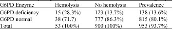 | |
| **Except sepsis, Rh and ABO incompatibility | |
| Table 2: | Hematological data of jaundiced newborn |
 | |
| Values are shown in mean±SD | |
(75%) and 34 females (25%) resulting in a male/female ratio of 3/1. The study could confirm the hemolysis criteria in 53 cases out of 953 neonates (5.6%), however in the 138 cases of G6PD deficiency we could confirm 15 cases (10.9%) of hemolysis and 123 cases (90.1%) without hemolysis. A total of 900 neonates out of 953 neonates did not have the criteria for hemolysis. In other words, the total frequency of G6PD deficiency with hemolysis in the hospitalized icteric newborns was 1.6% (Table 1). Mean total bilirubin concentration in the G6PD deficiency group compared with the normal G6PD group showed significant difference (p<0.05). But Mean hemoglobin concentration in G6PD deficient neonates compared with the normal G6PD group showed no significant difference (p>0.05). Furthermore, the study did not show a significant difference in the reticulocyte count between normal G6PD and deficient G6PD neonates (p>0.05) (Table 2). All G6PD deficient patients and other icteric neonates were treated with phototherapy; exchange transfusion is performed in 2 out of 138 G6PD deficient neonates (0.2%). None of the 138 G6PD deficient neonates had ABO and Rh incompatibility.
The etiological relationship between G6PD deficiency and neonatal hyperbilirubinemia has been confirmed by several studies. G6PD-deficient babies are 3-fold more prone to neonatal jaundice than G6PD-deficient infants (Al-Naama et al., 1987; Dawodu et al., 1998). G6PD deficiency is the most common red cell enzymopathy to cause neonatal hemolysis and jaundice. Good population data are available from West Africa (Bienzle, 1981), the Mediterranean (Milbauer et al., 1973) and the Far East (Tan, 1981). The presence of an additional hemolytic process such as ABO incompatibility was found to have little impact on the degree of hemolysis and hyperbilirubinemia (Kaplan et al., 1998). According to a WHO report, Iran is in a moderately high incidence area for G6PD deficiency (WHO Working Group, 1989). Favism is also common in some provinces of Iran, where fava beans are a common food. The objectives of this prospective study were to estimate the incidence of G6PD deficiency in newborns of Mazandaran province (north of Iran), to establish its relationship with hemolysis. The results showed that 13.6% of the study group affected by G6PD deficiency and that most of them (89.1%) were icteric without any signs of hemolysis. The results of the study confirmed the outcome of previous studies in Iran and other countries (Kaplan and Hammerman, 2004; Tanphaichitr et al., 2003; Ahmed et al., 1999; Verma et al., 1999; Eghbalian and Monsef, 2007). The hemolysis of G6PD is non-immune, therefore, direct Coombs test is negative, the results of our study did not show any positive Coombs test in the G6PD deficient group (Halmaek and Stevenson, 2002; Kaplan and Hammerman, 2004). Another study in Nigeria, showed that 40% of icteric newborns were suffering from G6PD deficiency, in most of them there was no without concomitant hemolysis (Ahmed et al., 1999). Madan et al. (2001) from India showed that in 12.2% of all icteric newborns suffered from G6PD deficiency and 48.7% of them had severe jaundice but without hemolysis (Madan et al., 2001). A study from Saudi Arabia showed 18.4% prevalence of G6PD deficiency in icteric newborns without any signs of hemolysis (Yaish et al., 1991). Present study showed a significant difference in the mean bilirubin level between G6PD deficient newborns and other icteric newborns. We performed exchange transfusion in 14.5% of G6PD deficient icteric neonates and 14.7% of neonates without G6PD deficiency. The results confirmed that jaundice in the G6PD deficient group is not more severe than in other causes of neonatal jaundice. A study in Saudi Arabia, showed severe jaundice without hemolysis in 18.4% of G6PD deficient neonates and in 15% of normal G6PD group neonates, they had blood exchange transfusion in 7% of G6PD deficient icteric newborns and in 8.2% of normal G6PD group (Eghbalian and Monsef, 2007). A screening program for G6PD enzyme in Malaysia on 8900 neonates revealed that the deficiency in 100 newborns and 17 out of them had exchange transfusion (17%). This study was in accord with our study (Hon and Balakrishnan, 1998). A study in Qazvin, Iran revealed that in 8% of hospitalized icteric newborns suffered from G6PD deficiency, 15.7% of them needed exchange transfusion (Eghbalian and Monsef, 2007).
Another study in Basrah, Iraq on 95 icteric newborns revealed that 51% of them suffered from G6PD deficiency and 27 out of them needed exchange transfusion (AL-Nama and Al-Sadoon, 1998). These findings were not compatible with our results; from the point of higher prevalence of G6PD deficiency and more cases exchange transfusion. We did not find a meaningful difference in the mean hemoglobin level and the reticulocyte count between normal and deficient G6PD enzyme groups. This confirms that the cause of jaundice in icteric newborns is not only the hemolysis, but also the concomitant mutation, leading to a decrease of G6PD and UDPGT activity that results jaundice. The texts also confirm these findings (Segel, 2004; Halmaek and Stevenson, 2002).
In this study the male/ female ratio was 3/1 in enzyme deficient neonates; it is predictable, because the disease is x-linked and more expressed in males. The affected females are homozygote or heterozygote, the later unfavorable lionization.
CONCLUSION
The results of the present study indicate that G6PD deficiency is a major cause of icterus in newborns (especially without hemolysis) in Iran and Mazandaran province, in Iran is a geographical area which the prevalence of Glucose-6-Phosphate Dehydrogenase Deficiency is high. So, We recommend that cord blood G6PD screening be considered in high-risk populations such as ours. This should be done in spite of laboratory findings for hemolysis as a routine test and it should be done as a screening test at birth time. Early diagnosis can reduce the number of complications in icteric neonate and prevents future acute hemolytic attacks. Due to a lack of funding and inavailability of quantitative G6PD enzyme assay kit, the program was applying qualitative methods which can lead to misdiagnosis of some cases of enzyme deficiency. So screening with quantitative tests at birth for every newborn is recommended.
ACKNOWLEDGMENT
This study supported by a grant from the Student Research Development Committee, Research Council of the Mazandaran University of Medical Sciences, Iran.
REFERENCES
- Ahmed, H., A. Yulubu and R. Hendricks, 1999. Neonatal jaundice in Zaria, Nigeria-A second prospective study. West Afr. J. Med., 18: 15-23.
PubMedDirect Link - Al-Naama, L.M., I.A. Al-Sadoon and M.M. Al-Naama, 1987. Neonatal jaundice and glucose-6-phosphate dehydrogenase deficiency in Basrah. Ann. Trop. Paediatr., 7: 134-138.
CrossRefPubMedDirect Link - Bienzle, U., 1981. Glucose-6-phosphate dehydrogenase deficiency. Trop. Afr. Clin. Haematol., 10: 785-799.
Direct Link - Dawodu, A., M.M. Qureshi, I.A. Moustafa and R.A. Bayoumi, 1998. Epidemiology of clinical hyperbilirubinaemia in Al Ain, United Arab Emirates. Ann. Trop. Paediatr., 18: 93-99.
PubMed - Eghbalian, F. and A.R. Monsef, 2007. Evaluation of glucose-6-phosphate dehydrogenase deficiency without hemolysis in icteric newborns. Iranian J. Pediatr., 17: 36-40.
Direct Link - Hon, A.T., S. Balakrishnan and Z. Ahmad, 1989. Hyperbilirubinaemia and erythrocytic glucose 6 phosphate dehydrogenase deficiency in Malaysian children. Med. J. Malaysia, 44: 30-34.
PubMed - Joseph, R., L.Y. Ho, J.M. Gomez, V.S. Rajdurai, S. Sivasankaran and Y.Y. Yip, 1999. Mass newborn screening for glucose-6-phosphate dehydrogenase deficiency in Singapore. Southeast Asian J. Trop. Med. Public., 30: 70-71.
PubMed - Kaplan, M. and A. Abramov, 1992. Neonatal hyperbilirubinemia associated with glucose-6-phosphate dehydrogenase deficiency in Sephardic-Jewish neonates: Incidence, severity and the effect of phototherapy. Pediatrics, 90: 401-405.
PubMed - Kaplan, M., H.J. Vreman, C. Hammerman, C. Leiter, B. Rudensky, M.G. MacDonald and D.K. Stevenson, 1998. Combination of ABO blood group incompatibility and glucose-6-phosphate dehydrogenase deficiency: Effect on hemolysis and neonatal hyperbilirubinemia. Acta Paediatr., 87: 455-457.
Direct Link - Kaplan, M., E. Beutler, H.J. Vreman, C. Hammerman, E. Levy-Lahad, P. Renbaum and D.K. Stevenson, 1999. Neonatal hyperbilirubinemia in glucose-6-phosphate dehydrogenase-deficient heterozygotes. Pediatrics, 104: 68-74.
PubMedDirect Link - Kaplan, M. and C. Hammerman, 2004. Glucose 6- phosphate dehydrogenase deficiency a hidden risk for kernicterus. Semin Perinatol., 28: 356-364.
Direct Link - Mallouh, A.A., G. Imseeh, Y.K. Abu-Osba and J.A. Hamdan, 1992. Screening for glucose-6-phosphate dehydrogenase deficiency can prevent severe neonatal jaundice. Ann. Trop. Pediatr. Int. Child Health, 12: 391-395.
CrossRefPubMedDirect Link - Milbauer, B., N. Peled and S. Svirsky, 1973. Neonatal hyperbilirubinemia and glucose-6-phosphate dehydrogenase deficiency. Isr. J. Med. Sci., 9: 1547-1552.
Direct Link - Slusher, T.M., H.J. Vreman, D.W. Melaren, L.J. Lewison, A.K. Brown and D.K. Stevenson, 2000. Glucose-6-phosphate dehydrogenase deficiency and carboxyhemoglobin concentrations associated with bilirubin-related morbidity and death in Nigerian infants. J. Pediatr., 126: 102-104.
CrossRefDirect Link - Tan, K.L., 1981. Glucose-6-phosphate dehydrogenase status and neonatal jaundice. Arch. Dis. Child., 56: 874-877.
PubMed - Tanphaichitr, V.S. , P. Pung-Amritt, S. Yodthong, J. Soongswang, C. Mahasandana and V. Suvatte, 2003. Glucose-6-phosphate dehydrogenase deficiency in the newborn: its prevalence and relation to neonatal jaundice. Southeast Asian J. Trop. Med. Public Health, 46: 137-144.
Direct Link - Valaes, T., 1994. Severe neonatal jaundice associated with glucose-6-phosphate dehydrogenase deficiency: Pathogenesis and global epidemiology. Acta Paediatr., 394: 58-76.
CrossRefDirect Link - WHO Working Group, 1989. Glucose-6-phosphate dehydrogenase deficiency. Bull. World Health Organ., 67: 601-611.
Direct Link - Yaish, H.M., G.A. Niazi, M. Al-Shaalan, S. Khan and G.S. Ahmed, 1991. Increased incidence of hyperbilirubinaemia in unchallenged glucose-6-phosphate dehydrogenase deficiency in term Saudi newborns. Ann. Trop. Paediatr., 11: 259-266.
PubMedDirect Link