
Lin Zhang
School of Science and Health, Parramatta Campus, Western Sydney University, Locked Bag 1797, Penrith NSW 2751, Australia
Narsimha Reddy
School of Science and Health, Parramatta Campus, Western Sydney University, Locked Bag 1797, Penrith NSW 2751, Australia
LiveDNA: 91.20335
Cheang Soo Khoo
WentworthInstitute, 302-306 Elizabeth Street, Surry Hills, NSW 2010, Australia
Sundar Rao Koyyalamudi
Institute of Endocrinology and Diabetes, The Children�s Hospital at Westmead, Sydney, NSW 2145, Australia
LiveDNA: 61.6258
Christopher E. Jones
School of Science and Health, Parramatta Campus, Western Sydney University, Locked Bag 1797, Penrith NSW 2751, Australia
Pharmacologia
Year: 2018 | Volume: 9 | Issue: 4 | Page No.: 157-168


ABSTRACT
Background and Objective: Lobelia chinensis Lour is an important anticancer herb used in traditional Chinese medicine. Many botanical polysaccharides are known to exhibit immunomodulatory and anticancer activities. This research aimed to analyze the L. chinensis polysaccharides (LCPs) for their biological activities relevant to their anticancer function. Materials and Methods: Water-soluble LCPs were extracted and purified using size-exclusion chromatography to obtain two dominant polysaccharides, LCP-1 and LCP-2 having molecular masses of 1899 kDa and 5.3 kDa, respectively. The antioxidant potentials of the isolated polysaccharides were evaluated by measuring radical scavenging activities against DPPH∙ (2,2-diphenyl-1-picrylhydrazyl radical), ABTS∙+(2,2'-azino-bis(3-ethylbenzothiazoline-6-sulphonic acid radical) and OH∙ (hydroxyl radical). Immunostimulatory activities of LCP-1 and LCP-2 were measured using mouse macrophages. Structure of the most active fraction (LCP-2) was determined using FT-IR and NMR spectroscopic techniques. Results: Two isolated polysaccharide fractions displayed significant antioxidant activities and stimulated the production of tumor necrosis factor-α (TNF-α) and interleukin-6 (IL-6), although LCP-2 is more effective. Detailed structural characterization by FT-IR and NMR was undertaken for the most active fraction (LCP-2) and confirmed that LCP-2 is(2,1)-β-fructan. Conclusion: The results suggested that the polysaccharides isolated from Lobelia chinensis Lour are potential candidates for immune-chemotherapy and suitable for the treatment of cancer.
PDF Abstract XML References Citation
How to cite this article
Lin Zhang, Narsimha Reddy, Cheang Soo Khoo, Sundar Rao Koyyalamudi and Christopher E. Jones, 2018. Antioxidant and Immunomodulatory Activities and Structural Characterization of Polysaccharides Isolated from Lobelia chinensis Lour. Pharmacologia, 9: 157-168.
URL: https://scialert.net/abstract/?doi=pharmacologia.2018.157.168
URL: https://scialert.net/abstract/?doi=pharmacologia.2018.157.168
INTRODUCTION
Lobelia chinensis Lour (Campanulaceae) is an important anticancer herb which has been widely used as diuretic, hemostat, antidote and anticancer agent in Traditional Chinese Medicine (TCM) for decades1,2. This herb is used in several traditional anticancer formulations to treat gastric cancer, lung cancer, colorectal cancer and liver cancer1,3. Modern scientific studies revealed that L. chinensis contains several important classes of bioactive compounds such as piperidine alkaloids, flavonoids, terpenoids and coumarins4-6. Several studies demonstrated that the extracts from this herb displayed a number of pharmacological activities such as antibacterial, anti-venom and anticancer activities4-6. The hot water extracts (decoction) from L. chinensis have been shown to display significant immunostimulatory and anticancer properties against liver cancer (H22) and Gastric cancer (BC-38)4,7,8. Preliminary scientific studies from authors’ laboratory indicated that the water extracts from L. chinensis have shown high antioxidant and immunostimulatory activities9-11. However, the literature on the polysaccharides from L. chinensis and their characterization is very limited1.
Literature demonstrates that botanical polysaccharides exhibit a variety of pharmacological activities that include anticancer, immune regulation and antioxidant activities12-21. In recent decades, herbal polysaccharides are proving to be ideal candidates for anticancer agents due to their relevant biological activities with minimal side effects1,14,21-26. Many polysaccharides such as lentinan, Polysaccharide Krestin (PSK), Polysaccharopeptide (PSP) and schizophyllan, isolated from medicinal mushrooms are in clinical use as anticancer agents due to their excellent anticancer properties along with immuno-regulatory effects12-14,21,27. Therefore, it is of enormous interest to study polysaccharides from traditional herbs that are promising candidates to develop novel therapeutics for the treatment of cancer as well as immune-regulation. To the best of our knowledge there is only one publication involving isolation of an α-Glucan from L. chinensis that displayed significant immunostimulatory activity1.
It is known in the literature that the plant extracts which show antioxidant and immunomodulatory potential, concurrently exhibit anticancer effects14,17,19-21,28-31. Therefore, the screening for potential immunomodulators is one of the important steps for the development of anticancer therapeutics. Hence, the objectives of this study were to isolate polysaccharides from L. chinensis and evaluate their antioxidant and immunostimulatory activities. It is also aimed to carry out structural characterization of L. chinensis polysaccharides using FT-IR and NMR spectroscopic techniques with a view to understand structure-activity relationship.
MATERIALS AND METHODS
This research was carried out between March 2015 to November 2017 as part of the program for the discovery of novel anticancer agents in Authors’ Laboratory.
Procurement of medicinal plant associated with this research: Lobelia chinensis Lour was purchased from Bei Jing Tong Ren Tang, a Chinese Herbal Medical Centre located in Sydney (Australia). This company has branches all over the world and is well known for their best practice in TCM. The herbs traded in Sydney centre have approvals from both Australian and Chinese Governments. The company undertakes stringent authentication and quality control procedures for all the herbal materials supplied by them.
Chemicals and materials: The 2,2-diphenyl-1-picrylhydrazyl (DPPH), 2,2'-azino-bis(3-ethylbenzothiazoline-6-sulphonic acid) (ABTS), dimethyl sulfoxide (DMSO), 1,10-phenanthroline, H2O2, ferrozine, 95% ethanol, ascorbic acid, sulfanilamide, N-(1-1-napthyl) ethylenediamine dihydrochloride, lipopolysaccharide (LPS) were purchased from Sigma (Australia) and Lomb Scientific Pvt., Ltd. (Australia). The Foetal Bovine Serum (FBS), antibiotics and Dulbecco’s modified Eagle’s medium (DMEM) with gluMax were purchased from BD Bioscience (USA). The tumour necrosis factor-α (TNF-α) and interleukin (IL-6) (mouse)-ELISA standards and antibodies were purchased from BD Bioscience (USA). Mouse macrophage cells (RAW 264.7) were purchased from Sigma-Aldrich.
Extraction and fractionation of polysaccharides from L. chinensis: To extract water-soluble compounds, 25 g of L. chinensis Lour was powdered then autoclaved (121°C, 2 h). The extract was cooled to laboratory temperature and the supernatant was separated by filtration and then crude compounds were precipitated by treating the supernatant with 95% ethanol (Fig. 1). The isolated polysaccharides were de-proteinated using Sevag method21 and purified using gel filtration (Sepharose CL-6B; 2.4×99 cm, flow rate: 0.51 mL min–1). Details of the procedure followed for the extraction and purification of polysaccharides is similar to that published previously16,17,19-21 and described in Fig. 1. Polysaccharide fractions were obtained based on the sugar profile of the eluted samples19-21. These fractions were collected, freeze-dried and then kept at -20°C until further studies.
Determination of average molecular mass: Molecular weights of these fractions were determined by calibrating the Sepharose CL-6B gel filtration column.
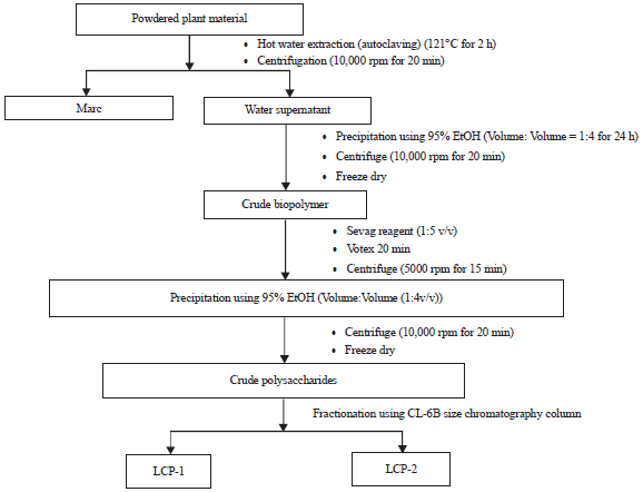 | |
| Fig. 1: | Flow chart for the extraction of polysaccharides from L. chinensis |
Standard dextrans with average molecular weight (from 2000 to 1 kDa) were used to obtain a calibration curve16,17,19-21. Regression of the standard curve gave a linear equation (with R² = 0.9915) represented by:
y = -0.2763x+1.8022
This equation was used to estimate the average molecular weight of polysaccharides obtained from L. chinensis (LCPs).
Determination of chemical composition: The total sugar content was measured using phenol-sulfuric acid method16,17,19-21,32. Glucose was used to produce a standard curve that was used to determine the sugar contents. Regression of the standard curve gave a linear equation (with R² = 0.9964) represented by:
y = 0.0018x+0.0374
The total protein contents were measured using modified Lowry’s method, using BSA to prepare standards17,19-21,33. BSA was used to build a standard equation that was used to determine the bound protein. Regression of the standard curve gave a linear equation (with R² = 0.9923) represented by:
y = 0.0017x-0.0212
The mono-sugar contents were determined by gas chromatography (Hewlett Packard 7890B) with FID detection16. The approach followed to prepare the samples for GC analysis was based on the procedure published previously16,21,34. Mannose, glucose, galactose, xylose, fucose, rhamnose, arabinose and ribose were used as mono-sugar standards.
FT-IR analysis: A TENSOR II FTIR Spectrometer (BRUKER) was used for structural characterization of LCP’s at room temperature (25°C)16. All spectra were recorded between the frequency range of 4000-450 cm–1.
NMR analysis: 1H, 13C, g-COSY and HSQC spectra were recorded using Bruker Avance 400 MHz NMR spectrometer using an inverse detection probe with pulsed field gradient capabilities. LCP-2 (25 mg) was dissolved in 600 μL of D2O (99.9%) containing 0.15% TSP (v/v ratio) and all NMR experiments were performed at 40°C.
Bioactivity tests
DPPH• scavenging assay: The Blois method35 was used to determine the DPPH• scavenging abilities of polysaccharides. The procedure employed for this assay was similar to previously published methods17,19,20,36. Ascorbic acid was employed as positive control and deionised water as blank. The absorbance values were determined using UV spectrophotometer at 492 nm (Multiskan 141 EX, Thermo Electron, USA). Regression of the data gave a linear standard curve (with R² = 0.9715) represented by the following equation:
y = -0.0026x+0.5578
DPPH• scavenging potential of LCPs was determined as the ascorbic acid equivalence using the above equation.
ABTS•+ radical scavenging assay: ABTS•+ scavenging abilities of polysaccharides were determined using published10,11,36,37. Ascorbic acid was employed as positive control with PBS buffer (pH 7.4) as blank. A standard curve was built using different concentrations of ascorbic acid solution (prepared in 60% methanol) in the range of 0-400 μM. Absorbance values were determined using a UV spectrophotometer at 734nm10,11 (Multiskan 141 EX, Thermo Electron, USA).
Regression of the data gave a linear standard curve (with R² = 0.9699) represented by the following equation:
y = -0.0017x+0.716
The ABTS•+ scavenging capacities of LCPs were determined as the ascorbic acid equivalence using the above equation.
OH• radical scavenging test: The OH• scavenging capacity of LCPs was measured employing the method described by De Avellar et al.38 and Pownall et al.39 with minor modification. Briefly, 50 μL of polysaccharide fractions or ascorbic acid (1 mg mL–1) were added with 50 μL of 3 mM 1,10-phenanthroline and 50 μL of 3 mM FeSO4 in a 96 well microtiter plate. About 50 μL of H2O2 (0.01% v/v) was then added and mixed to trigger the competition between polysaccharides38 and Fe2+ for OH•. The solution was incubated at ~37°C for 60 min. The absorbance values were measured at 536 nm using UV-spectrophotometer. The OH• scavenging ability of LCPs was determined employing the equation:
where, the negative control is the reaction mixture without sample and without ascorbic acid. The blank is the reaction mixture without sample, ascorbic acid and H2O2.
Immunostimulatory activity assays: Procedure for the preparation and maintenance of mouse macrophages (RAW 264.7) is similar to that published in the literature10,11,16,40.
IL-6 production: ELISA kit (IL-6, BD Biosciences, San Jose, CA, USA) is then used to measure the concentration of IL-6 as per the procedure provided in the manufacturer’s manual10,11,16,40. All experiments were conducted in triplicate. Standard IL-6 (mouse) was used to produce the calibration curve that gave a linear equation (with R² = 0.992):
y = 0.0019x+0.0248
The concentrations of IL-6 produced by the polysaccharides were calculated using the above equation.
TNF-α production: ELISA kit (TNF-α, BD Biosciences, San Jose, CA, USA) was then used to measure the concentration of TNF-α as per the method provided in the manual and as previously described10,11. Triplicate measurements were conducted.
Standard TNF-α (mouse) was used to produce the calibration curve that gave a linear equation (with R² = 0.9875):
y = 0.0017x+0.0706
The concentration of TNF-α produced by the polysaccharide extracts were calculated using the above equation.
Determination of toxicity by MTT test: Viability of macrophage cells (RAW 264.7) were measured employing the MTT assay as previously described10,11,16,41. The absorbance values were the measured at 595 nm and the fraction of live cells was determined using the following equation:
The positive control was mouse macrophages treated by only the DMEM medium (without LPS and sample).
Statistical analysis: All data were measured in triplicate and Mean±SD were determined. A one-way analysis of variance (ANOVA) and Duncan’s multiple range tests were used for analysis of data. Statistical calculations were performed using OriginPro 8.5 and Excel 2016. The data were considered to be statistically significant if p<0.05.
RESULTS AND DISCUSSION
Extraction and fractionation of polysaccharides from L. chinensis: Two polysaccharide fractions, namely, LCP-1 and LCP-2 were isolated from L. chinensis using hot water extraction and subsequent purification on a Sepharose CL-6B column (Fig. 2a). As could be seen from the sugar profile (Fig. 2a), LCP-2 is the major polysaccharide fraction. Calibration of the column with dextran standards revealed that the average molecular masses of LCP-1 and LCP-2 were 1899 and 5.3 kDa, respectively (Fig. 2b).
Chemical compositions of the fractions: Total carbohydrate and total protein compositions of isolated fractions (LCP-1 and LCP-2) were analyzed and these results are presented in Table 1. The carbohydrates are the chief constituents of each fraction (94% in LCP-1 and 96% in LCP-2). Table 1 also presented the mono-saccharide compositions of the isolated fractions. It can be seen from these results that LCP-1consisted mainly of Rhamnose (14.39%), Arabinose (24.39%), Galactose (33.8%) and Glucose (18.87%).
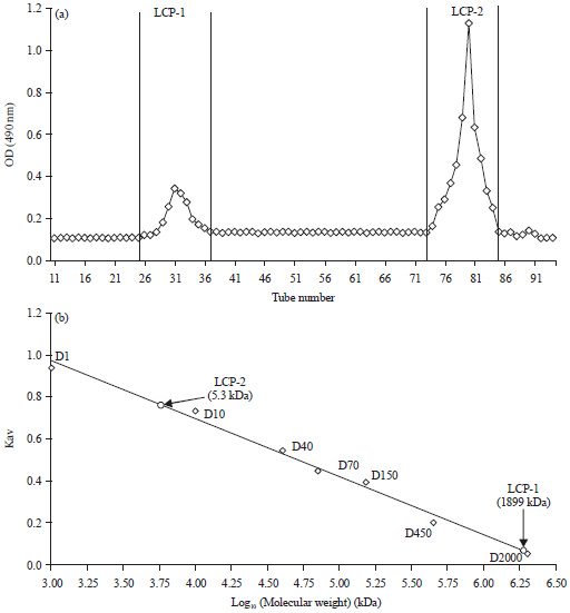 | |
| Fig. 2(a-b): | (a) Isolation and purification of LCPs using Sepharose CL-6B column (two fractions: LCP-1and LCP-2 were separated) and (b) Standard calibration curve for the calculation of average molecular masses of LCPs based on the elution volume and the molecular mass of standard dextran samples |
| D2000: 2,000 kDa, D450: 450 kDa, D150: 150 kDa, D70: 70 kDa, D40: 40 kDa, D10: 10 kDa and D1: 1 kDa; Kav: (Ve-Vo)/(Vt-Vo), Vo is void volume of the column, Vt is total column volume, Ve is elution volume of the fraction | |
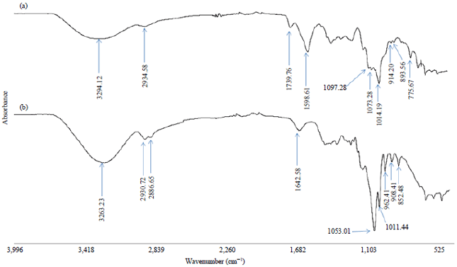 | |
| Fig. 3: | FTIR spectrum of (a) LCP-1 and (b) LCP-2 |
| Table 1: | Chemical composition and monosaccharide contents of LCPs |
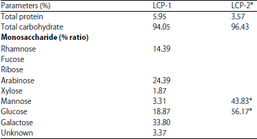 | |
*Fructose is the major monosaccharide present in LCP-2. It should be noted that, during the reduction step of GC sample preparation, fructose gets reduced to mannitol and glucitol. Therefore, mannose and glucose are seen in GC results instead of fructose | |
However, the main mono-saccharides in LCP-2 were mannose (43.83%) and glucose (56.17%) (Table 1). It is important to note at this point that, reduction of fructose during GC sample preparation yields mannitol and glucitol42-44. It is therefore expected that LCP-2 may possibly contain glucomannan and/or fructan. FT-IR and NMR spectroscopic investigations have provided further structural details of LCP-2 indicating the presence of fructose units in this polysaccharide.
Vibrational spectroscopy analysis: Figure 3 provided FT-IR spectra of L. chinensis polysaccharides LCP-1 and LCP-2. LCP-1 showed peaks (Fig. 3a) consistent with α-glycosidic linkage (755.6 cm–1) and β-glycosidic linkage (893 and 914 cm–1)16,21,45,46. The spectrum also showed three strong absorption bands at 1014.19, 1073.28 and 1097.28 cm–1 (corresponding to C-Ostretching vibrations related to glycosidic bonds) indicating the presence of a pyranose sugar in LCP-116,21,45,46. The broad peak centred at 3294.12 cm–1 corresponds to hydroxyl stretching vibrations of the polysaccharide and the peaks at 2934.58 cm–1 are due to C-H stretching vibrations21,46. Remaining peaks are consistent with polysaccharide structure. These observations lead to the conclusion that LCP-1 contains pyranose sugars with α- and β-glycosidic linkages. LCP-2 (Fig. 3b) has peaks at 908.41 and 852.48 cm–1 indicating the presence of β-glycosidic linkage16,21,45,46. The two strong absorption peaks in the range of 1011-1053 cm–1 (corresponding to C-O stretching vibrations related to glycosidic bonds) indicate the presence of furanose sugars in LCP-221,46. As can be seen from the vibrational bands in the range 1010-1100 cm–1 of the FT-IR spectrum (Fig. 3b), there are no pyranose type sugars21,46 in LCP-2. The presence of only furanose sugars indicates that LCP-2 might be a fructan42,43. The broad band centred on 3263.68 cm–1 corresponds to the hydroxyl stretching vibrations of the polysaccharide and the peaks at 2930.72 and 2886.65 cm–1 belong to C-H stretching vibrations. These observations confirm that LCP-2 contains furanose sugars with β-glycosidic linkages.
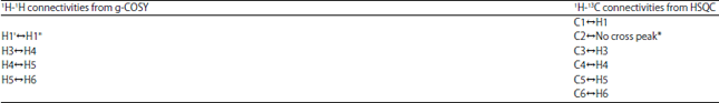
NMR spectroscopy analysis: Results demonstrated that LCP-2 is the most active and major polysaccharide fraction isolated from L. chinensis. Therefore, a detailed structural analysis of LCP-2 was carried out by NMR spectroscopy. 1H, 13C, g-COSY and HSQC spectra of LCP-2 were given in Fig. 4. Standard assignment protocols were used to obtain proton and carbon chemical shift assignments (Table 2) using 1D- and 2D-NMR spectra. Absence of intense proton resonances in 4.4-5.5 ppm range (Fig. 4a) indicated that there were no anomeric protons in LCP-2. In addition, LCP-2 showed six intense carbon peaks (Fig. 4b) indicating that it contains a single mono-saccharide unit in the main chain. These observations together with GC and FT-IR results strongly indicate that LCP-2 is a fructan with fructose units in the main chain. The weak anomeric doublet at 5.42 ppm in the proton spectrum (Fig. 4a) is likely due to the presence of a chain terminating glucose residue is the fructan. A set of weak resonances between 3.3-3.6 ppm confirms the terminal glucose residue.
The fructan backbone structure of LCP-2 can be confirmed from proton and carbon connectivities in g-COSY and HSQC spectra. The g-COSY spectrum showed several proton connectivities as shown in Table 3. Clearly, the protons H3 to H6 within the fructofuranosyl ring (Fig. 5) displayed correlations in the g-COSY spectrum. Also, the two allylic protons H1' and H1" attached to C1 showed intense cross peak in the g-COSY spectrum (Table 3, Fig. 4c). Five intense cross peaks are clearly observed for the five protonated carbons in the HSQC spectrum (Table 3, Fig. 4d). The anomeric carbon (C2) at 105.91 ppm (Table 2, Fig. 4b) did not show any cross peak as there are no protons attached to this carbon (Table 3, Fig. 5). Literature showed that β-anomeric 13C resonances are commonly located between21,46,47 103 and 105 ppm. Therefore, the large chemical shift value (105.91 ppm) observed for the anomeric carbon demonstrates that LCP-2has a β-glycosidic linkage21,46,47. Proton and carbon chemical shifts of LCP-2 (Table 2) are consistent with the correlations observed in 2D-NMR spectra. These assignments together with FT-IR and GC findings lead to following conclusions: (i) GC analysis revealed that LCP-2 may contain glucomannan and/or fructan, (ii) FT-IR results indicated that LCP-2 contains only furanose sugars with β-glycosidic linkage (and no pyranose sugars are present) indicating the presence of fructose units in the backbone with β-linkage and (iii) NMR results together with FT-IR findings confirm that LCP-2 is a β-D-(2→1) fructofuranoside. These results demonstrated that LCP-2 contains the following fructan backbone with glucose in the chain terminating position:
The chemical shift values of LCP-2 presented in Table 2 match well with inulin type β-fructans isolated from Artemisia japonica42, Saussurea costus43, Ophiopogon japonicas44 and Matrisia maritima48 confirming the structure proposed for LCP-2 (Fig. 5). This is a significant result reporting isolation of an immunostimulatory fructan for the first time from Lobelia chinensis.
Radical scavenging activities: The results of radical scavenging activities of LCPs (against three different radicals) are given in Table 4.
| Table 2: | 1H and 13C chemical shifts (ppm) of LCP-2 from L. chinensis |
| Table 3: | Proton and carbon correlations from 2D-NMR spectra data of LCP-2 from L. chinensis |
 | |
| *There is no proton on C2 (no H2) in fructan | |
| Table 4: | Radical scavenging activities of LCPs along with their average molecular mass |
 | |
| #ABTS and DPPH free radical scavenging activity was expressed as equivalent of ascorbic acid, *The percentage of inhibition of OH production after treatment with polysaccharides, Values: Mean±standard deviation (n = 3) | |
 | |
| Fig. 4(a-d): | NMR spectra of LCP-2 isolated from L. chinensis (a) 1H-NMR, (b) 13C-NMR, (c) g-COSY and (d) HSQC |
It was cleared from the results that, both LCP-1 and LCP-2 displayed significant radical scavenging activities against the three radicals tested in this research. LCP-1 exhibited slightly better scavenging activity than LCP-2 (Table 4). An interesting point to be noted here is that the only mono-saccharide present in LCP-2 is fructose.
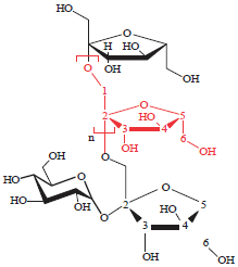 | |
| Fig. 5: | Structure of LCP-2 |
The antioxidant activities of LCP-2 observed in this research are consistent with literature studies showing that fructans have robust antioxidant activities49.
Immuno-stimulatory activities of L. chinensis polysaccharides: Immuno-stimulatory activities of LCP-1 and LCP-2 were measured by treating RAW 264.7 cells with purified LCPs.
Findings of this research indicated that LCP-1 and LCP-2 displayed immuno-stimulatory effects as demonstrated by increase in the production of TNF-α and IL-6 as a function of polysaccharide concentration (Fig. 6). It is extremely important to note that the results presented in Fig. 6a demonstrated that LCP-2 displays better activity than LPS (positive control) with respect to the production of TNF-α. As could be seen from Fig. 6c and d, immuno-stimulatory activities of LCP-1 and LCP-2 increase sharply when the polysaccharide concentration is greater than 30 μg mL–1.
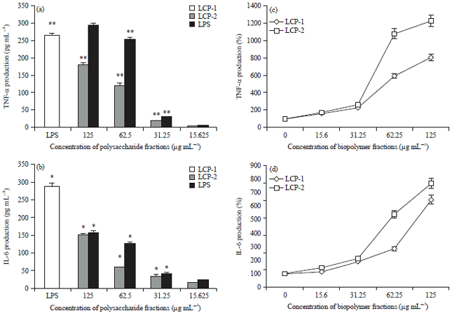 | |
| Fig. 6(a-d): | Effects of L. chinensis polysaccharides on murine RAW 264.7 macrophages, (a, c) Production of tumor necrosis factor-α (TNF-α) and (b, d) Production of interleukin 6 (IL-6) |
| LPS was the positive control (100 ng mL–1). ELISA assay was used for the quantification of IL-6 and TNF-α production, *Statistical difference for the positive control (LPS treated group) and the samples was significant, (p<0.02, n = 3), **Statistical difference for the positive control (LPS treated group) and the samples was significant, (p<0.01, n = 3) | |
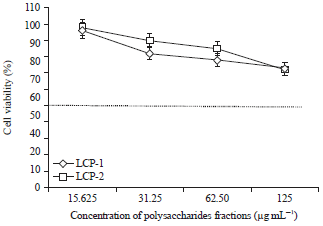 | |
| Fig. 7: | Cell viabilities of isolated polysaccharides from L. chinensis |
Excellent immuno-stimulatory activities have been observed for LCP-2 at 125 μg mL–1 as indicated by (i) More than 12 fold enhancement in the production of TNF-α in relation to the negative control (untreated macrophages) (Fig. 6c) and (ii) Nearly 8 fold increase in the production of IL-6 (Fig. 6d).
Cell viability: Effect of LCP-1 and LCP-2 on cell viabilities was given in Fig. 7. Results demonstrated that the polysaccharides from L. chinensis showed low toxicities even at the largest concentrations (125 μg mL–1) studied in this research (Fig. 7). These findings are in agreement with those reported in the literature that plant polysaccharides display less toxicity14,17,19-21,28-31.
The results demonstrated that Lobelia chinensis polysaccharides exhibit significant radical scavenging activities. An important point to be noted here is that LCP-2 is the major polysaccharide fraction present in this herb and the only mono-saccharide in this fraction is fructose. The antioxidant activities of LCP-2 observed in this research demonstrated the robust nature of antioxidant activities of fructans49.
Findings of this research demonstrate that LCP-2 exhibited excellent immuno-stimulatory activities (Fig. 6c, d) and the observed activity is superior to the activity of positive control (LPS) with respect to the production of TNF-α. These observations are very significant and demonstrate that LCP-2 is highly suitable herbal polysaccharide to stimulate the immune system. Literature demonstrates that fructans can stimulate immune cells by binding to Toll like Receptor (TLR)49. It is important to note from the literature that the immunomodulatory activity plays a critical role in anticancer activity14,17,19-21,28-31. It is therefore concluded that the major component of L. chinensis polysaccharides (LCP-2) is highly potential candidate for immuno-chemotherapy. FT-IR and NMR spectroscopic studies have confirmed the structure of this fructan.
CONCLUSION
In this study, two polysaccharide fractions were isolated from L. chinensis (LCP-1 and LCP-2). A novel immuno-stimulatory polysaccharide (LCP-2) with β-D-(2→1)-fructofuranoside structure has been identified from L. chinensis. LCP-1 and LCP-2 have shown highly significant immuno-stimulatory and antioxidant activities. Especially, LCP-2 has exhibited extremely high immuno-stimulatory activity and less toxicity demonstrating that it has huge potential for immuno-therapeutic applications. This is a significant result reporting the isolation of an immuno-stimulatory fructan for the first time from Lobelia chinensis. These findings suggested the potential of L. chinensis polysaccharides to develop effective formulations for immunotherapeutic and anticancer applications.
SIGNIFICANCE STATEMENT
Lobelia chinensis Lour is an important anticancer herb used in traditional Chinese medicine. Abundant literature demonstrates that plant/herbal polysaccharides display significant immunomodulatory and anticancer effects with minimal toxicity. This research was aimed to isolate pure polysaccharides from L. chinensis and study their biological activities with a view to discover strong immunostimulators. The results of this research suggested that the polysaccharides isolated from Lobelia chinensis Lour were highly potential candidates for immuno-chemotherapy. Especially, one of the fraction (LCP-2) is a fructan that displayed extremely high immunomodulatory activity that was superior to the activity of LPS (a known positive control). The structure of this most active fraction (LCP-2) was found to be (2,1)-β-fructan. Major significance of this research is the discovery of immune-enhancing fructan from L. chinensis. This polysaccharide has huge potential to develop novel formulations for immunotherapy that will aid the discovery of effective immuno-chemotherapeutic regimen to treat cancer. Cancer therapy is expensive and many patients cannot afford the cost. Hence, the discovery of novel therapeutics from medicinal herbs will provide great benefit.
ACKNOWLEDGMENTS
Lin Zhang acknowledges IPRS scholarship from the Western Sydney University and National Institute of Complementary Medicine (NICM) during his PhD candidature. Help from Dr Allan Torres with NMR spectroscopy studies is gratefully acknowledged. Author also acknowledges Professor Kai Yip Cho Memorial Scholarship from Prof. Cho’s family.
REFERENCES
- Li, X.J., W.R. Bao, C.H. Leung, D.L. Ma and G. Zhang et al., 2016. Chemical structure and immunomodulating activities of an α-glucan purified from Lobelia chinensis lour. Molecules, Vol. 21.
CrossRefDirect Link - Shibano, M., D. Tsukamoto, A. Masuda, Y. Tanaka and G. Kusano, 2001. Two new pyrrolidine alkaloids, radicamines A and B, as inhibitors of α-glucosidase from Lobelia chinensis Lour. Chem. Pharm. Bull., 49: 1362-1365.
CrossRefDirect Link - Chen, M.W., W.R. Chen, J.M. Zhang, X.Y. Long and Y.T. Wang, 2014. Lobelia chinensis: Chemical constituents and anticancer activity perspective. Chin. J. Natl. Med., 12: 103-107.
CrossRefDirect Link - Kuo, P.C., T.L. Hwang, Y.T. Lin, Y.C. Kuo and Y.L. Leu, 2011. Chemical constituents from Lobelia chinensis and their anti-virus and anti-inflammatory bioactivities. Arch. Pharm. Res., 34: 715-722.
CrossRefDirect Link - Yang, S., T. Shen, L. Zhao, C. Li, Y. Zhang, H. Lou and D. Ren, 2014. Chemical constituents of Lobelia chinensis. Fitoterapia, 93: 168-174.
CrossRefDirect Link - Shao, J.H. and H. Zhang, 2010. Influence of Lobelia chinensis lour. Decoction on expression of C-erbB-2 and P53 on H22 tumor-bearing mice. Chin. J. Clin. Pharm., 19: 372-375.
Direct Link - Ravipati, A.S., L. Zhang, S.R. Koyyalamudi, S.C. Jeong and N. Reddy et al., 2012. Antioxidant and anti-inflammatory activities of selected Chinese medicinal plants and their relation with antioxidant content. BMC Complement. Alternat. Med., Vol., 12.
CrossRefDirect Link - Zhang, L., C. Khoo, S.R. Koyyalamudi, N.D. Pedro and N. Reddy, 2017. Antioxidant, anti-inflammatory and anticancer activities of ethanol soluble organics from water extracts of selected medicinal herbs and their relation with flavonoid and phenolic contents. Pharmacologia, 8: 59-72.
CrossRefDirect Link - Zhang, L., C.S. Khoo, S.R. Koyyalamudi, N. de Pedro and N. Reddy, 2018. Immunostimulatory and anticancer activities of polysaccharides extracted from traditional anticancer chinese medicinal herbs. Pharmacologia, 9: 18-29.
Direct Link - Cheng, K.F. and P.C. Leung, 2008. General review of polysaccharopeptides (PSP) from C. versicolor: Pharmacological and clinical studies. Cancer Therapy, 6: 117-130.
Direct Link - Friedman, M., 2016. Mushroom polysaccharides: Chemistry and antiobesity, antidiabetes, anticancer and antibiotic properties in cells, rodents and humans. Foods, Vol. 5, No. 4.
CrossRefDirect Link - Schepetkin, I.A. and M.T. Quinn, 2006. Botanical polysaccharides: Macrophage immunomodulation and therapeutic potential. Int. Immunopharmacol., 6: 317-333.
CrossRefDirect Link - Thambiraj, S.R., M. Phillips, S.R. Koyyalamudi and N. Reddy, 2015. Antioxidant activities and characterisation of polysaccharides isolated from the seeds of Lupinus angustifolius. Ind. Crops Prod., 74: 950-956.
CrossRefDirect Link - Jeong, S.C., S.R. Koyyalamudi, Y.T. Jeong, C.H. Song and G. Pang, 2012. Macrophage immunomodulating and antitumor activities of polysaccharides isolated from Agaricus bisporus white button mushrooms. J. Med. Food, 15: 58-65.
CrossRefDirect Link - Yao, Y., Y. Zhu and G. Ren, 2016. Immunoregulatory activities of polysaccharides from mung bean. Carbohydr. Polym., 139: 61-66.
CrossRefDirect Link - Zhang, L., S.R. Koyyalamudi, S.C. Jeong, N. Reddy, P.T. Smith, R. Ananthan and T. Longvah, 2012. Antioxidant and immunomodulatory activities of polysaccharides from the roots of Sanguisorba officinalis. Int. J. Biol. Macromol., 51: 1057-1062.
CrossRefDirect Link - Zhang, L., K.S. Rao, S.C. Jeong, N. Reddy, T. Bailey and T. Longvah, 2013. Immunomodulatory activities of polysaccharides isolated from Taxillus chinensis and Uncaria rhyncophylla. Carbohydr. Polym., 98: 1458-1465.
CrossRefPubMedDirect Link - Zhang, L., N. Reddy and S.R. Koyyalamudi, 2014. Isolation, Characterization and Biological Activities of Polysaccharides from Medicinal Plants and Mushrooms. In: Studies in Natural Products Chemistry, Volume 42, Atta-ur-Rahman (Ed.). Chapter 5, Elsevier, UK., ISBN: 978-0-444-63281-4, pp: 117-147.
Direct Link - Kowalczewska, M., J. Piotrowski, T. Jedrzejewski and W. Kozak, 2016. Polysaccharide peptides from Coriolus versicolour exert differential immunomodulatory effects on blood lymphocytes and breast cancer cell line MCF-7 in vitro. Immunol. Lett., 174: 37-44.
CrossRefDirect Link - Liu, F., Z.Y. Zhu, X. Sun, H. Gao and Y.M. Zhang, 2017. The preparation of three selenium-containing Cordyceps militaris polysaccharides: Characterization and anti-tumor activities. Int. J. Biol. Macromol., 99: 196-204.
CrossRefDirect Link - Seedevi, P., M. Moovendhan, S. Vairamani and A. Shanmugam, 2016. Structural characterization and biomedical properties of sulfated polysaccharide from the gladius of Sepioteuthis lessoniana (Lesson, 1831). Int. J. Biol. Macromol., 85: 117-125.
CrossRefDirect Link - Shan, T., T. Shan, F. Liu, H. Zheng and G. Li, 2017. Effects of Lycium barbarum polysaccharides on the damage to human endometrial stromal cells induced by hydrogen peroxide. Mol. Med. Rep., 15: 879-884.
CrossRefDirect Link - Zhu, Z.Y., F. Dong, X. Liu, Q. Lv and F. Liu et al., 2016. Effects of extraction methods on the yield, chemical structure and anti-tumor activity of polysaccharides from Cordyceps gunnii mycelia. Carbohydr. Polym., 140: 461-471.
CrossRefDirect Link - Ina, K., T. Kataoka and T. Ando, 2013. The use of lentinan for treating gastric cancer. Anti-Cancer Agents Med. Chem. (Formerly Curr. Med. Chem.-Anti-Cancer Agents), 13: 681-688.
Direct Link - Bafna, A. and S. Mishra, 2010. Antioxidant and immunomodulatory activity of the alkaloidal fraction of Cissampelos pareira Linn. Sci. Pharm., 78: 21-31.
CrossRefDirect Link - Ayeka, P.A., Y. Bian, P.G. Mwitari, X. Chu, Y. Zhang, R. Uzayisenga and E.O. Otachi, 2016. Immunomodulatory and anticancer potential of Gan cao (Glycyrrhiza uralensis Fisch.) polysaccharides by CT-26 colon carcinoma cell growth inhibition and cytokine IL-7 upregulation in vitro. BMC Complement. Altern. Med., Vol. 16, No. 1.
CrossRefDirect Link - Wang, L., Y. Li, L. Zhu, R. Yin and R. Wang et al., 2016. Antitumor activities and immunomodulatory of rice bran polysaccharides and its sulfates in vitro. Int. J. Biol. Macromol., 88: 424-432.
CrossRefDirect Link - Yu, X.H., Y. Liu, X.L. Wu, L.Z. Liu, W. Fu and D.D. Song, 2017. Isolation, purification, characterization and immunostimulatory activity of polysaccharides derived from American ginseng. Carbohydr. Polym., 156: 9-18.
CrossRefDirect Link - DuBois, M., K.A. Gilles, J.K. Hamilton, P.A. Rebers and F. Smith, 1956. Colorimetric method for determination of sugars and related substances. Anal. Chem., 28: 350-356.
CrossRefDirect Link - Lowry, O.H., N.J. Rosebrough, A.L. Farr and R.J. Randall, 1951. Protein measurement with the folin phenol reagent. J. Biol. Chem., 193: 265-275.
CrossRefPubMedDirect Link - Jones, T.M. and P. Albersheim, 1972. A gas chromatographic method for the determination of aldose and uronic acid constituents of plant cell wall polysaccharides. Plant Physiol., 49: 926-936.
CrossRefDirect Link - Blois, M.S., 1958. Antioxidant determinations by the use of a stable free radical. Nature, 181: 1199-1200.
CrossRefDirect Link - Alam, M.N., N.J. Bristi and M. Rafiquzzaman, 2013. Review on in vivo and in vitro methods evaluation of antioxidant activity. Saudi Pharm. J., 21: 143-152.
CrossRefDirect Link - Jeong, S.C., R. Tulasi and S.R. Koyyalamudi, 2016. Antioxidant capacities of hot water extracts and endopolysaccharides of selected Chinese medicinal fruits. Cancers, Vol. 8.
CrossRefDirect Link - De Avellar, I.G., M.M. Magalhaes, A.B. Silva, L.L. Souza, A.C. Leitao and M. Hermes-Lima, 2004. Reevaluating the role of 1, 10-phenanthroline in oxidative reactions involving ferrous ions and DNA damage. Biochim. Biophys. Acta (BBA)-Gen. Subj., 1675: 46-53.
CrossRefDirect Link - Pownall, T.L., C.C. Udenigwe and R.E. Aluko, 2010. Amino acid composition and antioxidant properties of pea seed (Pisum sativum L.) enzymatic protein hydrolysate fractions. J. Agric. Food Chem., 58: 4712-4718.
CrossRefDirect Link - Ni, L.J., N.N. Wang, L.G. Zhang, Y.Z. Guo and W.Z. Shi, 2016. Evaluation of the effects of active fractions of Chinese medicine formulas on IL-1β, IL-6 and TNF-α release from ANA-1 murine macrophages. J. Ethnopharmacol., 179: 420-431.
CrossRefDirect Link - Dore, C.M.P.G., M.G.C.F. Alves, L.S.E.P. Will, T.G. Costa and D.A. Sabry et al., 2013. A sulfated polysaccharide, fucans, isolated from brown algae Sargassum vulgare with anticoagulant, antithrombotic, antioxidant and anti-inflammatory effects. Carbohydr. Polym., 91: 467-475.
CrossRefDirect Link - Li, N., C. Shi, S. Shi, H. Wang, J. Yan and S. Wang, 2017. An inulin-type fructan isolated from Artemisia japonica and its anti-arthritic effects. J. Funct. Foods, 29: 29-36.
CrossRefDirect Link - Fan, H., F. Liu, S.A. Bligh, S. Shi and S. Wang, 2014. Structure of a homofructosan from Saussurea costus and anti-complementary activity of its sulfated derivatives. Carbohydr. Polym., 105: 152-160.
CrossRefDirect Link - Xu, D.S., Y. Feng, X. Lin, H.L. Deng, J.N. Fang and Q. Dong, 2005. Isolation, purification and structural analysis of a polysaccharide MDG-1 from Ophiopogon japonicas. Acta Pharm. Sin., 40: 636-639.
Direct Link - Pawar, H.A. and K.G. Lalitha, 2014. Isolation, purification and characterization of galactomannans as an excipient from Senna tora seeds. Int. J. Biol. Macromol., 65: 167-175.
CrossRefDirect Link - Yang, L. and L.M. Zhang, 2009. Chemical structural and chain conformational characterization of some bioactive polysaccharides isolated from natural sources. Carbohydr. Polym., 76: 349-361.
CrossRefDirect Link - Bubb, W.A., 2003. NMR spectroscopy in the study of carbohydrates: Characterizing the structural complexity. Concepts Magn. Reson. Part A: Educ. J., 19: 1-19.
CrossRefDirect Link - Cerantola, S., N. Kervarec, R. Pichon, C. Magne, M.A. Bessieres and E. Deslandes, 2004. NMR characterisation of inulin-type fructooligosaccharides as the major water-soluble carbohydrates from Matricaria maritime (L.). Carbohydr. Res., 339: 2445-2449.
CrossRefDirect Link - Peshev, D. and W. van den Ende, 2014. Fructans: Prebiotics and immunomodulators. J. Funct. Foods, 8: 348-357.
CrossRefDirect Link