
Konate Kiessoun
Laboratory of Food Biochemistry, Enzymology, Biotechnology and Bioinformatic, University of Ouagadougou, P.O. Box 848, 03 Ouagadougou, Burkina Faso
LiveDNA: 226.15067
Mamounata Diao
Laboratory of Food Biochemistry, Enzymology, Biotechnology and Bioinformatic, University of Ouagadougou, P.O. Box 848, 03 Ouagadougou, Burkina Faso
Dibala I. Crepin
Laboratory of Food Biochemistry, Enzymology, Biotechnology and Bioinformatic, University of Ouagadougou, P.O. Box 848, 03 Ouagadougou, Burkina Faso
Yomalan Kassi
Laboratory of Animal Physiology, UFR Bioscience, University of Felix Houphouet Boigny of Abidjan, P.O. Box 582, 22 Abidjan, Ivory Cost
Alain Souza
Laboratory of Animal Physiology, Electrophysiology and Pharmacology, Faculty of Sciences, University of Science and Technology of Masuku, Franceville, Gabon
LiveDNA: 225.23753
Mamoudou H. Dicko
Laboratory of Food Biochemistry, Enzymology, Biotechnology and Bioinformatic, University of Ouagadougou, P.O. Box 848, 03 Ouagadougou, Burkina Faso
Pharmacologia
Year: 2018 | Volume: 9 | Issue: 4 | Page No.: 149-156


ABSTRACT
Background and Objective: Out of a large number of plant species used for curing various ailments, there are certain species which are not used widely but occur immensely in major parts of the state. Folklore claims also support the uses of such species for curing various diseases. In the present study one of such species Boswelli dalzielii Hutch have been selected to investigate the in vitro antibacterial and in vivo antipyretic activities of bioactive fraction (dichloromethane fraction) of stem bark from Boswelli dalzielii Hutch in order to confirm the ethnomedicinal use of Boswelli dalzielii Hutch stem bark. Materials and Methods: In vitro antibacterial of dichloromethane fraction of stem bark from Boswelli dalzielii Hutch was assessed using ten bacteria strains MDR (Gram-negative) with free separate methods. About to antipyretic activity, animal model by using albino rats was performed. Results: All test bacteria were susceptible to the dichloromethane fraction of stem bark from Boswelli dalzielii Hutch. Time-kill results showed that after 5 h exposition there was no viable microorganism in the initial inoculum. Moreover, the data analysis indicates that the tested of dichloromethane fraction has significant effects when compared with the standard antibiotic. Concerning antipyretic activity, it was noticed that oral administration of dichloromethane fraction of stem bark induced significant antipyretic activity at a dose of 300 mg kg–1 b. wt and the effect of the high dose 300 mg kg–1 b.wt., nearly similar to that of the standard metamizole sodium (50 mg kg–1 b.wt.) after 3 h. Conclusion: These results therefore justify the traditional use of Boswelli dalzielii Hutch., to treat infectious diseases.
PDF Abstract XML References Citation
How to cite this article
Konate Kiessoun, Mamounata Diao, Dibala I. Crepin, Yomalan Kassi, Alain Souza and Mamoudou H. Dicko, 2018. Antibacterial and Antipyretic Properties of Boswelli dalzielii Hutch (Burseraceae) Bioactive Fraction Against Multidrug-Resistant Bacteria. Pharmacologia, 9: 149-156.
URL: https://scialert.net/abstract/?doi=pharmacologia.2018.149.156
URL: https://scialert.net/abstract/?doi=pharmacologia.2018.149.156
INTRODUCTION
The world is currently experiencing challenges of increased resistance development against available antimicrobials. The AIDS pandemic has also resulted in large numbers of immunocompromised patients susceptible to opportunistic bacterial and fungal infections. Toxicity of currently used antimicrobial drugs, is a limiting factor in their use. Additionally, the cost of effective antimicrobials plays a vital role in their availability, mainly in developing countries1. Moreover, bacterial infections are the cause of a large burden of diseases and bacteria are listed in the first position among the common microorganisms responsible for opportunistic diseases occurring associated with AIDS. Therapy of bacterial infections is a frequent problem due to the emergence of bacterial strains resistant to numerous antibiotics2,3. Even though pharmaceutical companies have produced a number of new antibacterials in the last years, resistance to these drugs has increased and has now became a global concern4. Therefore, assist to the global emergence of multi-drug resistant (MDR) bacteria is increasingly limiting the effectiveness of current drugs and significantly causing treatment failure5. Due to the increase of resistance to antibiotics, there is serious need to develop new antimicrobial agents that are very effective with minimal unwanted side effects and higher plants represent a potential source of novel antibiotic prototypes6. Medical plants have shown a promising alternative for the treatment of infectious diseases. In the antibacterial research, the vast majority, 78% of the new chemical entities are natural or natural products derived molecules7. In effect, at last 35,000 plant species are used for medicinal purposes throughout the world8. The most important industrial medicines nowadays are based on about 90 species of herbs and in developing countries, traditional remedies are usually based on mixtures of herbs collected from nature9. Among such plants, Boswelli dalzielii Hutch, is a plant broadly distributed in tropical and subtropical areas. In Burkina Faso, the aqueous extracts of Boswelli dalzielii Hutch are mostly used in popular folk medicine for the treatment of several diseases10. Ethnobotanical investigations in the central region of Burkina Faso and certain recent studies performed in laboratory have shown that Boswelli dalzielii Hutch is frequently and widely used in traditional medicine to treat various kinds of diseases such as infectious diseases in children, malaria, fever, pain, variola, antibacterial, anti-inflammatory, analgesic activities and hepatoprotective10. According the scientific literature, different fractions of stem bark have not been systemically studied. The aim was to justify the traditional antimicrobial use of this species and to produce scientific data for a future project for manufacturing phytomedicines, for use in combination with conventional antimicrobial drugs to better manage resistant bacteria infectious diseases. So, the present investigation was performed to evaluate antimicrobial and antipyretic activities of bioactive fraction (dichloromethane fraction) of stem bark from Boswelli dalzielii Hutch.
MATERIAL AND METHODS
Plants material: The vegetable materials (Fresh stem barks) of Boswelli dalzielii Hutch (Burseraceae) were collected in August, 2014 in Dedougou, 230 Km West of Ouagadougou, capital of Burkina Faso. This plant was botanically identified by Dr. Traoré Lassina from the plants Biology Department of the University of Koudougou.
Bacterial strains and antibiotic: Microorganisms used in this study were isolated from clinical samples at Laboratory of the General Hospital of Ouagadougou in Burkina Faso. Commercially available antibiotic diffusion discs (Ciprofloxacin: 10 μg/disc) were purchased from Alkom Laboratories LTD. Clinical isolates were MDR Gram’negative bacteria: Shigella dysenteriae, Shigella boydii, Shigella flexneri, Salmonella thyphi, Klebsiella pneumonia, Klebsiella arogenes, Escherichia coli, Pseudomonas stuartii, Pseudomonas aeruginosa and Proteus mirabilis. The following microorganisms were all identified by the use of their biochemical profiles as recommended by the manual11.
Animals handling: Swiss NMRI mice (25-30 g) of both sexes were used for acute toxicity and Wister albino rats (180-240 g) of both sexes were used for antipyretic activity. All animals were housed in cages under controlled conditions of 12 h light and 12 h without light and 25°C. They received pellets of food enriched with 20% protein and water ad libitum. They were deprived of food for 15 h (but with access to drinking water) and weighed before the experiments. Experiments on the animals were performed according to the protocols already approved by the Institute of Health Sciences Research/University of Ouagadougou (Burkina Faso) and met the international standards for animal study12.
Preparation of aqueous acetone extract for acute toxicity study: The field grown fresh samples (stem barks) were washed with tap water followed by distilled water to remove the adhering dust particles. After blotting, samples were air dried in shade. The dried plant materials (stem barks) were ground to fine powder and stored in clean air tight containers. A sample of 50 g of stem barks was placed in the soxhlet and run by using 80% aqueous acetone (500 mL) in 1/10 ratio (w/v) for 24 h under mechanic agitation at room temperature. After filtration all the extracts were dried in vacuum rotary evaporator at 40°C under reduced pressure. Extracts were weighed and stored at 4°C for further analysis. Fractionation of bioactive fraction (dichloromethane fraction) Fifty grams (50 g) of powdered plant material were extracted with 80% aqueous acetone (500 mL) in 1/10 ratio (w/v) for 24 h under mechanic agitation (SM 25 shaker, Edmund BÜHLER, Germany) at room temperature. After filtration, acetone was removed under reduced pressure in a rotary evaporator (BÜCHI, Rotavopor R-200, Switzerland) at approximately 40°C. The aqueous extracts were subjected to sequential liquid-liquid extraction with oil ether to remove chlorophyll and other low molecular weight compounds and dichloromethane. This fraction (dichloromethane fraction) was then collected and concentrated to dryness under reduced pressure to obtain phenol acids (dichloromethane fraction). The fraction was freeze-dried by Telstar Cryodos 50 freeze-dryer. The fraction residues were packed in water proof plastic flasks and stored at 4°C until use. For the tests, lyophilized sample was dissolved with 10% DMSO in water at the desired concentration10.
In vitro antimicrobial profile
Preparation of inocula: The susceptibility tests were performed by Mueller Hinton agar-well diffusion method13. The bacterial strains grown on nutrient agar at 37°C for 18 h were suspended in a saline solution (0.9%, w/v) NaCl and adjusted to a turbidity of 0.5 Mac Farland standard (108 CFU mL kg–1). To obtain the inocula, these suspensions were diluted 100 times in Muller Hinton broth to give 106 colony forming units (CFU)/Ml14.
Preparation of discs: The stock solutions of dichloromethane fraction of stem bark from Boswelli dalzielii Hutch, was dissolved in 10% dimethyl sulfoxide (DMSO) in water15 at a final concentration of 100 μg mL–1 after a serial two-fold dilution. Each stock solution of phenol acid-rich fractions was sterilized by filtration through 0.22 μm sterilizing Millipore express filter. The sterile discs (6 mm) were impregnated with 10 μL of the sterile phenol acid-rich fractions. Negative controls were prepared using discs impregnated with 10% DMSO in water and commercially available antibiotic diffusion discs (Ciprofloxacin) from Alkom Laboratories LTD) were used as positive reference standards (10 μg/disc) for all bacterial strains.
Disc-diffusion assay: Petri plates (9 cm) were prepared with 20 mL of a base layer of molten Mueller Hinton agar (DIFCO, Becton Dickinson, USA). Each Petri plate was inoculated with 15 μL of each bacterial suspension (106 CFU mL–1). After drying in a sterile hood, 6 mm diameter discs soaked with 10 μL of the different dichloromethane fraction dilutions were placed on the agar.
Discs containing Ciprofloxacin (10 μg/disc) were used as positive controls and 10% DMSO was used as a negative control. The plates were incubated for 24 h at 37°C and at 44°C for Escherichia coli because this bacterium is thermoresistant. The diameters of the inhibition zones were evaluated in millimeters. The dichloromethane fraction inducing inhibition zone >3 mm around disc were considered as antibacterial. All tests were performed in triplicate and the bacterial activity was expressed as the mean of inhibition diameters (mm) produced16.
Micro-well dilution assay: Minimum Inhibitory Concentration (MIC) was determined by the microdilution method in culture broth as recommended by NCCLS17. Eight serial two-fold dilutions of dichloromethane fraction were prepared as described before, to obtain final concentration range of 400-3.125 μg mL–1. The 96 well micro-plates (NUNC, Danemark) containing 100 μL of Mueller Hinton (MH) broth were used. For each bacteria strain, three columns of eight wells to the micro-plate were used. Each well has getting: The culture medium+dichloromethane fraction+inoculum (10 μL of inocula) and INT (50 μL; 0.2 mg mL–1). The plates were covered and incubated at 37 and at 44°C for Escherichia coli for 24 h. All tests were performed in triplicate and the bacterial activity was expressed as the mean of inhibitions produced. Inhibition of bacterial growth was judged by rose or yellow colour. The MIC was defined as the lowest concentration of extract or fraction of extract at which no colony was observed after incubation. So, the MIC was defined as the lowest concentration at which no visible growth was observed.
Minimal Bactericidal Concentration (MBC): Minimum Bactericidal Concentration (MBC) was recorded as a lowest dichloromethane fraction concentration killing 99.9% of the bacterial inocula after 24 h incubation at 37°C. Each experiment was repeated at least three times. MBC values were determined by removing 100 μL of bacterial suspension from subculture demonstrating no visible growth and inoculating nutrient agar plates. Plates were incubated at 37°C for a total period of 24 h. The MBC is determined with the wells whose the concentrations are >MIC16, 18. The MBC were determined in Mueller Hinton (MH) agar (DIFCO, Becton Dickinson, USA) medium.
Evaluation of bactericidal and bacteriostatic capacity: The action of an antibacterial on the bacterial strains can be characterized with two parameters such as Minimum Inhibitory Concentration (MIC) and Minimum Bactericidal Concentration (MBC). According to the ratio MBC/MIC, we appreciated antibacterial activity. If the ratio MBC/MIC = 1 or 2, the effect was considered as bactericidal but if the ratio MBC/MIC = 4 or 16, the effect was defined as bacteriostatic19.
Time-kill assay: A bactericidal effect is defined as a 3 Log decrease in the CFU mL–1 or a 99.9% kill over a specified time20. The definition of kill for this study has been used as per21. Kill-time can be determined at 6 h22. A 90% kill at 6 h is equivalent to a 99.9% kill at 24 h23. In this study the kill measurement was determined by the actual reduction in viable counts at 6 h for each isolate. Bacteria strains possessing the bactericidal effect were chosen to perform time-kill assay. Thus, 0.5 Mac Farland standards suspensions of the microorganisms were diluted to have 50 mL of approximately 106 CFU mL–1 in nutriment broth and the concentration corresponding to the best MIC, were respectively added to the corresponding culture. The cultures were incubated at 37°C. At 0, 1, 2, 3, 4, 5 and 6 h, an aliquot of 100 μL was removed and diluted with 10 ml sterile broth. The obtained suspension was used to inoculate 9 cm diameter Petri plates with a sterile non toxic cotton swab on a wooden applicator as indicated before in the agar-well diffusion assay. After 24 h incubation at 37°C, the viability of the microorganisms was evaluated by the presence of colonies on the plates. The experiment was carried out twice following24 method with light modifications.
Acute toxicity study in mice of aqueous acetone extract: Healthy male and female Swiss mice (25-30 g) were randomly divided into 7 groups (1 control group and 6 treated assay groups) of 6 animals (3 male and 3 female). The control group received water containing 10% dimethyl sulfoxide (DMSO) administered intra-peritoneally. The water/acetone of extract of stem bark from Boswelli dalzielii Hutch, suspended in 10% DMSO was administered respectively intra-peritoneally at doses of 1, 2, 2.5, 3, 4, 5 and 6g kg–1. The general behaviour of the mice was observed for 120 min after the treatment. The animals were observed for morbidity and mortality once a day for 14 days. The number of survivors after the 14 days period was noted. The toxicological effect was assessed on the basis of mortality for 14 days, which was expressed as the median lethal dose (LD50) (Lethal Dose 50) was estimated from the regression of log-probit mortality rate25.
Antipyretic activity test: The method described by Alperman26 was used for studying the antipyretic effect of dichloromethane fraction of stem bark from Boswelli dalzielii Hutch. Thirty rats of both sex weighing 180-240 g were divided into five groups of six rats in each. All rats were made hyperthermic by subcutaneous injection of brewer’s yeast in a physiological saline in a dose of 1.5 g kg–1 b.wt. After 17 h, the initial body temperature of each rat was measured rectally using a medical thermometer. The first group was kept as control; the second group was given metamizole sodium 50 mg kg–1 b.wt., as a standard antipyretic. The third and fourth groups were used to reveal the antipyretic effect of the tested dichloromethane fraction of stem bark from Boswelli dalzielii Hutch, when given orally in a dose of 200 and 300 mg kg–1 b.wt., respectively. The body temperature of each rat was then recorded every hour for 3 successive hours.
Statistical analysis: Percentages reduction of hyperthermia were expressed as Mean±SEM. The significance of difference between the controls and treated groups were determined using ANOVA test. P (ANOVA with a statistical significance level set at p<0.05 and linear regression) with XLSTAT 7.1.
RESULTS
Antimicrobial profiles: In this present study, ten MDR bacteria strain (Gram-negative) were used. The antibacterial assays were performed by the agar-well diffusion and the broth micro dilution methods; so that they could be qualified and quantified by inhibition zone diameters, MIC, MBC, Time-kill assays. One noticed that the susceptibility of the bacteria to the phenol acid-rich fractions on the basis of inhibition zone diameters varied according to the microorganism, the results were reported in (Table 1). There is a significant variation in the diameters of inhibition zone values (DIZ) of dichloromethane fraction (Table 1).
As for the micro-well dilution assay (MIC) and Minimum Bactericidal Concentration (MBC) of dichloromethane fraction, result varied according to the microorganism (Table 2). The MIC values were ranged from 12.5-100 μg mL–1 and for the MBC values were ranged from 25-400 μg mL–1. The bactericidal and bacteriostatic effect of phenol acid-rich fractions was determined using the ratio MBC/MIC (Table 2). Concerning the time-kill assay of dichloromethane fraction (Table 3), the results showed that after 5 h exposition there was no viable microorganism in the initial inoculums. The effect of dichloromethane fraction was faster on Salmonella thyphi than the other bacteria strains (Table 3).
| Table 1: | Inhibition zone diameters (mm) recorder in agar well diffusion assay using dichloromethane fraction from Boswelli dalzielii Hutch and Ciprofloxacin (10 μg/disc) |
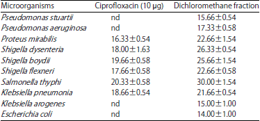 | |
| The results are the means of number of the colonies±Standard Deviations, nd: No detected activity | |
| Table 2: | Bacteriostatic (-) and Bactericidal (+) effects of dichloromethane fraction of from Boswelli dalzielii Hutch |
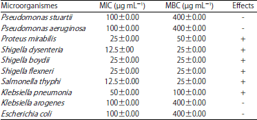 | |
| The results are the means of number of the colonies±standard deviations, +: Bactericidal effect, -: Bacteriostatic effect | |
| Table 3: | Viability of microorganisms after 6 h exposure of dichloromethane fraction of stem bark from Boswelli dalzielii Hutch |
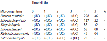 | |
The results are the means of number of the colonies±standard deviations, +: The presence of the colonies, -: Absence of colonies, UC: Uncountable | |
Acute toxicity study in mice: The effect of intraperitoneal treatment of the aqueous acetone extract from Boswelli dalzielii Hutch, on mortality, LD50 is 1872.6 mg kg–1 b.wt., for intraperitoneal administration. No significant difference in body weight gain of the treated assay groups over the period of observation. No statistical difference was observed between the organ weights in the control and the intraperitoneal route groups.
Antipyretic effects: The antipyretic effect of dichloromethane fraction of stem bark was studied in hyperthermic rats using brewer's yeast and data were recorded in (Table 4). The subcutaneous injection of brewer's yeast suspension markedly elevated rectal temperature after 17 h of administration. Oral administration of dichloromethane fraction of stem bark induced significant antipyretic activity at a dose of 300 mg kg–1 b. wt., while the low dose 200 mg kg–1 failed to decrease the raised body temperature. The effect of the high dose 300 mg kg–1 b.wt., nearly similar to that of the standard metamizole sodium (50 mg kg–1 b. wt.) after 3 h. Significance was indicated by lowering the body temperature were (37.62±0.11 and 37.67±0.15) after 2 h of administration of standard drug and stem bark fraction, respectively and at 3 h (37.48±0.15 and 37.72±0.14) when compared to the control non-treated group.
DISCUSSION
The discovery of effective antibiotics, vaccines and other products or methods has decreased the devastating impact of infectious diseases and improved quality of life. However, the efficacy of many antibiotics is being threatened by the emergence of microbial resistance to existing chemotherapeutic agents because of their indiscriminate and inappropriate use27. The use of some antibiotics is associated with side effects, including allergy, immune suppression and hypersensitivity28. Many populations who live in developing countries are deprived of the advantages of modern medicine because of its high cost; hence, poor people are more vulnerable to infectious diseases. Besides these, co-infection with multiple diseases is an obstacle to infection prevention and treatment.
| Table 4: | Antipyretic effect of dichloromethane fraction of stem bark from Boswelli dalzielii Hutch in hyperthermic rats (Mean±SE, N = 6) |
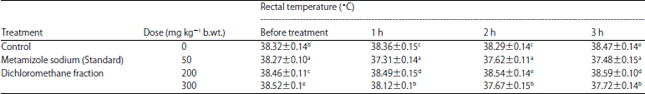 | |
| Values represent the Mean±SE of five animals for each group, values in each column with different superscript letters (a, b, c, d, e) are significantly different at p<0.05 | |
For all these reasons, there is a pressing need to identify new, safe and cost-effective antimicrobial agents that would help to alleviate the problems of infectious diseases. Plant-derived natural products represent an attractive source of antimicrobial agents because they are natural and affordable, especially for rural societies29. Acceptance of medicines from such plant origins as an alternative form of health care is increasing because they are serving as promising sources of novel antibiotic prototypes. Moreover, these compounds may have different mechanisms of action than conventional drugs and could be of clinical importance to improve health care21,30. Some of the phytochemical compounds e.g., glycoside, saponin, tannin, flavonoids, terpenoid and alkaloids, have been reported to have antimicrobial activity31,32. According to a recent study, Boswelli dalzielii contains saponins, glycosides, flavonoids, tannins, phenolics, alkaloids, quinones and terpenoids types10. Phytochemical screening of Boswelli dalzielii Hutch showed the presence of a number of bioactive constituents such as polyphenol compounds. The antimicrobial activity could be due to the presence of these phytoconstituents. The natural products were found to possess promising antimicrobial and the metabolites have been shown to be responsible for therapeutic activity of plants33. The data analysis indicates that the tested fraction showed the significant results when compared with the standard antibiotic. Indeed, the antibacterial activity profile of the isolated constituents (polyphenols) when compared with antibiotic effects showed that the activity depends on the pure form of the constituents. This may be due to the fact that the bioactive constituents such as polyphenol compounds were responsible for the antimicrobial activity. In Africa, for the treatment of several infections, indigenous medicinal plants are often the only means34. Infectious due to multidrug-resistant microorganisms, pose an important clinical problem. Many of bacterial strains are resistant to the standard antibiotic (Ciprofloxacin etc.) comparatively to the dichloromethane fraction. One could say that, the metabolites have been shown to be responsible for therapeutic activity of plants35. Also, Gram-negative bacteria possess an outer membrane and unique periplasm space not found in Gram-positive bacteria36. The resistance of Gram-negative bacteria towards antibacterial substances is related to the hydrophilic surface of their outer membrane which is rich in lipopolysaccharide molecules, presenting a barrier to the penetration of numerous antibiotic molecules and is also associated with the enzymes in periplasmic space, which are capable of breaking down the molecules introduced from outside37. Gram-positive bacteria do not have such an outer membrane and cell wall structure. Phenolic and terpenic antimicrobial activities are well documented16. Polyphenols, such as tannins and flavonoids, are important antibacterial activity34. Also, polyphenols have a good antimicrobial activity against the biggest number of bacterial as such Escherichia coli, Proteus mirabilis, Salmonella typhimurium, Bacillus cereus and Staphylococcus aureus38. The mechanism of toxicity of polyphenol against microbial could be explained by the hydrolytic (proteases and carbohydrolases) or to the other interactions whose can destroy microbial andesine (enzyme), sometimes the transport proteins26. In addition tannins can realize some polymerizations during oxidation reactions and this could have a toxicity effect again microbialstrains38,39. On the toxicity of the extract, according to Scalbert38, pharmacological substances whole LD50 is less than5 mg kg–1 b.wt., are classified in the range of highly toxic substances, those with a LD50 between 5 and 5000 mg kg–1 b.wt., are classified in the range of moderately toxic substances and those with the lethal dose is more than 5000 mg kg–1 b.wt., not toxic. In this fact, if refer to this classification it could say that the extract of Boswelli dalzielii is moderately toxic and would be regarded as being safe or of low toxicity40.
As concerning antipyretic potential, one could say that antipyretic activity may be attributed to the presence of phytochemical constituents such as β-sitosterol triterpenes, flavonoids, saponins, glycosides, tannins and alkaloids41. The β-sitosterol reduces PG and leukotrienes synthesis and in turn shows anti-inflammatory and antipyretic activity by inhibiting the pro-inflammatory cytokines and TNF-α42,43.
CONCLUSION
The present study showed that dichloromethane fraction of stem bark from Boswelli dalzielii possesses on a one hand significant antimicrobial activities to treat infectious diseases due to multi-resistant bacterial strains. On the other, study also proves the antipyretic activity of dichloromethane fraction of stem bark from Boswelli dalzielii. The study though supports the traditional claim; further studies are needed to identify the chemical constituents that are responsible for these properties.
SIGNIFICANCE STATEMENT
The present finding showed that the tested of dichloromethane fraction has significant effects when compared with the standard antibiotic. Concerning antipyretic activity, it was also noticed that oral administration of dichloromethane fraction of stem bark induced significant antipyretic activity. That can be explained by the solubility or insolubility of the active compound(s) in the solvent used for extraction. This study adequately justifies the ethno-medical use of this plant in the management of some infectious diseases. This study further suggests the researchers to isolate and identify the phytochemicals and active compounds in the plant.
ACKNOWLEDGMENTS
The authors thank Pr Karou of University of Lomé, Department of Biochemistry-Microbiology for the finalization of this manuscript article. Also, authors think Dr Traoré Lassina of University Nobert Zongo from Koudougou, for the botanically identified of plants.
REFERENCES
- Mokoka, T.A., L.J. McGaw, L.K. Mdee, V.P. Bagla, E.O. Iwalewa and J.N. Eloff, 2013. Antimicrobial activity and cytotoxicity of triterpenes isolated from leaves of Maytenus undata (Celastraceae). BMC complementary Altern. Med., Vol. 13.
CrossRefDirect Link - Morimoto, K. and M. Fujimoto, 1999. Report of questionnaire survey for methicillin-resistant Staphylococcus aureus and penicillin-resistant Streptococcus pneumoniae in the Kinki district Kansenshogaku zasshi. J. Jap. Assoc. Infect. Dis., 73: 584-592.
CrossRefDirect Link - Adwan, G. and M. Mhanna, 2008. Synergistic effects of plant extracts and antibiotics on Staphylococcus aureus strains isolated from clinical specimens. Middle-East J. Sci. Res., 3: 134-139.
Direct Link - Hancock, R.E.W., 2005. Mechanisms of action of newer antibiotics for gram-positive pathogens. Lancet Infect. Dis., 5: 209-218.
CrossRefPubMedDirect Link - Afolayan, A.J., 2003. Extracts from the shoots of Arctotis arctotoides inhibit the growth of bacteria and fungi. Pharm. Biol., 41: 22-25.
CrossRefDirect Link - Newman, D.J., G.M. Cragg and K.M. Snader, 2003. Natural products as sources of new drugs over the period 1981-2002. J. Nat. Prod., 66: 1022-1037.
CrossRefPubMedDirect Link - Kong, J.M., N.K. Goh, L.S. Chia and T.F. Chia, 2003. Recent advances in traditional plant drugs and orchids. Acta Pharmacol. Sin., 24: 7-21.
PubMedDirect Link - Ikpeme, E.V., O. Udensi, U.B. Ekaluo, E.A. Uyoh, B.O. Asuquo, F.V. Udoh and P.B. Udoh, 2007. Effect of crude extract of Carica papaja seeds on the reproductive efficiency of male albino rats. Global J. Pure Applied Sci., 13: 365-368.
Direct Link - Zimmermann, M., 1983. Ethical guidelines for investigations of experimental pain in conscious animals. Pain, 16: 109-110.
CrossRefPubMedDirect Link - Perez, C., M. Pauli and P. Bazerque, 1990. An antibiotic assay by the agar-well diffusion method. Acta Biol. Med. Exp., 15: 113-115.
Direct Link - Ezoubeiri, A., C.A. Gadhi, N. Fdil, A. Benharref, M. Jana and M. Vanhaelen, 2005. Isolation and antimicrobial activity of two phenolic compounds from Pulicaria odora L. J. Ethnopharmacol., 99: 287-292.
CrossRefDirect Link - Pujol, V., C. Seux and J. Villard, 1990. Research of antifungal substances secreted by higher fungi in culture. Ann. Pharm. Fr., 48: 17-22, (In French).
PubMedDirect Link - Rabe, T., D. Mullholland and J. van Staden, 2002. Isolation and identification of antibacterial compounds from Vernonia colorata leaves. J. Ethnopharmacol., 80: 91-94.
CrossRefDirect Link - Rabe, T. and J. van Staden, 1997. Antibacterial activity of South African plants used for medicinal purposes. J. Ethnopharmacol., 56: 81-87.
CrossRefPubMedDirect Link - White, R.L., D.S. Burgess, M. Manduru and J.A. Bosso, 1996. Comparison of three different in vitro methods of detecting synergy: Time-kill, checkerboard and E test. Antimicrob. Agents Chemother., 40: 1914-1918.
PubMedDirect Link - Karou, D., A. Savadogo, A. Canini, S. Yameogo and C. Montesano et al., 2006. Antibacterial activity of alkaloids from Sida acuta. Afr. J. Biotechnol., 8: 195-200.
Direct Link - Miller, L.C. and M.L. Tainter, 1944. Estimation of the LD50 and its error by means of logarithmic-probit graph paper. Proc. Soc. Exp. Biol. Med., 57: 261-264.
CrossRefDirect Link - Cowan, M.M., 1999. Plant products as antimicrobial agents. Clin. Microbiol. Rev., 12: 564-582.
CrossRefPubMedDirect Link - Ahmad, I., Z. Mehmood and F. Mohammad, 1998. Screening of some Indian medicinal plants for their antimicrobial properties. J. Ethnopharmacol., 62: 183-193.
CrossRefPubMedDirect Link - Ghosh, A., B.K. Das, A. Roy, B. Mandal and G. Chandra, 2008. Antibacterial activity of some medicinal plant extracts. J. Nat. Med., 62: 259-262.
CrossRefDirect Link - Okeke, M.I., C.U. Iroegbu, E.N. Eze, A.S. Okoli and C.O. Esimone, 2001. Evaluation of extracts of the root of Landolphia owerrience for antibacterial activity. J. Ethnopharmacol., 78: 119-127.
CrossRefDirect Link - Ebi, G.C. and S.I. Ofoefule, 1997. Investigations into the folkloric antimicrobial activities of Landolphia owrrience. Phytother. Res., 11: 149-151.
CrossRefDirect Link - Jazani, N.H., M. Zartoshti, S. Shahabi, Z. Yekta and S. Nateghi, 2007. Evaluation of the synergetic effect of water soluble extracts of green tea (Camellia sinensis) on the activity of ciprofloxacin in urinary isolated E. coli. J. Biol. Sci., 7: 1500-1503.
CrossRefDirect Link - Machado, T.D.B., I.C.R. Leal, A.C.F. Amaral, K.R.N. dos Santos, M.G. da Silva and R.M. Kuster, 2002. Antimicrobial ellagitannin of Punica granatum fruits. J. Braz. Chem. Soc., 13: 606-610.
Direct Link - Shrivastava, S.M., S. Kumar and M. Chaudhary, 2009. Time-kill curve studies of ampucare against Escherichia coli, Staphylococcus aureus, Klebsiella pneumoniae and Proteus vulgaris. Res. J. Med. Plant, 3: 116-122.
CrossRefDirect Link - Nikaido, H., 1996. Outer Membrane. In: Escherichia coli and Salmonella typhimurium: Cellular and Molecular Biology, Neidhardt, F.C. (Ed.). Amercian Society of Microbiology Press, Washington, DC., USA., pp: 29-47.
Direct Link - Gao, Y., M.J. van Belkum and M.E. Stiles, 1999. The outer membrane of Gram-negative bacteria inhibits antibacterial activity of Brochocin-C. Applied Environ. Microbiol., 65: 4329-4333.
PubMedDirect Link - Scalbert, A., 1991. Antimicrobial properties of tannins. Phytochemistry, 30: 3875-3883.
CrossRefDirect Link - Gibbons, S., 2003. An overview of plant extracts as potential therapeutics. Exp. Opin. Ther. Pat., 13: 489-497.
CrossRefDirect Link - Patel, J.D. and V. Kumar, 2008. Annona squamosa L.: Phytochemical analysis and Antimicrobial screening. J. Pharm. Res., 1: 34-38.
Direct Link - Gupta, M.B., R. Nath, N. Srivastava, K. Shanker, K. Kishor and K.P. Bhargava, 1980. Anti-inflammatory and antipyretic activities of β-sitosterol. Planta Med., 39: 157-163.
CrossRefDirect Link - Bouic, P.J. and J.H. Lamprecht, 1999. Plant sterols and sterolins: A review of their immune-modulating properties. Altern. Med. Rev., 4: 170-177.
PubMed