
S. Kameshwaran
Swamy Vivekanandha College of Pharmacy, Elayampalayam, Tiruchengode, Namakaal, Tamil Nadu, India
R. Senthilkumar
Swamy Vivekanandha College of Pharmacy, Elayampalayam, Tiruchengode, Namakaal, Tamil Nadu, India
S. Thenmozhi
Swamy Vivekanandha College of Pharmacy, Elayampalayam, Tiruchengode, Namakaal, Tamil Nadu, India
M. Dhanalakshmi
Swamy Vivekanandha College of Pharmacy, Elayampalayam, Tiruchengode, Namakaal, Tamil Nadu, India
Pharmacologia
Year: 2014 | Volume: 5 | Issue: 6 | Page No.: 215-221


ABSTRACT
Background: The entire wound healing process is a complex series of events that begins at the moment of injury and can continue for months to years. The stages of wound healing are inflammatory phase, proliferation phase, fibroblastic phase and maturation phase. The present study provides a précised valuation for the wound healing potential of ethanolic extract of Tecoma stans (EETS). Materials and Methods: Excision, incision and burn wounds were inflicted upon three groups of six rats each. Group I was assigned as control (ointment base). Group II was treated with 10% ethanolic extract of Tecoma stans (EETS) ointment. Group III was treated with standard silver sulfadiazine (0.01%) cream. The parameters observed were percentage of wound contraction, epithelialization period, hydroxyproline content, tensile strength including histopathological studies. Results: Result showed that the EETS ointment showed significant (p<0.01) healing in all wound models when compared with the control group. All parameters such as wound contraction, epithelialization period, hydroxyproline content, tensile strength and histopathological studies showed considerable (p<0.01) changes when compared with the control. Conclusion: Finally, it was accomplished that the EETS ointment efficiently stimulates wound contraction; increases tensile strength of excision, incision and burn wounds.
PDF Abstract XML References Citation
How to cite this article
S. Kameshwaran, R. Senthilkumar, S. Thenmozhi and M. Dhanalakshmi, 2014. Wound Healing Potential of Ethanolic Extract of Tecoma stans Flowers in Rats. Pharmacologia, 5: 215-221.
URL: https://scialert.net/abstract/?doi=pharmacologia.2014.215.221
URL: https://scialert.net/abstract/?doi=pharmacologia.2014.215.221
INTRODUCTION
Wounds may be defined as loss or breaking of cellular and anatomic or functional continuity of living tissue. It may be produced by physical, chemical, thermal, microbial, or immunological damage to the tissue. When skin is torn, cut, or punctured it is termed as an open wound and when force trauma causes a contusion, it is called closed wound, whereas the burn wounds are caused by fire, heat, radiation, chemicals, electricity, or sunlight (Charde et al., 2006; Reddy et al., 2012). Wound healing, or wound repair, is the critical physiological process by which the body repairs skin or organ tissue after injury. Wound healing is a complex process that results in the contraction and closure of the wound and restoration of functional barrier: Cutaneous wound repair is accompanied by an ordered and definable sequence of biological events starting with wound closure and progressing to the repair and remodeling of damaged tissue. Repair of injured tissues includes inflammation, proliferation and migration of different cell types. Inflammation, which constitutes a part of the acute response, results in a coordinated influx of neutrophils at the wound site (Kumar, 2012). Wounds are classified as open and closed wound on the underlying cause of wound creation and acute and chronic wounds on the basis of physiology of wound healing. Open wounds: In this case blood escapes the body and bleeding is clearly visible. It is further classified as: Incised wound, laceration or tear wound, abrasions or superficial wounds, puncture wounds, penetration wounds and gunshot wounds. Closed wounds: In closed wounds blood escapes the circulatory system but remains in the body. It includes Contusion or bruises, hematomas or blood tumor, crush injury etc. Acute wound: Acute wound is a tissue injury that normally precedes through an orderly and timely reparative process that result in sustained restoration of anatomic and functional integrity. Acute wounds are usually caused by cuts or surgical incisions and complete the wound healing process within the expected time frame. Chronic wounds: Chronic wounds are wounds that have failed to progress through the normal stages of healing and therefore enter a state of pathologic inflammation chronic wounds either require a prolonged time to heal or recur frequently. Local infection, hypoxia, trauma, foreign bodies and systemic problems such as diabetes mellitus, malnutrition, immunodeficiency or medications are the most frequent causes of chronic wounds (Kumar, 2012; Rawat et al., 2012). The healing process can be categorized into primary and secondary healing. Primary healing, or first intention, is the least complex as it refers to the healing together of the edges of clean, closely opposed wound edges. Secondary healing or second intention involves not only apposition of edges, but also the filling of a soft tissue defect as seen in traumatic, infection or disease induced wounds. Delayed primary closure or third intention is a combination of the first two (Harding et al., 2002). There are mainly 3 phases of wound healing there response to injury, either surgically or traumatically induced, is immediate and the damaged tissue or wound then passes through three phases in order to affect a final repair. Inflammatory phase: The inflammatory phase starts immediately after the injury that usually last between 24 and 48 hrs and may persist for up to 2 weeks in some cases the inflammatory phase launches the haemostatic mechanisms to immediately stop blood loss from the wound site. This phase is characterized by vasoconstriction and platelet aggregation to induce blood clotting and subsequently vasodilatation and phagocytosis to produce inflammation at the wound. Fibroplastic phase: The second phase of wound healing is the fibroplastic phase that lasts upto 2 days to 3 weeks after the inflammatory phase. This phase comprises of three steps viz., granulation, contraction and epithelialisation. In the granulation step fibroblasts form a bed of collagen and new capillaries are produced. Fibroblast produces a variety of substances essential for wound repair including glycosaminoglycans and collagen. Remodeling phase: This phase last for 3 weeks to 2 years. New collagen is formed in this phase. Tissue tensile strength is increased due to intermolecular cross-linking of collagen via vitamin-C dependent hydroxylation. The scar flattens and scar tissues become 80% as strong as the original (Reddy et al., 2012; Kumar, 2012).
Tecoma stans (common name yellow bell) also known as yellow trumpet bush belongs to the family bignoniaceae. It is an ornamental plant. It is an erect, branched, sparingly hairy or nearly smooth shrub two to four meters in height. The leaves are opposite, odd-pinnate, upto 20 centimeters in length with 5 to 7 leaflets. The leaflets are lanceolate to oblong- lanceolate, 6 to 13 centimeters long, pointed at both ends and toothed on the margins. Trumpet shaped flowers are yellow faintly scented and borne in short, dense, terminal clusters. The calyx is green. 5 to 7 millimeters long and 5 toothed. Flowering can begin as early as April and continue in to fall. The flowers are followed by 6 inch long, tan pods that are filled with small, papery winged seeds (Parrotta, 2001).
Leaves of Tecoma stans contain the alkaloids tecomin and tecostamine are potent hypoglycaemic agent when given intravenously. Anthranilic acid is responsible for the anti diabetic activity. Roots are powerful diuretic and vermifuge (Rao, 2010). Tecoma is not a toxic because this plant is used in Latin America as a remedy for diabetes and moreover for feeding cattle and goats in Mexico (Khare, 2007). The preliminary phytochemical screening of methanolic extract of flower extract of Tecoma stans showed the presence of flavaniods, phenol, alkaloids, tannins, steroids, triterpenes, anthraqunones and saponins etc. The present study is performed to evaluate the wound healing capability of ethanolic extract of Tecoma stans (EETS) flowers.
MATERIALS AND METHODS
Plant extraction: The flowers of Tecoma stans were collected in the month of May 2011 from Rasipuram (Namakkal District) Tamil Nadu. A herbarium specimen of the plant was deposited in the Department of Pharmacognosy. The plant was identified by Dr.G.V.S.Murthy, Joint Director of the Botanical Survey of India, Southern circle, TNAU Campus, Coimbatore, who authenticated the plant from information available in the literature. The flower petals were dried in the shade and then powdered and 100 g of the dried powder was extracted with ethanol using a soxhlet apparatus. The solvent was removed under reduced pressure and controlled temperature using a rotary flash evaporator. Suspension EETS in 2% (v/v) tween-80 was prepared for oral administration by gastric intubation method. The different identification tests were performed to detect the presence of phytoconstituent (Horbone, 1998). The screening revealed the presence of oils and fats, org. acids, flavonoids, triterpenes, steroids, sterols and proteins.
Preparation of formulation and standard used: Simple ointment was geared up from the 10% EETS by titration method in a ceramic pestle and mortar using White soft paraffin obtained from S.D. Fine Chemical, India (Cooper, 1987). About 10 g of semisolid extract was incorporated into the 100 g of simple ointment base (Anonymous, 1953). Simple ointment base was used as the control group and was applied twice per day. Extract ointment was used two times per day to indulgence different groups of animals. Silver sulfadiazine (0.01%) obtained from Rexin Pharmaceutical Pvt. Ltd. was worn as standard drug for comparing the wound healing prospective of extract in diverse animal models and was applied twice per day.
Animals: Wistar albino rats (150-180 g) of either sex were selected for the experiment. They were housed individually in standard laboratory environment for 7 days of period, fed with commercial pellets and water ad libitum. The procedures were reviewed and approved by the institutional animal ethics committee.
Wound healing activities
Grouping of animals: For excision, incision and burn wound model, animals were separated into three groups in every group consisting of six animals as follows: group I-simple ointment base; Group II-10% EETS and Group III-silver sulfadiazine (0.01%) cream was used as standard.
Excision wound model: Excision wound model was performed by dividing animal in to three groups containing six rats in each group were anesthetized by open mask method with anesthetic ether. The rats were placed on the back and a predetermined area of 500 mm2 occupied thickness skins was excised in the dorsal interscapular region. Rats were left undressed to the open atmosphere. The formulation ointment and standard drug were applied every day until the complete healing. In this model, wound contraction and epithelialization period was monitored. Wound contraction was deliberate as percent contraction in each 2 days later than wound formation. From the healed wound, a specimen sample of tissue was collected from each rat for histopathological examination.
Incision wound model: In incision wound model, 6 cm long paravertebral incision were made through the full thickness of the skin on either side of the vertebral column of the rats, after all the animals of each group were anesthetized under light ether anesthesia (Ehrlich and Hunt, 1969). No local or systemic antimicrobials were used throughout the experiment. All groups were treated same as in excision model, the both edges kept together and stitched with black silk surgical thread (No. 000) and a curved needle (No. 11) was used for stitching. The continuous threads on both wound edges were tightened for good closure of the wound. After stitching, wound was left undressed then ointment base, standard ointment and extracts ointment were applied daily until 10 days; when wounds were cured thoroughly the sutures were removed on the day 10 and tensile strength of cured wound skin was measured using tensiometer (Hemalatha et al., 2001).
Burn wound model: Burn wounds were created on dorsal part of shaved rat’s skin surface using concentrate sulfuric acid, exposed for 10 s. After 24 h, dead tissues were excised using sterile surgical blade through a template designed to produce a third degree burn (Nakae and Inaba, 2000). All groups were treated same as in excision model. In this model, wound contraction and epithelialization period was monitored. A specimen sample from the healed wound of tissue was collected from each rat for histopathological examination (McManus and Mowry, 1965).
Wound healing evaluation parameters
Measurement of wound contraction: An excision wound margin was traced subsequent to wound creation by using transparent paper and area measured by graph paper. Wound contraction was deliberate in each 2 days hiatus, until absolute wound healing and uttered in percentage of healed wound area (Sadaf et al., 2006). The evaluated surface area was then engaged to determine the percentage of wound contraction, taking initial size of wound, 300 mm2, as 100%, by using the following formula as:
Epithelialization period: It was evaluated by noting the number of day’s requisite for the Escher to fall off from the wound surface exclusive of leaving a raw wound behind (Rashed et al., 2003).
Measurement of tensile strength: The tensile strength of a wound represents the degree of wound healing. It indicates how much the repaired tissue resists to breaking under tension and may designate in part the quality of repaired tissue. The sutures were removed on the 9th day after wounding and the tensile strength of uninvolved tissue was measured on the 10th day with the help of tensiometer (Kuwano et al., 1994). In this method, wound breaking strength was deliberated as the weight of water at the time of wound breaking per area of the specimen.
Hydroxyproline estimation: Wound tissues were analyzed for hydroxyproline content, a basic element of collagen. Tissues were dried in a hot air oven at 60-70°C to constant weight and hydrolyzed in 6 N HCl at 130°C for 4 h in sealed tubes. The hydrolysate was neutralized to pH 7 then subjected to chloramine-T oxidation for 20 min (Woessner, 1961). The rejoinder was terminated by the addition of 0.4 M perchloric acid and developed color with Ehlrich reagent at 60°C was read at 557 nm in ultraviolet (Cintra) spectrophotometer.
Histopathological studies: Wound tissue specimens from control, test and standard groups were taken after absolute healing of excision, incision and dead burn wound and after customary processing 6 mm thick sections were cut and stained with hematoxylin and eosin (Sutradhar et al., 2008). Sections were qualitatively assessed under the light microscope and observed in respect of fibroblast proliferation, collagen formation, angiogenesis and epithelialization.
Statistical analysis: All treated groups were compared with the control groups. The outcomes were analyzed statistically using one-way analysis of variance (ANOVA). The results were found to be significantly at p<0.01. All tests were conducted using Graph Pad Software, Inc., a privately held California corporation.
RESULT
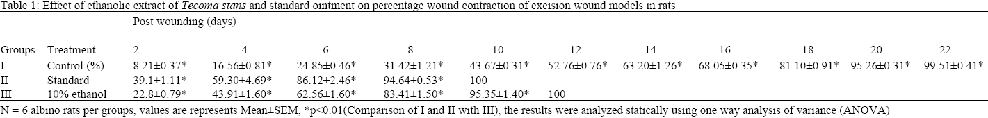
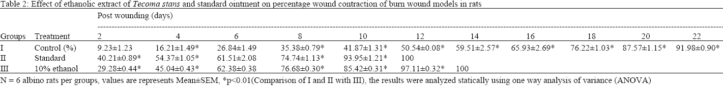
Wound contraction: A healthier healing pattern with inclusive wound closure was observed in both methods. In excision wound model inclusive wound closure occurs on day 10 in standard group but in control group it was about 43.67±0.31% wound healed, also only 95.35±1.4% of wound healed in the treated group. The complete wound healing occurred on day 12 in treated group. While it was about 22 days in control rats (Table 1). In burn wound model wound contraction occurs on day 12 in standard drug, where it was about 97.11± 0.32 percentage wound only healed in the treated group. The complete wound healing occurred on day 14 in treated group (Table 2).
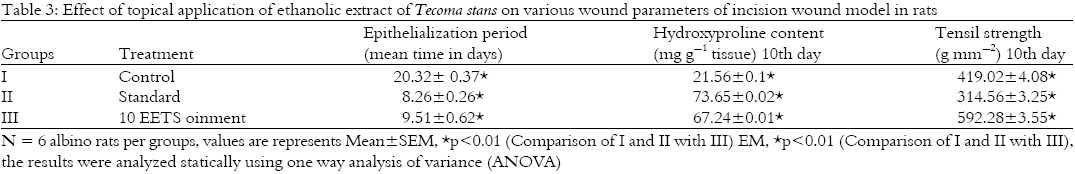
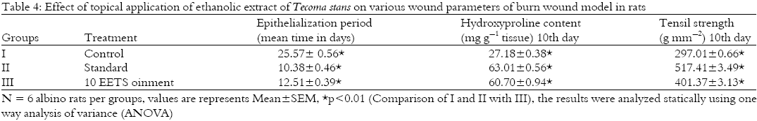
Epithelialization period: The epithelialization time was considered from the 1st day. The epithelialization time was found to be drastically (p<0.01) reduced in group II it is about 8.26±0.26 days. In group III the epithelialization time was 9.51±0.62 days (Table 3 and 4) for incision and burn models. The treatments with ethanolic extract as well as conservative wound treatment cream were finer to control groups, which received ointment base.
Tensile strength of incision and burn wound model: Tensile strength of the treated group of incision and burn wound model on day 10 was 592.28±3.55, 401.37±3.13 g-1 mm2, respectively. It showed a significant (p<0.01) value than the control group of both incision and burn wound model on day 10, 419.02±4.08, 297.01±0.66 g-1 mm2, respectively. (Table 2 and 4).
Hydroxyproline estimation: Treated group showed momentous augment in hydroxyproline level in both incision and burn wound model on day 10 67.24±0.01 tissue, 60.70±0.94 mg g-1 tissue, respectively when compared with the control group of both incision and burn wound model when it was 21.56±0.1, 27.18±0.38 mg g-1 tissue, respectively.
 |
 |
 |
 |
The EETS showed a significant (P < 0.01) activity which is comparable with the standard. (Table 2 and 4).
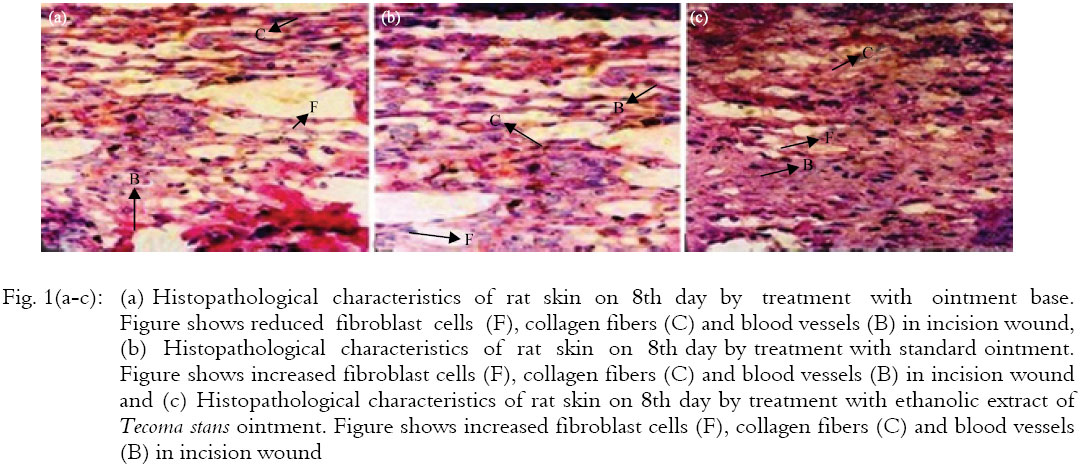
Histopathological studies: The histopathological studies of the tissue of the excision, incision and burn wound area was performed and histopathological characters of incision wound treated group with control, standard and extract ointment are shown in Fig. 1 (a-c). Group I (control) showed compact fibroblast cells, collagen fibers and blood vessels (Fig. 1a). While group II (standard) showed enlarged in fibroblast cells, collagen fibers and blood vessels in both incision and burn wound models (Fig. 1b). Group III (extract ointment) also showed improved in fibroblast cells, collagen fibers and blood vessels, but not more than group II (Fig. 1c) in both incision and burn wound models. The histopathology examination showed that the original tissue regeneration was much better on ethanolic extract and standard. Fibroblast cells, collagen fibers and blood vessels are outstandingly present in standard and extract treated group as compared with the control.
The results showed that ethanolic extract ointment possesses a definite prohealing action. This was confirmed by a significant raise in the rate of wound contraction and by enhanced epithelialization. Momentous augment (p<0.01) in tensile strength and hydroxyproline content were observed, which was further supported by histopathological studies. Since incisional wound treated with EETS showed greater tensile strength, it might be inferred that the EETS not only increased collagen synthesis per cell, but also aided in cross-linking of the protein.
DISCUSSION
The chemical constituents of EETS and their medicinal values have been reviewed. Its chemical constituents mainly consist of oils and fats, organic acids, flavonoids, triterpenes, steroids, sterols and proteins. Biological activities in the skin are due to its interaction with various binding protein. In this tissue repair process inflammatory cells promote the migration and proliferation of endothelial cells, which produce extracellular matrices including collagen and of keratinocytes resulting to re-epithelialization of the wounded tissue. No such a wound healing study was perfomed previously in this Tecoma stans plant. The wound healing action of EETS may probably be due to the phytoconstituents present in the plant or could be a role of either the individual or the additive effects of the phytoconstituents. The current result also indicated a important decrease in wound area from day 8 onward representing early healing. In incision wound, an boost in tensile strength of treated wounds was observed and this may be due to the raise in collagen concentration and stabilization of the fibers (Udupa et al., 1995).
The EETS increased cellular proliferation and collagen synthesis at the wound site as evidenced by the augment in total protein and total collagen contents reflected by hydroxyproline content of granulation tissues. In our study, hydroxyproline content was appreciably increased (p<0.01) when compared with the control. Since EETS has increased levels of these compounds considerably it is liable that the observed amplify in tensile strength was not only due to increased collagen synthesis but also due to its proper deposition and alignment.
Increased cellular proliferation may be due to the mitogenic activity of the plant extract, which might have significantly contributed to healing process. Early dermal and epidermal regeneration in treated mice also confirmed that the extract had a positive effect towards cellular proliferation, granular tissue formation and epitheliasation. Tannins and anthraquinones are the major phytoconstituent present in this plant which may be responsible for wound healing action. The plant Portulaca oleracea containing the tannins possesses wound healing activity as that of the R. cordifolia (Rashed et al., 2003).
 |
The gel of ethanolic extract of the plant Vernonia scorpioides possess wound healing action by improving regeneration and organization of the new tissue due to the presence of tannins (Leite et al., 2002). The embellin isolated from the ethanol extract of plant Emblica officinalis containing condensed tannins when formulated as a gel possess significant wound healing property (Kumara Swamy et al., 2007) as that of gel prepared by R. cordifolia ethanol extract. A number of secondary metabolites/active compounds isolated from plants have been demonstrated in animal models (in vivo) as active principles responsible for facilitating healing of wounds. Some of the most important ones include tannins from Terminalia arjuna, (Chaudhari and Mengi 2006), oleanolic acid from Anredra diffusa (Moura-Letts et al., 2006), polysaccharides from Opuntia ficus-indica (Trombetta et al., 2006), gentiopicroside, sweroside and swertiamarine from Gentiana lutea (Ozturk et al., 2006), shikonin derivatives (deoxyshikonin, acetyl shikonin, 3-hydroxy-isovaleryl shikonin and 5,8-Odimethyl acetyl shikonin) from Onosma argentatum (Ozgen et al., 2006), asiaticoside, asiatic acid and madecassic acid from Centalla asiatica (Maquart et al., 1999; Shukla et al., 1999; Hong et al., 2005), quercetin, isorhamnetin and kaempferol from Hippophae rhamnoides (Fu et al., 2005), curcumin from Curcuma longa (Jagetia and Rajanikant, 2004).
Collagen is the principal extracellular protein in the granulation tissue of healing wound and there is a hasty granulation tissue of healing wound and there is wound rapid augment in the synthesis of this protein in the wound area soon after an injury which provides strength and integrity to tissue matrix capacity of this hydroxyproline, which comes from the breakdown of collagen, has been used as an index of collagen turnover. The hydroxyproline content of the granulation tissue indicates the presence of higher collagen content and its turnover foremost to rapid healing with a simultaneous raise in the tensile strength of the treated wounds.
CONCLUSION
The fallout of the study showed that the ethanolic extract ointment of Tecoma stans successfully stimulates wound contraction; increases tensile strength of excision, incision and burn wound as compared with the control group. This verdict could justify the inclusion of this plant in the management of wound healing.
REFERENCES
- Charde, M.S., S.V. Fulzele, P. Satturwar, S.B. Joshi and A.V. Kasture, 2006. Wound healing and antiinflammatory potential of madhu ghrita. Indian J. Pharm. Sci., 68: 26-31.
CrossRefDirect Link - Ehrlich, H.P. and T.K. Hunt, 1969. The effects of cortisone and anabolic steroids on the tensile strength of healing wounds. Ann. Surg., 170: 203-206.
Direct Link - Fu, S.C., C.W.C. Hui, L.C. Li, Y.C. Cheuk, L. Qin, J. Gao and K.M. Chan, 2005. Total flavones of Hippophae rhamnoides promotes early restoration of ultimate stress of healing patellar tendon in a rat model. Med. Eng. Phys., 27: 313-321.
CrossRefDirect Link - Harding, K.G., H.L. Morris and G.K. Patel, 2002. Healing chronic wounds. Br. Med. J., 324: 160-163.
CrossRef - Hemalatha, S., N. Subramanian, V. Ravichandran and K. Chinnaswamy, 2001. Wound healing activity of Indigofera enneaphylla Linn. Indian J. Pharm. Sci., 63: 331-333.
Direct Link - Hong, S.S., J.H. Kim, H. Li and C.K. Shim, 2005. Advanced formulation and pharmacological activity of hydrogel of the titrated extract ofC. asiatics. Arch. Pharmacol. Res., 28: 502-508.
CrossRefDirect Link - Kumara Swamy, H.M., V. Krishna, K. Shankarmurthy, B. Abdul Rahiman and K.L. Mankani et al., 2007. Wound healing activity of embelin isolated from the ethanol extract of leaves of Embelia ribes Burm. J. Ethnopharmacol., 109: 529-534.
CrossRef - Kuwano, H., K. Yano, S. Ohno, M. Ikebe and K. Kitamura et al., 1994. Dipyridamole inhibits early wound healing in rat skin incisions. J. Surg. Res., 56: 267-270.
CrossRefDirect Link - Moura-Letts, G., L.F. Villegas, A. Marcalo, A.J. Vaisberg and G.B. Hammond, 2006. In vivo wound-healing activity of oleanolic acid derived from the acid hydrolysis of Anredera diffusa. J. Nat. Prod., 69: 978-979.
CrossRefDirect Link - Maquart, F.X., F. Chastang, A. Simeon, P. Birembaut, P. Gillery and Y. Wegrowski, 1999. Triterpenes from Centella asiatica stimulate extracellular matrix accumulation in rat experimental wounds. Eur. J. Dermatol., 9: 289-296.
PubMed - Nakae, H. and H. Inaba, 2000. Effectiveness of electrolyzed oxidized water irrigation in a burn-wound infection model. J. Trauma-Injury Infect. Crit. Care, 49: 511-514.
Direct Link - Ozgen, U., M. Ikbal, A. Hacimuftuoglu, P.J. Houghton, F. Gocer, H. Dogan and M. Coskun, 2006. Fibroblast growth stimulation by extracts and compounds of Onosma argentatum roots. J. Ethnopharmacol., 104: 100-103.
CrossRef - Ozturk, N., S. Korkmaz, Y. Ozturk and K.H.C. Baser, 2006. Effects of gentiopicroside, sweroside and swertiamarine, secoiridoids from Gentian (Gentiana lutea ssp. symphyandra), on cultured chicken embryonic fibroblasts. Planta Med., 72: 289-294.
CrossRefPubMedDirect Link - Parrotta, J.A., 2001. Healing Plants of Peninsular India. CABI Publishing, Wallingford, UK, ISBN: 9780851995014, Pages: 917.
CrossRefDirect Link - Sutradhar, R.K., M. Rahman, M.U. Ahmad and S.C. Bachar, 2008. Bioactive flavones of Sida cordifolia. Phytochem. Lett., 1: 179-182.
CrossRefDirect Link - Rashed, A.N., F.U. Afifi and A.M. Disi, 2003. Simple evaluation of the wound healing activity of a crude extract of Portulaca oleracea L. (growing in Jordan) in Mus musculus JVI-1. J. Ethnopharmacol., 88: 131-136.
CrossRefPubMedDirect Link - Rawat, S., R. Singh, P. Thakur, S. Kaur and A. Semwal, 2012. Wound healing agents from medicinal plants: A review. Asian Pacific J. Trop. Biomed., 2: S1910-S1917.
CrossRefDirect Link - Sadaf, F., R. Saleem, M. Ahmed, S.I. Ahmad and Navaid-ul-Zafar, 2006. Healing potential of cream containing extract of Sphaeranthus indicus on dermal wounds in Guinea pigs. J. Ethnopharmacol., 107: 161-163.
CrossRef - Shukla, A., A.M. Rasik, G.K. Jain, R. Shankar, D.K. Kulshrestha and B.N. Dhawan, 1999. In vitro and in vivo wound healing activity of asiaticoside isolated from Centella asiatica. J. Ethnopharmacol., 65: 1-11.
PubMed - Trombetta, D., C. Puglia, D. Perri, A. Licata and S. Pergolizzi et al., 2006. Effect of polysaccharides from Opunta ficus-indica cladodes on the healing of dermal wounds in the rat. Phytomedicine, 13: 289-294.
CrossRefDirect Link - Udupa, A.L., D.R. Kulkarni and S.L. Udupa, 1995. Effect of Tridax procumbens extracts on wound healing. Int. J. Pharmacognosy, 33: 37-40.
CrossRef - Woessner Jr. J.F., 1961. The determination of hydroxyproline in tissue and protein samples. Arch. Biochem. Biophys., 93: 440-447.
PubMed - Chaudhari, M. and S. Mengi, 2006. Evaluation of phytoconstituents of Terminalia arjuna for wound healing activity in rats. Phytother. Res., 20: 799-805.
CrossRefPubMedDirect Link