
R. Malathi
Department of Biochemistry, Thanthai Hans Roever College, Perambalur, India
M. Patrick Gomez
Department of Biotechnology, St. Josephs College, Tiruchirappalli, Tamil Nadu, South India
Journal of Pharmacology and Toxicology
Year: 2007 | Volume: 2 | Issue: 8 | Page No.: 737-742


ABSTRACT
The aim of the present study was to evaluate the hepatoprotective and antioxidant properties of methanolic leaves extracts of Tylophora asthmatica (META) on paracetamol-induced (1 g kg-1 body weight, i.p.,) hepatotoxicity in wistar strain of rats. The extract produced significant hepatoprotective effects as evidenced by decreased (p< 0.05) serum enzyme activities, ALT, AST, ALP, LDH and serum bilirubin (200 mg kg-1 body weight orally for 21 days) compared with control group. For antioxidant activity, META exhibited significant (p< 0.05) hepatoprotective effect by increasing the levels of superoxide dismutase (SOD), catalase (CAT) and glutathione peroxidase (GPx) and reducing lipid peroxidation (LPO). These results suggest that META may have hepatoprotective potential, probably by its antioxidant properties on hepatocytes and also support the use of the plant as a hepatoprotective agent.
PDF Abstract XML References
How to cite this article
R. Malathi and M. Patrick Gomez, 2007. Hepatoprotective Effect of Methanolic Leaves Extracts of Tylophora asthmatica Against Paracetamol-Induced Liver Damage in Rats. Journal of Pharmacology and Toxicology, 2: 737-742.
DOI: 10.3923/jpt.2007.737.742
URL: https://scialert.net/abstract/?doi=jpt.2007.737.742
DOI: 10.3923/jpt.2007.737.742
URL: https://scialert.net/abstract/?doi=jpt.2007.737.742
INTRODUCTION
Liver is one of the most important organs in the biotransformation of food, drugs, endogenous and exogenous substances. Profuse supply of blood and the presence of many redox systems (e.g., cytochromes and various enzymes) enable liver to convert these substances into different kinds of inactive, active or even toxic metabolites. The burden of metabolism and exposure to dangerous chemicals make liver vulnerable to a variety of disorders, such as acute or chronic inflammation, toxin-/drug-induced hepatitis, cirrhosis and hepatitis after viral infection (Sherlock and Dooley, 2002). Drugs can cause toxic effects which can mimic almost every naturally occurring liver disease. Normally, the toxins and drugs are absorbed from the intestinal tract gain access first to the liver resulting in a variety of liver ailments. Thus liver diseases remain one of the serious heath problems (Lin et al., 2002).
Oxidative stress is one out of several etiological and pathophysiological factors implicated in various diseases. Exposure of biological systems to xenobiotics, ionizing radiations and certain normal metabolic processes in the body leads to overproduction of Reactive Oxygen Species (ROS) that overwhelms the antioxidant armory. Recent studies have demonstrated that overproduction of ROS can further aggravate the oxidative stress and the result is a unifying mechanism of injury that occurs in many developments of clinical disease processes, such as heart disease, diabetes, liver injury, cancer, aging, etc. (Giordano, 2005; Rolo and Palmeira, 2006; Jaeschke, 2000; Klaunig and Kamendulis, 2004; Bokov et al., 2004). Maintaining the balance between ROS and antioxidant enzymes [especially superoxide dismutase (SOD), catalase (CAT) and glutathione peroxidase (GPx)] is therefore crucial and could serve as a major mechanism in preventing damage by oxidative stress. This balance has been suggested to play an important role in drug toxicity, such as from paracetamol (Jaeschke et al., 2003).
Paracetamol is an antipyretic analgesic drug that is available over-the-counter and an overuse of paracetamol can cause overproduction of ROS during formation of N-acetyl-p-benzoquinoneimine (NAPQI) by cytochrome P450 (Dahlin et al., 1984). This mechanism has been suggested to participate in the development of oxidative stress and injury in paracetamol-induced hepatotoxicity (James et al., 2003).
Modern medicines have little to offer for alleviation of hepatic diseases and it is chiefly the plant based preparations which are employed for their treatment of liver disorders. But there are not much drugs available for the treatment of liver disorders (Karan et al., 1999; Chaterrjee, 2000). Therefore, many folk remedies from plant origin are evaluated for its possible antioxidant and hepatoprotective effects against different chemical-induced liver damage in experimental animals. Paracetamol-induced hepatotoxicity model is frequently used for the investigation of hepatoprotective effects of drugs and plant extracts. The changes associated with paracetamol-induced liver damage are similar to that of acute viral hepatitis (James et al., 2003).
Tylophora asthmatica, a wild indigenous plant, belongs to the family Asclepidaceae and is commonly called as Indian ipecac. It is an endangered plant species, endemic to the state of Tamilnadu in India. The powdered leaves, stems and root contains 0.2-0.3% alkaloids, of these, tylophorine, tylophorinine and tylophorinidine are important alkaloids. Various studies have confirmed the anti-inflammatory activity (Gopalakrishnan et al., 1980), direct stimulant of adrenal cortex (Udupa et al., 1991), anti-asthmatic (Shivpuri et al., 1972) and the treatment of bronchitis, rheumatism and dermatitis (Nadkarni, 1976). The extract from the leaves and stems of T. asthmatica have been reported to possess anti-feedent, insecticidal and anti-tumour properties (Verma et al., 1986).
Review of the literature revealed that this rare medicinal plant remained unexplored for many of its claimed pharmacological activities. The aim of the present study was to evaluate the hepatoprotective and antioxidant properties of the methanolic leaves extracts of Tylophora asthmatica using rat model.
MATERIALS AND METHODS
Collection and Processing of Plant Material
The fresh leaves of T. asthmatica were collected during the month of February-2006 in the Banks of Cauvery River, Tiruchirappalli, South India. It was botanically identified and authenticated. A voucher specimen (TAL-12) has been kept in our laboratory for future reference. The leaves were shade dried, powdered, sieved through 40 mesh and stored in a tightly closed container for further use.
Preparation of the Plant Extract
The powdered plant material (500 g) was extracted with petroleum ether (60-80°C) using soxhlet apparatus to remove lipids. It was filtered and the filtrate was discarded. The residue was extracted with methanol by soxhlet apparatus. The extract was completely dried in vauco, stored in refrigerator at 4°C and protected from sunlight until the time for extract administration. The yield of methanolic dried extract was 8.63% (w/w).
Animals Used
Adult healthy male Wistar strain rats weighing 180 to 200 g were procured from Fredrick Institute of Plant Protection and Toxicology, Padappai, Chennai, India. They were maintained under uniform laboratory conditions in standard steel cages and provided with food (Hindustan Lever Ltd., Bangalore) and water ad libitum. The animals were maintained under laboratory conditions for 2 weeks to acclimatize before performing the experiment.
Experimental Protocol
Paracetamol (Acetaminophen; Sigma Chemical Company, USA) was suspended in saline solution in boiling water and was administered intraperitoneally (i.p.) after cooling (37°C) at a dose of 1 g kg-1 body weight. This dose is known to cause liver damage in rats (Akintonwa et al., 1990). The experimental animals were divided into four groups, each group comprising six animals.
| Group 1: | Control rats fed with standard diet. |
| Group 2: | Intoxication with 2 mL of paracetamol suspension (1 g kg-1 body weight, i.p.,). |
| Group 3: | Treatment with META extracts (200 mg kg-1 body weight for 21 days). |
| Group 4: | Co-treatment with META extracts (200 mg kg-1 body weight for 21 days; orally) prior to induction of liver damage with 2 mL of paracetamol suspension (1 g kg-1 body weight, i.p.,). |
Biochemical Investigations
At the end of the experimental period (21 days), the animals were killed by cervical dislocation. Blood was collected immediately and the sera were separated by centrifugation. Liver was excised, weighed and homogenized in Tris-HCl buffer (0.1M, pH 7.4). Serum and liver homogenate were used for the estimation of proteins (Lowry et al., 1951) and serum was used for the estimation of aminotransferases (King, 1965a), lactate dehydrogenase (King, 1965b), alkaline phosphatase (King, 1965c) and bilirubin (Malloy and Evelyn, 1937) levels. Estimation of lipid peroxidation (Ohkawa et al., 1979), superoxide dismutase (Misra and Fridovich, 1972), catalase (Sinha, 1972), glutathione peroxidase (Rotruck et al., 1973) and reduced glutathione (GSH) (Maron et al., 1979) were carried out in the liver homogenate.
Statistical Analysis
All data were evaluated with SPSS/10 software. Hypothesis testing methods included one-way analysis of variance (ANOVA) followed by least significant difference (LSD) test. p-values of <0.05 were considered to indicate statistical significance. All the results were expressed as mean±SD for six animals in each group.
RESULTS
An increase (p<0.05) in AST, ALT, ALP, LDH and bilirubin in the serum of experimental animals were observed in the sera of animals treated with only paracetamol (p<0.05) compared with control animals (Group 1). Serum marker enzymes and bilirubin levels were decreased (p<0.05) in the animals co-treated with META at the dose (200 mg kg-1 body weight) tested, against the hepatotoxicity induced by paracetamol (Group 2) administered animals. Animals treated with only META (Group 3) at the dose of 200 mg kg-1 body weight showed no significant variation (p<0.05) of the marker enzymes and bilirubin levels in comparison to the control animals (Group 1) (Table 1).
Table 2 shows the values of lipid peroxidation (LPO) products and antioxidant status in the liver of experimental animals. A significant (p<0.05) decrease in the levels of superoxide dismutase (SOD), catalase, reduced glutathione (GSH), glutathione peroxidase (GPx) and significant (p<0.05) increase of LPO products were seen in the animals that received only paracetamol (Group 2) compared to control animals (Group 1). Protection to the liver, by increasing the antioxidant enzymes and decreasing the LPO products were noted in the animals co-treated with META against the toxic doses of paracetamol (Group 4). In comparison to the control animals (Group 1), no significant alterations (p<0.05) in the LPO and antioxidant enzyme levels were observed in animals treated with only META (Group 3).
| Table 1: | Hepatoprotective effect of META on liver function indices in rats |
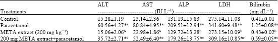 | |
| Values are given as mean±SD of 6 rats from each group, Values are statistically significant at *: p<0.05, ALT: Alanine transaminase; AST: Aspartate transaminase; ALP: Alkaline phosphatase; LDH: Lactate dehydrogenase, a: Paracetamol treated rats were compared with control rats, b: META extract treated rats were compared with control rats, c: META extract and paracetamol co-treated rats were compared with paracetamol treated rats | |
| Table 2: | Antioxidant properties of META on paracetamol-induced hepatotoxicity in rats |
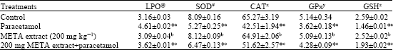 | |
| Values are given as mean±SD of 6 rats from each group, Values are statistically significant at *: p<0.05, LPO: Lipid peroxidation; SOD: Superoxide dismutase; CAT: Catalase; GPx: Glutathione peroxidase; GSH: Reduced glutathione @: μmoles of MDA liberated/mg protein; #: 50% inhibition of epinephrine autoxidation/min/mg protein in tissues; x: μmoles of H2O2 utilized/min/mg protein; y: mmoles of GSH oxidized/min/mg protein; z: μg mg-1 protein, a: Paracetamol treated rats were compared with control rats, b: META extract treated rats were compared with control rats, c: META extract and paracetamol co-treated rats were compared with paracetamol treated rats | |
DISCUSSION
In the present study, hepatotoxicity model in Wistar rats was successfully produced by induction with 2 mL of paracetamol (1 g kg-1 body weight, i.p.,) suspension (Akintonwa et al., 1990). We did not study the metabolites of administered dose of META extract in the blood of paracetamol treated rats because absorption and metabolism of phenanthroindolizidine (tylophorine, tylophorinine and tylophorinidine) alkaloids derived compounds are only partially understood.
Hence, META exhibit hepatoprotective effect as demonstrated by significant decreases in ALT, AST and ALP concentrations in rats induced with paracetamol. Moreover, the META enhanced the activities of antioxidant enzymes and diminished the amount of LPO products against the paracetamol-induced hepatotoxicity in these animals. This suggests that the reduction of oxidative stress in this scenario likely plays a role in the mechanism of its hepatoprotective effects.
Paracetamol is an effective and safe antipyretic and analgesic at pharmacological doses. However, acute over dosage can lead to liver and kidney failure and death in severe cases (Boyd and Bereczky, 1966; Boyer and Rouff, 1971; Thomsen et al., 1995). This hepatotoxicant is primarily metabolized by sulfation and glucuronidation to reactive metabolites and is activated by the Cytochrome P-450 system to produce liver injury (Moldeus, 1978). Conversion of paracetamol to the highly reactive metabolite N-acetyl-p-benzoquinoneimine (NAPQI) by the cytochrome P-450 system plays a central role in triggering cellular necrosis and organ failure (Savides and Oehm, 1983; Dahlin et al., 1984; Vermeulen, 1992).
Oxidative stress has been postulated to be the most important in the development of paracetamol-induced hepatotoxicity. Thus, increased formation of superoxide would lead to hydrogen peroxide and peroxidation reactions by Fenton-type mechanisms. It has been shown that N-acetyl-p-benzoquinoneimine (NAPQI) reacts very rapidly with GSH (Coles et al., 1988). Under conditions of NAPQI formation due to toxic doses of paracetamol, GSH concentration may be very low in the centrilobular cells and the major peroxide detoxification enzymes (GPx) that functions very ineffectively under conditions of GSH depletion (Nakamura et al., 1974) is expected to be inhibited. In addition, during formation of NAPQI by cytochrome P-450, the superoxide anion is formed with dismutation leading to hydrogen peroxide formation (Dai and Cederbaum, 1995). Due to the inhibition of GPx, catalase is overburdened and this may have resulted in the depletion of catalase observed in this study. Hepatocellular damage has been correlated to the poorer clearance of hydrogen peroxide due to catalase depletion and GPx inhibition. The fall in antioxidant enzymes were very well correlated to the increased lipid peroxidation, to the raised liver marker enzymes and bilirubin in the serum. Elevated levels of serum enzymes are indicative of cellular leakage and loss of functional integrity of cell membrane (Drotman and Lawhorn, 1978; Plaa and Hewitt, 1982), in line with this an increase in the levels of marker enzymes in the serum was noted in animals induced with higher doses of paracetamol.
The observed hepatoprotective/antioxidant activity of META against paracetamol-induced hepatotoxicity might be due to the synergestic action of various components in the META and mainly of the phenanthroindolizidine (tylophorine, tylophorinine and tylophorinidine) alkaloids. In accordance with these results, an increase in the antioxidant enzymes, decrease in the LPO products, serum marker enzymes and serum bilirubin levels in the animals co-treated with META were observed in this study. This indicated that treatment with META (200 mg kg-1) may represent the best hepatoprotective effect in serum enzyme examination compared with paracetamol treated group (Group 2).
REFERENCES
- Akintonwa, A. and A.R. Essien, 1990. Protective effects of Garcinia kola seed extracts against paracetamol-induced hepatotoxicity in rats. J. Ethnopharmacol., 29: 207-211.
Direct Link - Bokov, A., A. Chaudhuri and A. Richardson, 2004. The role of oxidative damage and stress in aging. Mech. Ageing Dev., 125: 811-826.
CrossRefDirect Link - Drotman, R.B. and G.T. Lawhorn, 1978. Serum enzymes as indicators of chemically induced liver damage. Drug Chem. Toxicol., 1: 163-171.
CrossRefPubMedDirect Link - Giordano, F.J., 2005. Oxygen, oxidative stress, hypoxia, and heart failure. J. Clin. Invest., 115: 500-508.
CrossRefDirect Link - Jaeschke, H., 2000. Reactive oxygen and mechanisms of inflammatory liver injury. J. Gastroenterol. Hepatol., 15: 718-724.
CrossRefDirect Link - Jaeschke, H., T.R. Knight and M.L. Bajt, 2003. The role of oxidant stress and reactive nitrogen species in acetaminophen hepatotoxicity. Toxicol. Lett., 144: 279-288.
Direct Link - James, L.P., P.R. Mayeux and J.A. Hinson, 2003. Acetaminophen-induced hepatotoxicity. Drug Metab. Dispos., 31: 1499-1506.
CrossRefPubMedDirect Link - Klaunig, J.E. and L.M. Kamendulis, 2004. The role of oxidative stress in carcinogenesis. Annu. Rev. Pharmacol. Toxicol., 44: 239-267.
CrossRefDirect Link - Lin, C.C., L.T. Ng, J.J. Yang and Y.F. Hsu, 2002. Anti-inflammatory and hepatoprotective activity of Peh-Hue-Juwa-Chi-Cao in male rats. Am. J. Chin. Med., 30: 225-234.
CrossRefPubMedDirect Link - Moron, M.S., J.W. Depierre and B. Mannervik, 1979. Levels of glutathione, glutathione reductase and glutathione S-transferase activities in rat lung and liver. Biochim. Biophys. Gen. Subj., 582: 67-78.
CrossRefPubMedDirect Link - Misra, H.P. and I. Fridovich, 1972. The role of superoxide anion in the autoxidation of epinephrine and a simple assay for superoxide dismutase. J. Biol. Chem., 247: 3170-3175.
CrossRefPubMedDirect Link - Ohkawa, H., N. Ohishi and K. Yagi, 1979. Assay for lipid peroxides in animal tissues by thiobarbituric acid reaction. Anal. Biochem., 95: 351-358.
CrossRefPubMedDirect Link - Rolo, A.P. and C.M. Palmeira, 2006. Diabetes and mitochondrial function: Role of hyperglycemia and oxidative stress. Toxicol. Applied Pharmacol., 212: 167-178.
CrossRefDirect Link - Savides, M.C. and F.W. Oehme, 1983. Acetaminophen and its toxicity. J. Applied Toxicol., 3: 96-111.
CrossRefDirect Link - Shivpuri, D.N., S.C. Singhal and D. Parkash, 1972. Treatment of asthma with an alcoholic extract of Tylophora indica: A cross-over, double-blind study. Ann. Allergy, 30: 407-412.
PubMed - Sinha, A.K., 1972. Colorimetric assay of catalase. Anal. Biochem., 47: 389-394.
CrossRefPubMedDirect Link - Rotruck, J.T., A.L. Pope, H.E. Ganther, A.B. Swanson, D.G. Hafeman and W.G. Hoekstra, 1973. Selenium: Biochemical role as a component of glutathione peroxidase. Science, 179: 588-590.
CrossRefPubMedDirect Link