
K. S. Vidyalakshmi
School of Chemical and Biotechnology, SASTRA University, Thanjavur, India
A.I. Charles Dorni
CARISM, SASTRA Deemed University, Thanj avur, India
Hannah R. Vasanthi
Sri Ramachandra Chandra Medical College and Research Institute,
Porur, Chennai, India
Journal of Pharmacology and Toxicology
Year: 2007 | Volume: 2 | Issue: 7 | Page No.: 660-665


ABSTRACT
Mussaenda queensirkit (MQ), an ornamental plant grown in tropical countries is shown to exhibit cytotoxic property. The 70% methanolic extract of fresh flowers of Mussaenda queensirkit (MQF) was studied for its cytotoxic and anti-mitotic activity with Brine Shrimp Lethality assay and Allium cepa Root Tip mitosis experiment, respectively. The LC50 value for MQF was found to be 83.33 μg mL-1 in Brine Shrimp Lethality Assay. The extract showed maximum anti-mitotic activity at the highest concentration studied (2000 μg mL-1) with a mitotic index of 9.0. In the MTT Assay for cytotoxic activity, MQF at 500 μg mL-1, showed a maximum of 71.44% inhibition against fibroblast cultured from skin compared to control whereas the standard, quercetin (50 μg mL-1) exhibited a maximum of 92.7% inhibition. The HPLC profile obtained had characteristic peaks due to flavonol glycosides which might be responsible for the observed cytotoxic and anti-mitotic activity of MQF. The observed cytotoxic and anti-mitotic activity of the extract shows the potential of the plant in the exploration of anti-cancer molecules.
PDF Abstract XML References
How to cite this article
K. S. Vidyalakshmi, A.I. Charles Dorni and Hannah R. Vasanthi, 2007. Anti-Mitotic and Cytotoxic Effect of Mussaenda queensirkit . Journal of Pharmacology and Toxicology, 2: 660-665.
DOI: 10.3923/jpt.2007.660.665
URL: https://scialert.net/abstract/?doi=jpt.2007.660.665
DOI: 10.3923/jpt.2007.660.665
URL: https://scialert.net/abstract/?doi=jpt.2007.660.665
INTRODUCTION
Mussaendas are increasingly popular for the showy color they provided during much of the year in South Florida landscapes. They are members of the Rubiaceae (madder or coffee family) and are native to the old world tropics, from West Africa through the Indian sub continent, South East Asia and into Southern China. There are more than 200 known species, of which ten are found in cultivation, with three of these being widely used for landscaping. Mussaendas are relatively pest and disease free (Huxley et al., 1999). In a previous study conducted in our laboratory the 70% methanolic extract of fresh flowers of Mussaenda has been shown to possess in vitro NO inhibition (Vidyalakshmi et al., 2006). In the present study the cytotoxic and anti-mitotic properties of 70% methanolic extract of the fresh flowers of Mussaenda queensirkit has been evaluated.
The cytotoxic effect of plant polyphenols is shown to be mediated through apoptosis. Considering the ability of the natural polyphenols especially the flavonoids to bind with proteins and metal ions, there is a possibility that they can elicit apoptosis signals through various receptors or proteins (Taraphdar, 2001). Apart from this, they are excellent antioxidants and they can prevent the free radical attack on DNA by acting as scavengers of these free radicals. A number of polyphenols are acting as topo II poisons through inhibition of topo I/topo II isomerases, enhancing the DNA cleavage (Ferguson, 2001).
In the present study cytoxicity has been tested against fibroblast cultured from skin by MTT assay. The Brine Shrimp (Artemia salina) Lethality bioassay (BSLB) is considered as an useful tool for preliminary assessment of toxicity (Solis et al., 1993) and it has been used for the detection of fungal toxins (Harwig and Scott, 1971), plant extract toxicity, heavy metals (McLaughlin et al., 1991) Cyanobacteria toxins (Martinez et al., 1990), pesticides (Jaki et al., 1999) and cytotoxicity testing of dental materials (Barahona and Sanchez, 1999). The BSLB is used for testing cytotoxicity of MQF, as it appears to be a convenient, rapid and inexpensive assay (Pelka et al., 2000).
The anti-mitotic activity was screened using Allium cepa root tip cells which have been used extensively in screening of compounds and extracts with anti-mitotic activity (Latha et al., 1998; Abhang et al., 1991). The region beyond root cap is capable of undergoing repeated cell division.The rate of cell division is higher in this region compared to that of the other tissues. This division is similar to the cancer cell division in humans. Hence, these meristamatic cells can be used for preliminary screening of compounds and extracts with anticancer activity. Even though doubts can be raised about extrapolation of results from plant tissues to animals and finally to humans. Thus, it is possible that molecules or chemical entities which affect plant chromosomes will also affect animals (Williams and Omoh, 1996).
Phytochemical evaluation plays a vital role in the process of standardization of herbal drugs. Hence HPLC profile has been derived which can be used for standardization of the active ingredient of the extract.
The present study has been done to evaluate the cytotoxicity of Mussaenda queensirikit as the extract of the flower showed good antioxidant property.
MATERIALS AND METHODS
Chemicals
Acetocarmine, Quercetin, 3-[4, 5-dimethylthiazol-2-yl]-2, 5-diphenyl tetrazolium bromide (MTT) and DMEM (Dulbecco’s Modified Eagle Medium) were purchased from M/s Sigma Laboratories, Monsanto, U.S.A. Brine Shrimp (Artemia salina) Eggs were purchased from Tuticorin local market and the Nauplii were hatched in artificial sea water before experiment. Allium cepa bulb was purchased from the local market and stored throughout the study. All other chemicals used were of analytical grade and were procured from E.Merck, Mumbai.
Plant Material
Fresh flowers of Mussaenda queensirkit have been collected in and around Thanjavur during the month of December 2005. The plant was taxonomically authenticated by the Botanists at Rabinat Herbarium, St. Joseph’s College (Autonomous), Tiruchirapalli and a voucher specimen has been deposited in the CARISM herbarium. The extract was prepared by soaking 100 g of the flowers (pink variety) in 500 mL of 70% methanol and concentrated in vacuuo. The yield obtained was found to be 0.48% (w/w). The concentrated extract was dried and stored in a dessicator.
HPLC Profile
The extract was qualitatively assessed for the presence of flavonoids (Shinoda Test). The profile was developed at λ max 280 nm. Ten milligram of methanolic extract of MQF was dissolved in methanol and was defatted using petroleum ether. The profile was obtained using RP-C18 (Waters) column monitored by PDA detector. The chromatogram was obtained using the gradient, methanol: water (30:70, 15 min) → methanol: water (50:50, 20 min) → methanol: water (80:20, 25 min).
Brine Shrimp Lethality Bioassay
The test was performed according to the method of Meyer et al. (1993). The extract was tested at concentrations of 1000, 100 and 10 μg mL-1 dispensing 100 μL in six replicates into wells of a 24 well microplate.
Anti-Mitotic Activity
This activity was evaluated using A. cepa root meristamatic cells (Williams and Omoh, 1996). A. cepa bulbs were sprouted in tap water for 48 h at room temperature. The bulbs that developed uniform roots were used for the experiment. These roots (Three roots per concentration) were treated with the extracts at concentrations of 2000, 1000, 500 and 250 μg mL-1. Water was used as medium/vehicle for dilution. A control was set with three roots from the same bulb and water was used as medium. After 3 h of treatment, the root tips were fixed in a fixing solution of acetic acid and alcohol. Squash preparations were made by staining with acetocarmine stain. The mitotic index was calculated as-
Cytotoxicity by MTT Assay
The fibroblasts cultured from skin were seeded on to the 48 well plate with a known count of cells (2500 cells/well) suspended in DMEM (Dulbecco’s Modified Eagle Medium) medium containing 10% Fetal Bovine Serum (FBS), Penicillin G (100 U mL-1) and Streptomycin (100 μg mL-1). The cells were maintained at 37°C in (5:95; CO2: Air) incubator. The MTT assay was performed according to the method of Skehan et al. (1990) Cells (100 cells/well) were incubated for 24 h with the extract at the concentrations of (100, 250 and 500 μg mL-1) and the standard, quercetin at concentrations of 10, 25 and 50 μg mL-1). At various concentrations 20 μL of MTT (5 mg mL-1) were added to each well and incubated for 4 h at 37°C. The formazan product was dissolved by adding 100 μL dimethylsulfoxide to each well and the plates were read at 550 nm. All measurements were performed in triplicate and each experiment was repeated at least three times.
Statistics
The values obtained for cytotoxic and anti-mitotic activities were expressed as mean of triplicate analyses as earlier studies. The LC50 value for Brine Shrimp Lethality Bioassay was calculated using Graph Pad Prism Software and Student t-test was performed for MTT assay using SPSS software.
RESULTS
HPLC Profile
The HPLC profile shows two distinct peaks with Rt at 9.65 and 44.54 min. The UV spectrum of compound at Rt 44.54 showed bands similar to that of a flavonol glycoside.
Brine Shrimp Lethality
The results from brine shrimp lethality test were analysed using mortality data. The LC50 value was found to be 83.33 μg mL-1 (Table 1). The results were expressed as mean of six replicates.
Anti-Mitotic Activity
Anti mitotic activity of the MQF was comparable to that of Quercetin. The extract showed highest activity at the concentration of 2000 μg mL-1, with the lowest mitotic index (Table 2). The maximal inhibition produced by MQF was considerably lower than that exhibited by quercetin.
Cytotoxicity
The results showed that 70% methanolic extract of MQF exhibited 71.44% activity at the concentration of 500 μg mL-1. The reference compound (Quercetin) displayed maximum activity at the concentration of 10 μg mL-1 (Table 3). The results were mean of three replicates.
| Table 1: | Brine shrimp lethality bioassay of MQF (n = 6) |
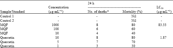 | |
| * An average of six replicates | |
| Table 2: | Anti-mitotic activity of MQF (n = 3) |
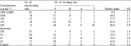 | |
| Values are expressed as mean±SD; P-Prophase; M-Metaphase; A-Anaphase; T-Telophase | |
| Table 3: | Cytotoxicity of MQF against fibroblast culture (MTT assay) |
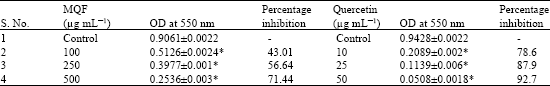 | |
| *MQF/Quercetin when compared with respective controls (p<0.5) | |
DISCUSSION
The results from brine shrimp lethality, antimitotic and cytotoxicity assay show that the 70% methanolic extract of fresh flowers of Mussaenda queensirkit (MQF) has considerable cytotoxic effect.
The preliminary phytochemical analysis suggested the flowers of MQ are found to be rich in flavonoids. The critical relationship of fruit and vegetable intake and cancer prevention has been thoroughly documented in a review (Block et al., 1992). Flavonoids of citrus fruits are known to have anticancer properties. Several flavonols, flavones and flavonones as well as the isoflavone biochanin A, were highly active; a carbonyl function at C-4 of the flavone nucleus was found to be essential for anti-mutagenic activity (Edenharder et al., 1999). Flavone -8 acetic acid was also shown to have antitumour effects (Thomsen et al., 1991).
The MQF seems to inhibit at prophase stage of mitoic cell division where the DNA duplication occurs. Generally, compounds inhibiting mitosis (M phase) affect microtubule configuration which results in an increased proportion of cells arrested in the metaphase (Armbruster et al., 1991; Lazareva et al., 2003). However in the extract treated cells, there was no increase in the number of cells in all mitotic phases except prophase when compared to control roots. This suggests that extract might not directly affect the process in mitosis but inhibit prophase of the cell cycle, resulting in the prevention of the cells entry into mitosis. The possible factors related to this action are the inhibition of DNA synthesis in the S phase, blocking the cell cycle progress in the G1/S or G2/M interface, fragmentation of DNA, malfunction of cyclin dependent kinase activity or proteolysis of cell cycle regulator (Planchais et al., 2000). The mitotic index does not increase considerably in the case of the extract when compared to the reference compound, Quercetin. This may be due to the fact that the extract may be mediating its effect through other mechanisms also. The extract may be binding with cell proteins responsible for cell division.
ACKNOWLEDGMENT
The authors wish to thank Prof. R. Sethuraman, The Vice Chancellor, SASTRA University, Thanjavur, for his constant support and encouragement for our research activities. Our thanks are due to Dr. G. Victor Rajamanickam Dean, CARISM, SASTRA University for providng the laboratory facilities of CARISM. Our sincere thanks are due to Mr. N. Shanmughasundaram, Department of Biotechnology, Central Leather Research Institute, Chennai for his assistance in the study. Financial support for the experiments from the Drugs and Pharmaceuticals Division, Department of Science and Technology, Govt. of India are gratefully acknowledged.
REFERENCES
- Abhang, R.Y., P.P. Joglekar and P.H. Kulkarni, 1991. Preliminary study on the effect of T. cordifolia on mitosis. Ancient Sci. Life, 2: 7-8.
Direct Link - Armbruster, B.L., W.T. Molin and M.W. Budd, 1991. Effect of the herbicide dithiopyr on celldivision in wheat root tips. Pest. Biochem. Physiol., 39: 110-120.
Direct Link - Barahona, M.V. and S. Sanchez-Fortun, 1999. Toxicity of carbamates to the brine shrimp Artemia salina and the effect of atropine, BW284c51, iso-OMPA and 2-PAM on carbaryl toxicity. Environ. Pollut., 104: 469-476.
CrossRefDirect Link - Block, G., B. Patterson and A. Subar, 1992. Fruit, vegetables and cancer prevention: A review of the epidemiological evidence. Nutr. Cancer, 18: 1-29.
CrossRefDirect Link - Edenharder, R., I. von Petersdorff and R. Rauscher, 1993. Antimutagenic effects of flavoniods, chalcones and structurally related compounds on the activity of 2-amino-3-methylinidazo[4,5-ƒ]quinoline (IQ) and other heterocyclic amine mutagens from cooked food. Mutat. Res./Fund. Mol. Mech. Mutagen., 287: 261-274.
CrossRefDirect Link - Ferguson, L.R., 2001. Role of plant polyphenols in genomic stability. Mutat. Res./Fundam. Mol. Mech. Mutagen., 475: 89-111.
CrossRefPubMedDirect Link - Harwig, J. and P.M. Scott, 1971. Brine shrimp (Artemia salina L.) larvae as a screening system for fungal toxins. Applied Microbiol., 21: 1011-1016.
PubMedDirect Link - Jaki, B., J. Orjala, H.R. Burgi and O. Sticher, 1999. Biological screening of cyanobacteria for antimicrobial and molluscicidal activity, brine shrimp lethality and cytotoxicity. Pharm. Biol., 37: 138-143.
CrossRefDirect Link - Latha, P.G., C.T. Chandralekha, G. Vilasini and K.R. Panikkar, 1998. Effects of the flower extract of Ixora coccinea linn. on the meristamatic cells A. cepa. Ancient Sci. Life, 17: 262-267.
Direct Link - Lazareva, E.M., V.Y. Polyakov, Y.S. Chentsov and E.A. Smirnova, 2003. Time and cell cycle dependent formation of heterogenous tubulin arrays induced by colchicines in Triticum aestivum root meristem. Cell Biol. Int., 27: 633-646.
Direct Link - Martinez, M., J.D. Ramo, A. Torreblanca and J. Diaz-Mayans, 1999. Effect of cadmium exposure on zinc levels in the brine shrimp Artemia parthenogenetica. Aquaculture, 172: 315-325.
CrossRefDirect Link - Pelka, M., C. Danzl, W. Distler and A. Petschelt, 2000. A new screening test toxicity testing of dental materials. J. Dent., 28: 341-345.
Direct Link - Planchais, S., N. Clat, D. Inze and C. Bergouniour, 2000. Chemical inhibitions: A tool for plant cell cycle studies. FEBS Lett., 476: 78-83.
Direct Link - Skehan, P., R. Storeng, D. Scudiero, A. Monks and J. McMahon et al., 1990. New colorimetric cytotoxicity assay for anticancer-drug screening. J. Nat. Cancer Inst., 82: 1107-1112.
CrossRefPubMedDirect Link - Solis, P.N., C.W. Wright, M.M. Anderson, M.P. Gupta and J.D. Phillipson, 1993. A microwell cytotoxicity assay using Artemia salina (brine shrimp). Planta Med., 59: 250-252.
CrossRefDirect Link - Taraphdar, A.K., M. Roy and R.K. Bhattacharya, 2001. Natural products as inducers of apoptosis: Implication for cancer therapy and prevention. Curr. Sci., 80: 1387-1396.
Direct Link - Vidyalakshmi, K.S., A.I. Charles Dorni, H.R. Vasanthi, G.V. Rajamanickam and D. Sukumar, 2006. Free radical scavenging activity of mussaenda glabra. J. Applied Sci., 6: 2251-2256.
CrossRefDirect Link - Williams, G.O. and L.E. Omoh, 1996. Mitotic effects of aqueous leaf extract of Cymbopogon citratus in Allium cepa root tips. Cytobios, 87: 161-168.
Direct Link