
Z.A. Zakaria
School of Biotechnology and Life Sciences, Universitl Industri Selangor, Jalan Zirkon A7/A, Seksyen 7, 40000 Shah Alam, Selangor, Malaysia
M.R. Sulaiman
Department of Biomedical Science, Faculty of Medlcme and Health Science, Universiti Putra Malaysia
A.K. Arifah
Department of Veterinary Pathology and Microbiology,
Faculty of Veterinary Medicine, Universiti Putra Malaysia,
43400 t_JPM Serdang, Selangor, Malaysia
A.M. Mat Jais
Department of Biomedical Science, Faculty of Medlcme and Health Science, Universiti Putra Malaysia
M.N. Somchit
Department of Biomedical Science, Faculty of Medlcme and Health Science, Universiti Putra Malaysia
K. Kirisnaveni
School of Biotechnology and Life Sciences, Universitl Industri Selangor, Jalan Zirkon A7/A, Seksyen 7, 40000 Shah Alam, Selangor, Malaysia
D. Punnitharrani
School of Biotechnology and Life Sciences, Universitl Industri Selangor, Jalan Zirkon A7/A, Seksyen 7, 40000 Shah Alam, Selangor, Malaysia
M. Safarul
School of Biotechnology and Life Sciences, Universitl Industri Selangor, Jalan Zirkon A7/A, Seksyen 7, 40000 Shah Alam, Selangor, Malaysia
C.A. Fatimah
School of Biotechnology and Life Sciences, Universitl Industri Selangor, Jalan Zirkon A7/A, Seksyen 7, 40000 Shah Alam, Selangor, Malaysia
R. Johari
Department of Veterinary Pathology and Microbiology,
Faculty of Veterinary Medicine, Universiti Putra Malaysia,
43400 t_JPM Serdang, Selangor, Malaysia
Journal of Pharmacology and Toxicology
Year: 2006 | Volume: 1 | Issue: 2 | Page No.: 139-146


ABSTRACT
The objective of this study was to elucidate the effects of C. olitorius as agent for relieving inflammation and fever. The aqueous extract of C. olitorius, in the concentration of 10, 50 and 100%, was used throughout the studies. The carrageenan-induced paw edema and brewer`s yeast-induced pyrexia assays were used as the anti-inflammatory and anti-pyretic assays, respectively. The extract, in the concentrations ranging from 50 to 100% and 10 to 100%, also exhibited significant (p<0.05) anti-inflammatory and anti-pyretic activities that lasted until the end of the experiments, respectively. In conclusion, the present studies provide scientific proof for the folklore medicinal used of C. olitorius as agent in the treatment of inflammation and fever.
PDF Abstract XML References
How to cite this article
Z.A. Zakaria, M.R. Sulaiman, A.K. Arifah, A.M. Mat Jais, M.N. Somchit, K. Kirisnaveni, D. Punnitharrani, M. Safarul, C.A. Fatimah and R. Johari, 2006. The Anti-inflammatory and Antipyretic Activities of Corchorus olitorius in Rats. Journal of Pharmacology and Toxicology, 1: 139-146.
DOI: 10.3923/jpt.2006.139.146
URL: https://scialert.net/abstract/?doi=jpt.2006.139.146
DOI: 10.3923/jpt.2006.139.146
URL: https://scialert.net/abstract/?doi=jpt.2006.139.146
INTRODUCTION
Corchorus olitorius L., locally known in the Malay language as ‘Senaung betina’, is an annual herb plant from the family Tiliaceae. Its leaves and roots are used as herbal medicine and eaten as vegetable by local people in East Malaysia, India, Egypt and Philippines (Zeghichi et al., 2003). C. Olitorius, also better known as jute, is a fiber crop. Its dried leaves are used in soups under the Arabic name "Molukhyia” (Zeghichi et al., 2003). Traditionally, its leaves are used in the treatment of pain, fever, chronic cystitis and tumours (Zeghichi et al., 2003; Abu-Hadid et al., 1994). Furthermore, the cold infusion is said to restore appetite and strength (Kirtikar and Basu, 1975).
Earlier studies have reported that the seeds C. olitorius contain oil (Watt and Breyer-Brandwijk, 1962), which was later found to possess estrogenic activity (Sharaf et al., 1979). In addition, the seeds were also reported to contain high content of hydrogen cyanide and several cardiac glycosides, for examples Corchoroside A and Corchoroside B (Negm et al., 1980). Recent studies have reported on C. olitorius moderate and weaker inhibitory effect towards the mutagenicity of 2-amino-3-methyl- midazo[4,5-f]quinoline or benzo[a]pyrene in S. typhimurium TA98 and TA100, respectively (Yen et al., 2001). Preliminary studies by Zakaria et al. (2005) have demonstrated that the aqueous extract of C. olitorius possessed peripherally and centrally mediated antinociceptive, which were both mediated, at least in part, via the opioid receptor. Based on the fact that there are no scientific papers reporting on the other pharmacological properties of this plant, the aim of this study were to evaluate the anti-inflammatory and antipyretic activities of the aqueous extract of C. olitorius leaves (AECO).
MATERIALS AND METHODS
Plant Material
The leaves of C. olitorius were collected from Shah Alam (Selangor, Malaysia) between January and February, 2004 and a voucher specimen (SK 963/04) was deposited at the Herbarium of Institute of Bioscience, Universiti Putra Malaysia, Malaysia.
Preparation of the Aqueous Extract of Corchorus olitorius Leaves (AECO)
The preparation of aqueous extract of C. olitorius leaves was carried out as described by Zakaria et al. (2005).
Preparation of Drugs
Acetylsalicylic acid (ASA; Bayer, Singapore), used as positive control, was prepared in the dose of 100 mg kg-1 (Sulaiman et al., 2004) by dissolving it in DH2O. Carrageenan (Sigma, USA) and Brewer’s yeast (BY) (Sigma, USA) were prepared to the required doses by dissolving them in DH2O.
Experimental Animals
Male ICR mice (25-30 g; 5-7 weeks old) were used in the analgesic study and male Sprague Dawley rats (180-250 g; 7-8 weeks old) were used in the anti-inflammatory and antipyretic studies. The animals were obtained from the Veterinary Animal Unit (UPM, Malaysia) and kept under room temperature (27±2°C; 70- 80% humidity; 12 h light/darkness cycle) in the Animal Holding Unit (UPM) for at least 48 h before use. Food and water were supplied ad libitum up to the beginning of the experiments. At all times the animals were handled in accordance with current UPM guidelines for the care of laboratory animals and the UPM ethical guidelines for investigations of experimental pain in conscious animals. All experiments were conducted between 09.30 and 12.30 h to minimize the effects of environmental changes.
Anti-inflammatory Assay
The carrageenan-induced paw edema method described by Chakraborty et al. (2004) was used with slight modification. Acute inflammation was produced by subplantar injection of 0.1 mL of 1% suspension of carrageenan in DH2O, in the right hind paw of the rats, 30 min after SC administration of the DH2O, ASA or AECO. The thickness of paw was measured using the calipher at 0, 30, 60, 90, 120, 180, 240, 300, 360 and 420 min after the carrageenan injection. The results obtained for the ASA-or AECO-treated groups were compared with the DH2O-treated group and the difference between the readings was taken as the volume of edema. ASA (100 mg kg-1; SC) was used as the standard drug.
Antipyretic Assay
Antipyretic actvity of AECO was measured according to the method described by Reanmongkol et al. (2002) with slight modifications. Rats were supplied with food and water ad libitum up to the beginning the experiments. Pyrexia was induced by subcutaneously injecting 10% (w/v) BY suspension (10 mL kg-1) into the animals’ dorsal region 30 min after the DH2O, ASA or AECO administration. The rectal temperature of each rat was measured before, immediately after (0 min) and at 30, 60, 120, 180, 240, 300, 360, 420 and 480 min after the BY administration using a digital thermometer (SK-1250MC, Sato Keiryoki Mfg. Co., Ltd., Japan).
Statistical Analysis
The results are presented as Mean±Standard Error of Mean (SEM). The one-way ANOVA test followed by Dunnet post-test was used to analyze and compare the data, with p<0.05 as the limit of significance.
RESULTS
The Anti-inflammmatory Activity
The anti-inflammatory activity was observed at the 50% concentration AECO, which appeared 240 min after its administration. The highest concentration of AECO (100%) exhibited the said activity 120 min after its administration (Table 1). Unexpectedly, the lowest concentration of AECO (10% concentration) was found to induce inflammation 30 to 120 min after its administration. The 50 and 100% concentrations AECO were found to produce significant anti-inflammatory activity, which lasted until the end of the experiments (420 min). ASA was found to produce anti-inflammatory effect 300 min after its administration.
| Table 1: | The anti-inflammatory activity of aqueous extract of Corchorus olitorius leaves in rats |
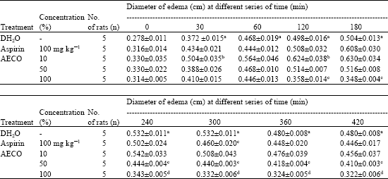 | |
| aSignificant (p<0.05) when compared against the DH2O-treated group of the same row at 0 min interval c, dSignificant (p<0.05) when compared against the DH2O-treated group of the respective column b Low dose of AECO that induced inflammation | |
| Table 2: | The anti-pyretic activity of aqueous extract of Corchorus olitorius leaves in rats |
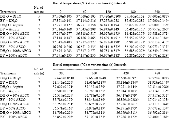 | |
| aSignificant (p<0.05) when compared to the (BY+ DH2O)-treated group of the same row at 0 h intervals, bSignificant (p<0.05) when compared to the (DH2O + DH2O)-treated group of the respective column, cSignificant (p<0.05) when compared to the (BY + DH2O)-treated group of the respective column | |
The Antipyretic Activity
From the Table 2 the AECO was found to show antipyretic activity at all concentrations (10, 50 and 100% concentration) used. All concentrations of extracts were found to produce significant antipyretic activity, which lasted until the end of the experiments (420 min). ASA was found to produce antipyretic effect 180 min after its administration.
DISCUSSION
In the present study, the anti-inflammatory and antipyretic activities of AECO were investigated. Earlier study has scientifically proven that the AECO possesses peripheral and central analgesic properties, which occur in a concentration-independent manner (Zakaria et al., 2005). Interestingly, the highest concentration of AECO was found to exhibit a total loss of analgesic activity when assessed using the abdominal constriction test, but not the hot plate test. The observed concentration-response relationship is in line with statement made by Tripathi (2001) who claimed that the presence of high concentrations of drugs active principle can sometimes lead to the reduction in its effectiveness. Furthermore, according to Tripathi (2001) the observed activity or relationship of AECO also indicates that the concentrations used in the abdominal constriction test have to be within the therapeutic window of AECO in which certain drugs exert their maximum curative effect. In addition, our observation can also be supported by Katzung (1995) who states that there is a possibility that the lost in analgesic activity of certain drugs, at their highest concentration used, may be due to the deactivation of the antinociceptive-inducing receptors within the peritoneal cavity as a result of the presence of high concentration of the respective extract bioactive compounds. However, further studies need to be carried out before we can clarify on the observed activity of AECO.
One of the tests widely used for screening of new anti-inflammatory agents is the carrageenan-induced rat paw edema (Chan et al., 1995; Amanlou et al., 2005). The production of edema is attributed to the participation of kinins and polymorphonuclear leucocytes with their pro-inflammatory factors, including prostaglandins (Amanlou et al., 2005; Di Rosa et al., 1971; Ferreira et al., 1973). Although in this anti-inflammatory study we only measured thickness of the hind paw after intraplantar injection of carrageenan, as previously used by many researchers (Niemegeer et al., 1964; Joseph et al., 2005; Freshwater et al., 2002) and did not involve the use of plethysmograph that involve measurement of volume difference between before and after carrageenan administration (Sulaiman et al., 2004), the significant results obtained after the administration of carrageenan when compared to that of DH2O do indicate the presence of edema and should not be excluded from the study. The method we used to assess the inflammatory activity of AECO has been used by other researchers recently (Di Meglio et al., 2005) and our finding does indicate the presence of anti-inflammatory activity in AECO. Present study has showed that the AECO possess concentration-dependent anti-inflammatory activity indicated by its ability to reduce thickness of rats paw edema. The significant anti-inflammatory activity was observed at the 25% concentration AECO with the lowest concentration used (10% concentration) induced inflammatory activity between 30 to 120 min.
Our study on AECO antipyretic activity has demonstrated that the extract possesses a concentration-independent activity, which was observed at the lowest concentration used (10% concentration). The activity in all concentration of extract used were observed after 120 min, which lasted until the end of the experiments. The ability of the extracts to lower the temperature at the interval of 240 min when brewer’s yeast (BY) alone failed to significantly increase the rectal temperature might be due to its ability to induce hypothermia on its own. This suggestion is supported by our additional finding on the AECO ability to reduce normal rats’ body temperature when given alone at all doses tested (DH2O + AECO). Furthermore, as can be seen from Table 2 this phenomenon can be observed after 120 min of the extracts administration. This finding seems to suggest that the AECO possesses an ability to affect the temperature regulating center in the brain and thus bringing down the normal body temperature or, in other words, it can act as a body cooling solution. In many part of the world, including Malaysia, coconut juice is drunk to give a body cooling effect to fight hot and humid condition (Fergus and Trijntje, 2005). Thus, our finding might also suggest the used of C. olitorius extract as body cooling drink.
Although isolation and purification of different fraction of AECO and assaying anti-inflammatory and antipyretic, as well as analgesic, activities of each fraction was not the main objective of this study, nevertheless, based on the results of the study, we can suggest that the anti-inflammatory and antipyretic activities of AECO may be attributed to inhibition of prostaglandin release or blocking of the enzyme, cyclo-oxygenase, that is responsible for prostaglandin production and similar mediators involved in this test (Di Rosa et al., 1971; Spector, 1962). In addition, the said activity may also be related partly to the presence of various types of bioactive compounds, such as flavonoids, steroids, essential oil and tannin, which have been reported to exhibit the said activities (Peres et al., 1998; Calixto et al., 2000).
There are many reports on the various dosages of reference drugs, in this case morphine and ASA, used for comparison against the tested solutions, either crude extracts (Somchit et al., 2004; Shanmugasundaram and Venkataraman, 2005), isolated active compounds (Idid et al., 1992) or synthetic drugs (Swedberg et al., 1997; Gray et al., 1998), respectively. Depending on the route of administration, Somchit et al. (2004) used 10 mg kg-1 morphine and 100 mg kg-1 ASA (IP) while Idid et al. (1992) and Shanmugasundaram and Venkataraman (2005) used the respective 5 mg kg-1 morphine (SC) and 100 mg kg-1 ASA (PO) as positive controls in the abdominal constriction test. On the other hand, Suaudeau et al. (1998) and Swedberg et al. (1997) used 2 and 30 mg kg-1 morphine (SC), respectively, as their positive control in the hot plate test. In the anti-inflammatory study, reports on used of ASA as positive control also differs based on the route of administration. For examples, Raji et al. (2002) and Sulaiman et al. (2004) used 150 mg kg-1 ASA (IP) and 100 mg kg-1 ASA (IP), respectively, while Chakraborty et al. (2004) used 100 mg kg-1 ASA (PO). In the antipyretic study, several different dosages of ASA have also been reported to be used by researchers like Reanmongkol et al. (2002) and Ahmadiani et al. (2001) who used 200 mg kg-1 (PO) and 300 mg kg-1 (IP) ASA, respectively. The reason for us to use 0.8 and 5 mg kg-1 morphine in the abdominal constriction and hot plate tests, respectively and 100 mg kg-1 ASA in the anti-inflammatory and antipyretic tests, which differs from previous reports was because we are trying to establish our own laboratory control group data. In our study, morphine at the dose of 0.8 mg kg-1 only produced significant antinociceptive activity in abdominal constriction test but not hot plate test, which leads us to change the dose of morphine to 5 mg kg-1 for the latter study. The reason for us to give ASA subcutaneously, but not intraperitoneally, as reported by Raji et al. (2002) and Ahmadiani et al. (2001) and Sulaiman et al. (2004), or orally, as reported by Reanmongkol et al. (2002), was because we would like to minimize contact with and thus to avoid injury of internal organ since acetic acid and brewer’s yeast were also given intraperitoneally. Furthermore, by giving all drugs and extract through one route (SC) can give us the opportunity to compare their effects together and show their relative potency.
In any case, the consistent of the result of this study with ASA activity supports the folklore use of C. olitorius as anti-inflammatory and antipyretic agent. In conclusion, present finding suggests that the C. olitorius extract possesses anti-inflammatory and anti-pyretic activities, which may explain the traditional basis of using this plant in the treatment of various ailments. Further studies are being carried out in our laboratory to elucidate the exact mechanism behind its traditional effects and to analyze its chemical constituents.
ACKNOWLEDGMENTS
This study was supported by the Research Grant of Universiti Industri Selangor, Malaysia. (Project Code No: 03013; Project Vote No: 3090103013). The authors would like to thank Universiti Putra Malaysia for the facilities and Mr. Hanan Kumar for his skillful help in the preparation of the manuscript.
REFERENCES
- Ahmadiani, A., M. Javan, S. Semnanian, E. Barat and M. Kamalinejad, 2001. Anti-inflammatory and antipyretic effects of Trigonella foenum-graecum leaves extract in the rat. J. Ethnopharmacol., 75: 283-286.
CrossRefDirect Link - Chakraborty, A., R.K.B. Devi, S. Rita, K.H. Sharatchandra and T.I. Singh, 2004. Preliminary studies on anti inflammatory and analgesic activities of Spilanthes acmella in experimental animal models. Indian J. Pharacol., 36: 148-150.
Direct Link - Chen, Y.F., H.Y. Tsai and T.S. Wu, 1995. Anti-inflammatory and analgesic activities from roots of Angelica pubescens. Planta Medica, 61: 2-8.
CrossRefPubMedDirect Link - Di Meglio, P., A. Ianaro and S. Ghosh, 2005. Amelioration of acute inflammation by systemic administration of a cell-permeable peptide inhibitor of NF-κB activation. Arthritis Rheum., 52: 951-958.
Direct Link - Di Rosa, M., J.P. Giroud and D.A. Willoughby, 1971. Studies of the mediators of the acute inflammatory response induced in rat in different site by carrageenan and turpentine. J. Pathol., 104: 15-29.
PubMed - Ferreira, S.H., S. Moncada and J.R. Van, 1973. Some effects of inhibiting endogenous prostaglandinformation on the responses of the cat spleen. Br. J. Pharmacol., 47: 48-58.
PubMedDirect Link - Freshwater, J.D., C.I. Svensson, A.B. Malmberg and N.A. Calcutt, 2002. Elevated spinal cyclo-oxygenase and prostaglandin release during hyperalgesia in diabetic rats. Diabetes, 51: 2249-2255.
Direct Link - Gray, A.M., P.S.J. Spencer and R.D.E. Sewell, 1998. The involvement of the opioidergic system in the antinociceptive mechanism of action of antidepressant compounds. Br. J. Pharmacol., 124: 669-674.
CrossRef - Negm, S., O. EI-Shabrawy, M. Arbid and A.S. Radwan, 1980. Toxicological study of the different organs of Corchorus olitorius L. plant with special reference to their cardiac glycosides content. Z. Ernahrungswiss., 19: 28-32.
CrossRef - Peres, M.T.L.P., F. Delle-Monache, M.G. Pizollatti, A.R.S. Santos, A. Beirith, J.B. Calixto and R.A. Yunes, 1998. Analgesic compounds of Croton urucurana Baillon. Pharmacochemical criteria used in their isolation. Phytother. Res., 12: 209-211.
Direct Link - Raji, Y., U.S. Udoh, O.O. Oluwadara, O.S. Akinsomisoye, O. Awobajo and K. Adeshoga, 2002. Anti-inflammatory and analgesic properties of the rhizome extract of Zingiber officinale. Afr. J. Biomed. Res., 5: 121-124.
Direct Link - Reanmongkol, W., S. Subhadhirasakul, C. Pairat, C. Poungsawai and W. Choochar, 2002. Analgesic activity of Dyera costulata extract in experimental animals. Songklanakarin J. Sci. Technol., 24: 227-234.
Direct Link - Shanmugasundaram, P. and S. Venkataraman, 2005. Antinciceptive activity of Hygrophila auriculata (Schum). Heine. Afr. J. Trad. CAM, 2: 62-69.
Direct Link - Sulaiman, M.R., M.N. Somchit, D.A. Israf, Z. Ahmad and S. Moin, 2004. Analgesic effect of Melastoma malabathricum ethanolic extract in mice. Fitoterapia, 75: 667-672.
Direct Link - Swedberg, M.D.B., M.J. Sheardown, P. Sauerberg, P.H. Olesen and P.D. Suzdak et al., 1997. Butylthio[2.2.2] (NNC 11-1053/LY297802): An orally active muscarinic agonist analgesic. J. Pharmacol. Exp. Ther., 281: 876-883.
Direct Link - Zakaria, Z.A., M. Safarul, R. Valsala, M.R. Sulaiman, C.A. Fatimah and A.M. Mat-Jais, 2005. Influence of temperature on the opioid-mediated antinociceptive activity of Corchorus olitorius L. in mice. Naunyn Schmiedeberg's Arch. Pharmacol., 372: 55-62.
CrossRefDirect Link